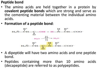
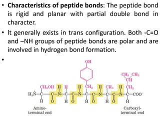
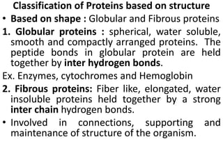
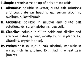
• Proteins are the most abundant organic molecules
of the living system.
• They occur in every part of the cell and constitute
about 50% of the cellular dry weight.
• Proteins form the fundamental basis of structure
and function of life.
• Functions of proteins
• Proteins perform a great variety of specialized and
essential functions in the living cells.
• These functions may be broadly grouped as static
(structural) and dynamic.
• Structural functions : are primarily responsible for
structure and strength of body.
• These include collagen and elastin found in bone
matrix, vascular system and other organs and α-
keratin present in epidermal tissues.
• Dynamic functions : include proteins acting as
enzymes, hormones, blood clotting factors,
immunoglobulins, membrane receptors, storage
proteins, besides their function in genetic control,
muscle contraction, respiration etc.
Elemental composition of Proteins
• Five major elements in the following proportion.
• Besides the above, proteins may also contain othere
lementss uch as P, Fe,C u, l, Mg, Mn, Zn etc.
• Proteins on complete hydrolysis( with concentrated
HCI for several hours) yield L— α—amino acids.
• Therefore, proteins are the polymers of L -α-amino
acids.
• Amino acids are building blocks of proteins.
• STANDARD AMINO ACIDS
• As many as 300 amino acids occur in nature
• Of these, only 20 known as standard amino acids
are repeatedly found in the structure of proteins,
isolated from different forms of life animal, plant
and microbial.
• This is because of the universal nature of the
genetic code available for the incorporation of only
20 amino acids
• After the synthesis of proteins, some of the
incorporated amino acids undergo modifications to
form their derivatives.
• Amino acids are a group of organic compounds
containing two functional groups amino and
carboxyl.
• The amino group (-NH2) is basic while the carboxyl
group (-COOH) is acidic in nature.
General structure of amino acids
• The amino acids are termed as α-amino acids, if
both the carboxyl and amino groups are attached to
the same carbon atom.
• The α - carbon atom binds to a side chain
represented by R which is different for each of the
20 amino acids found in proteins.
• The amino acids mostly exist in the ionized form in
the biological system
Optical isomers of amino acids
• lf a carbon atom is attached to 4 different groups, it
is asymmetric and therefore exhibits optical
isomerism.
• The amino acids (except glycine) possess four
distinct groups (R, H, COO-, NH;) held by α-carbon.
• Thus all the amino acids (except glycine) have
optical isomers.
• The structures of L- and D-amino acids are written
based on the configuration of L- and D-
glyceraldehyde.
R and S nomenclature
• For compounds with more than one chiral center,
the most useful system of nomenclature is the RS
system.
• In this system, each group attached to a chiral
carbon is assigned a priority.
• The priorities of some common substituents are
• For naming in the RS system, the chiral atom is
viewed with the group of lowest priority pointing
away from the viewer.
• If the priority of the other three groups decreases in
clockwise order, the configuration is (R) (Latin
rectus, “right”);
• if in counterclockwise order, the configuration is (S)
(Latin sinister, “left”).
Classification of amino acids
• There are different ways of classifying the amino
acids based on the structure and chemical nature,
nutritional requirement, metabolic fate etc.
A. Amino acid classification based on the structure :
• A comprehensive classification of amino acids is
based on their structure and chemical nature.
• Each amino acid is assigned a 3 letter or 1 letter
symbol.
• The 20 amino acids found in proteins are divided
into seven distinct groups.
B. Classification of amino acids based on polarity :
classified into 4 groups
• The polarity in turn reflects the functional role of
amino acids in protein structure.
1. Non-polar amino acids( neutral) : also referred to
as hydrophobic .
• They have no charge on the 'R' group.
• Ex. alanine, leucine, isoleucine, valine, methionine,
phenylalanine, tryptophan and proline.
2. Polar amino acids with no charge on 'R‘ group :
• These as such, carry no charge on the 'R'group. But
possess groups such as hydroxyl, sulfhydryl and
amide and participate in hydrogen bonding of
protein structure.
• Ex. glycine, serine, threonine, cysteine, glutamine,
asparagine and tyrosine.
3. Polar amino acids with positive 'R' group or basic
amino acids :
• Ex. lysine, arginine and histidine
4. Polar amino acids with negative 'R'group or acidic
amino acids :
• Ex. The dicarboxylic monoamino acids aspartic acid
and glutamic acid
C. Nutritional classification of amino acids :
• The 20 amino acids are required for the synthesis of
variety of proteins, besides other biological
functions.
• However, all these 20 amino acids need not be
taken in the diet.
• Based on the nutritional requirements amino acids
are grouped into two classes Essential and
nonessential
1. Essential or indispensable amino acids :
• The amino acids which cannot be synthesized by the
body and, therefore, need to be supplied through the
diet are called essential amino acids.
• They are required for proper Growth and maintenance
of the individual.
• 10 essential amino acids are Arginine, Valine, Histidine,
lsoleucine, Leucine, Lysine, Methionine, Phenylalanine,
Threonine, Tryptophan
• HILL, MAPV, TT.
• The 2 aa namely arginine and histidine can be
synthesized by adults and not by growing children,
hence these are considered as semi-essential aa.
• Thus, 8 amino acids are absolutely essential while 2
are semi-essential.
2. Non-essential or dispensable amino acids :
• The body can synthesize these amino acids to meet
the biological needs, hence they need not be
consumed in the diet.
• These are-glycine, alanine, serine, cysteine,
aspartate,asparagine, glutamate, glutamine,
tyrosine and proline.
D. Amino acid classification based on their metabolic
fate :
• The carbon skeleton of amino acids can serve as
precursor for the synthesis of glucose(glycogenic) or
fat (ketogenic) or both.
• Are divided into 3 groups.
1.Glycogenic amino acids : can serve as precursors for
the formation of glucose or glycogen.
• e.g. alanine,aspartate, glycine, methionine etc.
2. Ketogenic amino acids : Fat can be synthesized from
these amino acids.
• 2 amino acids leucine and lysine are exclusively
ketogenic.
3. Glycogenic and ketogenic amino acids : The 4 amino
acids isoleucine, Phenyl alanine, tryptophan, tyrosine
are precursors for synthesis of glucose as well as fat.
Properties of amino acids
• The amino acids differ in their physico - chemical
properties which ultimately determine the
characteristics of proteins.
A. Physical properties
1. Solubility : Most of the amino acids are usually
soluble in water and insoluble in organic solvents.
2. Melting points: Amino acids generally melt at
higher temperatures often above 200oC.
3. Taste: Amino acids may be sweet (Gly, Ala, Val),
tasteless (Leu) or bitter (Arg, lle).
• Monosodium glutamate (ajinomoto) is used as a
flavoring agent in food industry
4. Optical properties: All the amino acids except
glycine possess optical isomers due to the presence
of asymmetric carbon atom.
• Some amino acids also have a second asymmetric
carbon e.g. isoleucine, threonine.
5. Amino acids as ampholytes : Amino acids contain
both acidic (-COOH) and basic (-NH2) groups and
can donate or accept a proton, hence regarded as
ampholytes
• Zwitterion or dipolar ion : It is a hybrid molecule
containing both +ve and -ve ionic groups.
• In strongly acidic pH (low pH), the amino acid is
+vely charged (cation)
• while in strongly alkaline pH (high pH), it is -vely
charged( anion).
• Each amino acid has a characteristic pH (e.g.
leucine, pH 6.0) at which it carries both positive and
negative charges and exists as zwitterion
• Isoelectric pH is defined as the pH at which a
molecule exists as a zwitterion and carries no net
charge and Thus the moleculei s electrically neutral
Titration of amino acids : The existence of different
ionic forms of amino acids can be more easily
understood by the titration curves.
• At low pH, leucine exists in a fully protonated form
as cation.
• As the titration proceeds with NaOH, leucine loses
its protons and at isoelectric pH (pl), it becomes a
zwitterion.
• Further titration results in the formation of anionic
form of leucine.
B. Chemical properties
• The general reactions of aa are mostly due to the
presence of 2 functional groups carboxyl (-COOH) and
amino (-NH2) group.
• Reactions due to -COOH group
1. Amino acids form salts (-COONa) with bases and
esters (-COOR') with alcohols.
2. Decarboxylation: Amino acids undergo
decarboxylationto produce corresponding amines.
• This reaction assumes significance in the living cells due
to the formation of many biologically important
amines.
• These include histamine, tyramine and GABA from the
amino acids histidine, tyrosine and glutamate,
respectively.
Reactions due to -NH2 group
1. The amino groups behave as bases and combine
with acids (e.g. HCI) to form salts (-NH3Cl-).
2. Reaction with ninhydrin : The α-amino acids react
with ninhydrin to form a purple, blue or pink colour
complex (Ruhemann's purple).
3. Reaction with Formaldehyde(Formal titration)
• A test tube of neutral formaldehyde is added to the
conical flask containing aa and titrated against
alkali.
• Neutral formaldehyde blocks the amino group of aa
in the form of Bishydroxy methyl derivative. This
allows the reaction b/n COOH group and NaOH
solution.
4. Reaction with Nitrous acid
Amino group reacts with Nitrous acid to liberate
Nitrogen and α – hydroxy acid is formed
5. Reaction with CO2
Amino group reacts with CO2to form N – Carboxy
amino acid.
• This reaction is of physiological significance as CO2
combines with free amino group of Hb tranporting
20% of CO2.
6. Reaction with 2,4 - Dinitro Fluorobenzene(DNFB)
• This reagent is also known as sanger’s reagent used
to determine the N – terminal residues of peptide
chain.
7. Reaction with Edman reagent
• Phenyl isothiocyanate, used to determine the N –
terminal residues of peptide chain.
+
• Colour Reactions of amino acids
Peptide bond
• The amino acids are held together in a protein by
covalent peptide bonds which are strong and serve as
the cementing material between the individual amino
acids.
• Formation of a peptide bond:
• A dipeptide will have two amino acids and one peptide
bond.
• Peptides containing more than 10 amino acids
(decapeptide) are referred to as polypeptides.
• Characteristics of peptide bonds: The peptide bond
is rigid and planar with partial double bond in
character.
• lt generally exists in trans configuration. Both -C=O
and –NH groups of peptide bonds are polar and are
involved in hydrogen bond formation.
•
 The conformational flexibility of peptide
chains is limited only to rotation about C - C
bond, due to the rigidity of peptide bond which
keeps the peptide links relatively planar and
resistant to conformational change
Classification of Proteins based on structure
• Based on shape : Globular and Fibrous proteins
1. Globular proteins : spherical, water soluble,
smooth and compactly arranged proteins. The
peptide bonds in globular protein are held
together by inter hydrogen bonds.
Ex. Enzymes, cytochromes and Hemoglobin
2. Fibrous proteins: Fiber like, elongated, water
insoluble proteins held together by a strong
inter chain hydrogen bonds.
• Involved in connections, supporting and
maintenance of structure of the organism.
Fibrous proteins
(i) Collagens are connective tissue proteins lacking
tryptophan. Collagens, on boiling with water or
dilute acids, yield gelatin which is soluble and
digestible.
(ii) Elastins: These proteins are found in elastic tissues
such as tendons and arteries.
(iii) Keratins: These are present in exoskeletal
structures. ex. hair, nails, horns.
Human hair keratin contains as much as 14% cysteine.
Classification based on composition into 3 classes
1. Simple proteins: are made up of only α-amino
acids
2. Conjugated proteins: amino acids and non amino
acids.
• The non–amino acid part of a conjugated protein is
usually called its prosthetic group.
1. Derived proteins : These are the hydrolytic
products of protein hydrolysis.
• These include peptones, polypeptides and peptide
l. Simple proteins: made up of only amino acids
I. Albumins: Soluble in water, dilute salt solutions
and coagulate on heating. ex. serum albumin,
ovalbumin, lactalbumin.
II. Globulins: Soluble in neutral and dilute salt
solutions. ex. serum globulins, egg yolk.
III. Glutelins: soluble in dilute acids and alkalies and
are coagulated by heat, mostly found in plants. Ex.
Glutelins, oryzenin (rice).
IV. Prolamines: soluble in 70% alcohol, insoluble in
water, rich in proline. Ex. gliadin( wheat),zein
(maize).
V. Histones: Strongly basic proteins, soluble in water
and dilute acids but insoluble in dilute alkali. Ex.
codfish sperm. They are associated with nucleic
acids.
VI. Protamines: They are smaller in size, strongly
basic and soluble in water and dilute acids and
alkalies. Not coagulated by heating. Basic nature
due to the presence of lysine and arginine.
• Also found in association with nucleic acids e.g.
sperm proteins.
VII. Scleroproteins: insoluble in water, salt slutions
and organic solvents.
• Form supporting tissues: collagen, cartilage
2. Coniugated proteins
(a) Nucleoproteins: Nucleic acid (DNA or RNA) is the
prosthetic group. ex. Nucleohistones,
nucleoprotamnies.
(b)Glycoproteins: The prosthetic group is
carbohydrate, which is less than 4% of protein, ex.
mucin(saliva), ovomucoid( egg white), antibodies .
(c) Lipoproteins: Protein found in combination with
lipids as the prosthetic group. ex. Serum
lipoproteins, membrane lipoproteins.
(d) Phosphoproteins: Phosphoric acid is the
prosthetic group e.g. casein (milk), vitelline (egg
yolk).
(e)Chromoproteins: The prosthetic group is coloured
in nature e.g. hemoglobins, cytochromes.
(f) Metalloproteins: These proteins contain metal ions
such as Fe, Co, Zn, Cu, Mg etc., ex. Ceruloplasm(Cu),
carbonic anhydras(Zn)
(g) Flavoproteins: contain Flavin group FMN or FAD.
Succinate dehydrogenase, NADH dehydrogenase.
3. Derived proteins: These are the derived products
during degradation or hydrolysis of simple and
conjugated proteins.
• From the nutritional point of view, proteins are
classified into 3 categories.
1. Complete proteins: These proteins have all the 10
essential aa in the required proportion to promote
good growth. e.g. egg albumin, milk casein.
2. Partially incomplete proteins: These proteins are
partially lacking one or more essential aa and hence
can promote moderate growth. e.g. wheat and rice
proteins (limiting Lys, Thr).
3. Incomplete proteins: These proteins completely lack
one or more essential aa. Hence they do not promote
growth at all Ex. gelatin( lacks Trp), zein (lacks Trp, Lys).
Functional classification
Class Function Example
Enzymes Catalytical All enzymes
St. proteins Support and shape collagen
contractile movement Actin, myosin
defence defence antibodies
transport transport Hb, albumin
Blood protein Clotting, osmolarity Fibrinogen, thrombin
Hormones signalling insulin
respiratory ETC Cytochromes a,b,c
Toxin toxic Venoms
Storage storage Casein, ferritin
vision vision Rhodopsin
• BIOTOGICALLY IMPORTANT PEPTIDES
• Several peptides display a wide spectrum of
biological functions
• Generally the term peptides applied when the
number of constituent amino acids is less than 10.
1. Glutathione: lt is a tripeptide composed of 3
amino acids.
• Chemically glutathione is glutamyl-cysteinyl-
glycine widely distributed in nature and exists in
reduced or
oxidized states.
• Glutathione (reduced) performs specialized
functions in erythrocytes
( i ) lt maintains RBC membrane structure and
integrity.
(ii) lt protects hemoglobin from getting oxidized by
agents such as H2O2.
• Glutathione is involved in the transport of amino
acids in the intestine and kidney tubules
• Glutathione is involved in the detoxication process.
2. Thyrotropin releasing hormone (TRH) : lt is a
tripeptide secreted by hypothalamus.
Pyroglutamate + Histidine + Prolinamide
• TRH stlmulates pituitary gland to release
thyrotropic hormone.
3. Oxytocin: lt is a hormone secreted by posterior
pituitary gland and contains 9 amino acids(
nonapeptide).
• It causes contraction of uterus.
4. Vasopressin (antidiuretic hormone, ADH) : It is also
a nonapeptide produced by posterior pituitary
gland.
• lt stimulates kidneys to retain water and thus
increases the blood pressure.
5. Angiotensin II : Angiotensin I is a decapeptide (10
amino acids) which is converted to angiotensin ll (8
amino acids).
• It is responsible for more hypertensive effect.
• It stimulates the release of aldosterone from
adrenal gland.
Asp-Arg-Val-Tyr-Ile-His-Pro-Phe
Angiotensin II
6. Methionine enkephalin : lt is a pentapeptide found
in the brain and has opiate like function.
• lt inhibits the sense of a pain.
7. Bradykinin and kallidin : They are nona and
decapeptides respectively.
• Both of them act as powerful vasodilators.
• They are produced from plasma proteins by snake
venom enzymes.
8. Peptide antibiotics : Antibiotics such as gramicidin,
bacitracin, tyrocidin and actinomycin are peptide in
nature.
9. Aspartame : lt is a dipeptide (aspartyl
phenylalanine methyl ester), produced by a
combination of aspartic acid and phenylalanine.
• Aspartame is about 200 times sweeter than
sucrose, and is used as a low-calorie artificial
sweetner in soft drink industry.
10. Gastrointestinal hormones : Gastrin, secretin etc.
are the gastro intestinal peptides which serve as
hormones.
• The structure of proteins is rather complex which can
be divided into 4 levels of organization.
1. Primary structure : The linear sequence of amino acids
forming the backbone of proteins (polypeptides).
2. Secondary structure: The spatial arrangement of
protein by twisting of the polypeptide chain.
3. Tertiary structure: The three dimensional structure of a
functional Protein formed by the interaction of
secondary st.
4. Quaternary structure : Some of the proteins are
composed of two or more polypeptide chains referred
to as subunits.
• The spatial arrangement of these subunits is known as
quaternary structure.
• The structural hierarchy of proteins is comparable
with the structure of a building.
• The amino acids may be considered as the bricks,
the wall as the primary structure,
• the twists in a wall as the secondary structure,
• a full-fledged self-contained room as the tertiary
structure.
• A building with similar and dissimilar rooms will be
the quaternary structure.
1. Primary structure of proteins :
• The primary structure comprises the identification
of constituent amino acids and the order in which
they are joined together by peptide bonds.
• H2N – aa1 – aa2 – aa3 – aa4 – aa5…….aan - COOH
• Insulin was the first polypeptide whose primary st.
was elucidated by Frederick Sanger in 1953 along
with many scientists over a period of a decade and
won Nobel prize.
• The primary structure influences the three
dimensional structure and biological activity of the
protein.
• Even a change in the single aa residue may affect
the st and function of the protein.
• Ex. Sickle cell anemia, a inherited disease cause low
oxygen binding capacity.
• 2 – α and 2 – β chains.
• In β chain 6th aa (Glutamate) from N terminal is
replaced by Valine.
• Hb has 574 aa and change in 2 aa changes the st
and fn.
Determination of primary structure
• It involves 3 stages :
1. Determination of amino acid composition: The
protein or polypeptide is completely hydrolysed to
aa by acid or alkali treatment.
• Resulting mixture of aas can be separated by
chromatography and also are quantitatively
estimated.
2. Degradation of protein into smaller fragments: As
Protein is a large molecule with polypeptide chains.
• Separation of polypeptides is essential before
degradation using enzymes or chemical cleavage
(Cyanogen bromide)
3. Determination of amino acid sequence:
• The smaller fragments are utilized for the
determination of sequencoef amino acid.
• This is done in a stepwise manner to finally build up
the order of aa in a protein.
• Sanger's reagent or Edman's reagent or
Sequenator are employed for sequence
determination.
• Sequenator: an automatic machine to determine
the amino acid sequence in a polypeptide.
• Uses Edman's degradation. Aa are determined
sequentially from N-terminal end.
• The PTH aminoacid liberated is identified by HPLC.
SECONDARY STRUCTURE OF PROTEIN
• It refers to the spatial orientation of the polypeptide
chain i.e the folding of polypeptide backbone rather
than the side chains.
• Secondary st is due to the following properties of
peptide bonds
1. Peptide bond is planar and rigid, side chains projecting
from the axis.
2. Partial double bond property which restricts the
rotation around C-N bonds but rotation possible Cα-C.
• Distance b/n C – N bond – 1.325 Ao
• Distance of double bond – 1.24 Ao
• Distance of single bond – 1.47 Ao
3. Hydrogen bonds exist b/n oxygen of C=O and
hydrogen of N-H
• Three common types of secondary structures, α-
helix, β-Pleated structures and Triple helix.
• lndian scientist Ramachandran made a significant
contribution in understanding the spatial
arrangement of polypeptide chains.
• α-Helix : It is the most common spiral sttructure of
protein.
• lt has a rigid arrangement of polypeptide chain
• Proposed by Pauling and Corey(1951) regarded as one
of the milestones in the biochemistry research.
The salient features of α-Helix are
1. α-Helix is tightly packed coiled structure with aa side
chains extending outward from the central axis to avoid
stearic hinderance.
2. α-Helix is stabilized by extensive hydrogen bonding
formed between H atom attached to N , and O atom
attached to C .
• The hydrogen bonds individually weak but collectively
strong enough to stabilize the helix.
3. Each turn of α-helix contains 3 .5 aa and travels a
distance(pitch) of 5.4Ao.
• The spacing of each aa is 0.15nm and the diameter
of helix is 10Ao.
4. α -Helix is a stable conformation formed
spontaneously with the lowest energy.
5. The right handed α -helix is more stable than left
handed helix
6. Certain amino acids( particularly proline and
hydroxy proline) disrupt the α –helix called as helix
breakers.
• Alanine and Cysteine promote helix formation.
• Ex. Myosin, keratin etc.
β – Pleated structures
• This is the second type of structure proposed by
Pauling and Corey.
• β -Pleated sheets are composed of two or more
segments of fully extended peptide chains.
• ln the β -sheets, the hydrogen bonds are formed
between the neighbouring segments of polypeptide
chain(s).
• β -Pleated sheet may be formed either by separate
polypeptide chains (H-bonds are interchain)or a
single polypeptide chain folding back on to itself( H-
bonds are intrachain).
Salient features are:
• Composed of 2 or more peptide chains
• The hydrogen bonds between adjacent strands are
perpendicular to long axis of strands.
• β -Pleated sheet is favoured by non polar as well as
small aas like Val, Ile, Trp, serine etc.
• Proline and Hydroxy Proline serves as Pleated sheet
breakers.
• The polypeptide chains in the β –sheets may be
arranged either in parallel (the same direction) or
anti-parallel(opposite direction)
• Occurrence of β -sheets:
• Many proteins contain β –pleated sheets.
• As such the α-helix and β –sheets are commonly
found in the same protein structure .
• In the globular proteins, β –sheets form the core
structure.
• Ex. Silk fibroin, Enzymes like SOD, Triose phosphate
isomerase etc.
• Triple helix of Collagen: collagen provide strength.
• It is found in connective tissue such as tendons,
cartilage, the organic matrix of bone, and the
cornea of the eye.
• Its secondary structure quite distinct from the α
helix.
• It is left-handed and has 3 aa residues per turn.
• Collagen is also a coiled coil, with three separate
polypeptides supertwisted about each other
• The superhelical twisting is right-handed in
collagen, opposite in sense to the left-handed helix
of the chains.
• They contain about 35% Gly, 11% Ala, and 21% Pro
and 4-Hyp (4-hydroxyproline)
The left-handed polypeptide helices
are twisted together to form a
right-handed superhelical structure.
An individual collagen polypeptide
helix has 3.3 residues per turn and a
pitch of 10.0 Å
The collagen triple helix has 10
Gly-X-Proline/HP units per turn and
a pitch of 86.1 Å.
TERTIARY STRUCTURE OF PROTEIN
• It is the three-dimensional arrangement of
polypeptide chains of the protein.
• Amino acids that are far apart in different types of
secondary structure polypeptides may interact
resulting completely folded structure of a protein.
• lt is a compact structure with hydrophobic side
chains held interior while the hydrophilic groups
are on the surface of the protein molecule.
• This type of arrangement ensures stability of the
molecule.
• Secondary st. often constitute distinct domains and
the tertiary st. describes the relationship b/n
different domains of a protein.
• Bonds of tertiary structure: Besides the hydrogen
bonds, disulfide bonds( -S-S), ionic interactions
(electrostatic bonds), vander waals forces and
hydrophobic interactions also contribute to the
tertiary structure of proteins.
• Hydrogen bonds: The hydrogen bonds are formed
by sharing of hydrogen atoms between the nitrogen
and carbonyl oxygen of different peptide bonds –
peptidal hydrogen bonds.
• Non peptidal hydrogen bonds are formed b/n polar
side chains.
• Each hydrogen bond is weak but collectively they
are strong
• The disulfide bonds may be intra or inter. These
bonds contribute to the structural conformation
and stability of proteins.
• Hydrophobic bonds: The non-polar side chains of
neutral amino acids tend to be closely associated
with each other in Proteins.
• The occurrence of hydrophobic forces is observed
in aqueous environment wherein the molecules are
forced to stay together .
• Electrostatic bonds: These bonds are formed by
interactions between negatively charged R groups
(COO-) of acidic amino acids with positively charged
R groups (NH+
3) of basic amino acids.
• Vander Waals forces: These are the non-covalent
associations formed by the electrostatic interactions
due to permanent or induced dipoles.
QUATERNARY STRUCTURE OF PROTEIN
• A great majority of the proteins are composed of
single polypeptide chains.
• Some of the proteins, however, consist of two or
more polypeptides which may be identical or
unidentical.
• Such proteins are termed as oligomers and posses
quaternary structure.
• The individual polypeptide chains are known as
monomers, protomers or subunits.
• A dimer consists of two polypeptides while a
tetramer has four.
• Bonds in quaternary structure: The monomeric
subunits are held together by hydrogen bonds,
hydrophobic interactions, Vander walls forces and
Disulphide bonds.
• Importance of oligomeric proteins: These proteins
play a significant role in the regulation of
metabolism and cellular function.
• Ex. Hemoglobin, aspartate transcarbomylase,
lactate dehydrogenase
• DENATURATION
• The phenomenon of disorganization of native
protein structure
• Denaturation results in the loss of secondary (but
not primary), tertiary and quaternary structure of
proteins.
• This involves a change in physical, chemical and
biological properties of protein molecules.
• Agents of denaturation
• Physical agents : Heat, violent shaking, X-rays, UV
radiation.
• Chemical agents : Acids, alkalies, organic solvents
(ether, alcohol), salts of heavy metals (Pb, Hg), urea,
salicylate.
Characteristics of denaturation
1. The native structure of protein is lost
2. The primary structure remains intact i.e., peptide
bonds are not hydrolysed.
3. The protein loses its biological activity.
4. Denatured protein becomes insoluble in the
solvent in which it was originally soluble.
5 The viscosity of denatured protein (solution)
increases while its surface tension decreases.
6. Denaturation is associated with loss of hydrogen
and disulfide bonds.
7. Denatured protein is more easily digested due to
increased exposure of peptide bonds to enzymes.
Cooking causes protein denaturation and,
therefore, cooked protein is more easily digested.
8. Denaturation is usually irreversible but sometimes
reversible (renaturation).
• Hb undergoes denaturation in the presence of
salicylate but removal of salicylate renatures Hb.
10. Denatured protein cannot be crystallized

Aminoacids.pptx

  • 1.
    • Proteins arethe most abundant organic molecules of the living system. • They occur in every part of the cell and constitute about 50% of the cellular dry weight. • Proteins form the fundamental basis of structure and function of life. • Functions of proteins • Proteins perform a great variety of specialized and essential functions in the living cells. • These functions may be broadly grouped as static (structural) and dynamic.
  • 2.
    • Structural functions: are primarily responsible for structure and strength of body. • These include collagen and elastin found in bone matrix, vascular system and other organs and α- keratin present in epidermal tissues. • Dynamic functions : include proteins acting as enzymes, hormones, blood clotting factors, immunoglobulins, membrane receptors, storage proteins, besides their function in genetic control, muscle contraction, respiration etc.
  • 3.
    Elemental composition ofProteins • Five major elements in the following proportion. • Besides the above, proteins may also contain othere lementss uch as P, Fe,C u, l, Mg, Mn, Zn etc.
  • 4.
    • Proteins oncomplete hydrolysis( with concentrated HCI for several hours) yield L— α—amino acids. • Therefore, proteins are the polymers of L -α-amino acids. • Amino acids are building blocks of proteins.
  • 5.
    • STANDARD AMINOACIDS • As many as 300 amino acids occur in nature • Of these, only 20 known as standard amino acids are repeatedly found in the structure of proteins, isolated from different forms of life animal, plant and microbial. • This is because of the universal nature of the genetic code available for the incorporation of only 20 amino acids • After the synthesis of proteins, some of the incorporated amino acids undergo modifications to form their derivatives.
  • 6.
    • Amino acidsare a group of organic compounds containing two functional groups amino and carboxyl. • The amino group (-NH2) is basic while the carboxyl group (-COOH) is acidic in nature. General structure of amino acids • The amino acids are termed as α-amino acids, if both the carboxyl and amino groups are attached to the same carbon atom.
  • 7.
    • The α- carbon atom binds to a side chain represented by R which is different for each of the 20 amino acids found in proteins. • The amino acids mostly exist in the ionized form in the biological system
  • 8.
    Optical isomers ofamino acids • lf a carbon atom is attached to 4 different groups, it is asymmetric and therefore exhibits optical isomerism. • The amino acids (except glycine) possess four distinct groups (R, H, COO-, NH;) held by α-carbon. • Thus all the amino acids (except glycine) have optical isomers. • The structures of L- and D-amino acids are written based on the configuration of L- and D- glyceraldehyde.
  • 9.
    R and Snomenclature • For compounds with more than one chiral center, the most useful system of nomenclature is the RS system. • In this system, each group attached to a chiral carbon is assigned a priority. • The priorities of some common substituents are • For naming in the RS system, the chiral atom is viewed with the group of lowest priority pointing away from the viewer. • If the priority of the other three groups decreases in clockwise order, the configuration is (R) (Latin rectus, “right”);
  • 10.
    • if incounterclockwise order, the configuration is (S) (Latin sinister, “left”).
  • 11.
    Classification of aminoacids • There are different ways of classifying the amino acids based on the structure and chemical nature, nutritional requirement, metabolic fate etc. A. Amino acid classification based on the structure : • A comprehensive classification of amino acids is based on their structure and chemical nature. • Each amino acid is assigned a 3 letter or 1 letter symbol. • The 20 amino acids found in proteins are divided into seven distinct groups.
  • 19.
    B. Classification ofamino acids based on polarity : classified into 4 groups • The polarity in turn reflects the functional role of amino acids in protein structure. 1. Non-polar amino acids( neutral) : also referred to as hydrophobic . • They have no charge on the 'R' group. • Ex. alanine, leucine, isoleucine, valine, methionine, phenylalanine, tryptophan and proline.
  • 20.
    2. Polar aminoacids with no charge on 'R‘ group : • These as such, carry no charge on the 'R'group. But possess groups such as hydroxyl, sulfhydryl and amide and participate in hydrogen bonding of protein structure. • Ex. glycine, serine, threonine, cysteine, glutamine, asparagine and tyrosine.
  • 21.
    3. Polar aminoacids with positive 'R' group or basic amino acids : • Ex. lysine, arginine and histidine 4. Polar amino acids with negative 'R'group or acidic amino acids : • Ex. The dicarboxylic monoamino acids aspartic acid and glutamic acid
  • 22.
    C. Nutritional classificationof amino acids : • The 20 amino acids are required for the synthesis of variety of proteins, besides other biological functions. • However, all these 20 amino acids need not be taken in the diet. • Based on the nutritional requirements amino acids are grouped into two classes Essential and nonessential
  • 23.
    1. Essential orindispensable amino acids : • The amino acids which cannot be synthesized by the body and, therefore, need to be supplied through the diet are called essential amino acids. • They are required for proper Growth and maintenance of the individual. • 10 essential amino acids are Arginine, Valine, Histidine, lsoleucine, Leucine, Lysine, Methionine, Phenylalanine, Threonine, Tryptophan • HILL, MAPV, TT. • The 2 aa namely arginine and histidine can be synthesized by adults and not by growing children, hence these are considered as semi-essential aa. • Thus, 8 amino acids are absolutely essential while 2 are semi-essential.
  • 24.
    2. Non-essential ordispensable amino acids : • The body can synthesize these amino acids to meet the biological needs, hence they need not be consumed in the diet. • These are-glycine, alanine, serine, cysteine, aspartate,asparagine, glutamate, glutamine, tyrosine and proline.
  • 25.
    D. Amino acidclassification based on their metabolic fate : • The carbon skeleton of amino acids can serve as precursor for the synthesis of glucose(glycogenic) or fat (ketogenic) or both. • Are divided into 3 groups. 1.Glycogenic amino acids : can serve as precursors for the formation of glucose or glycogen. • e.g. alanine,aspartate, glycine, methionine etc. 2. Ketogenic amino acids : Fat can be synthesized from these amino acids. • 2 amino acids leucine and lysine are exclusively ketogenic. 3. Glycogenic and ketogenic amino acids : The 4 amino acids isoleucine, Phenyl alanine, tryptophan, tyrosine are precursors for synthesis of glucose as well as fat.
  • 26.
    Properties of aminoacids • The amino acids differ in their physico - chemical properties which ultimately determine the characteristics of proteins. A. Physical properties 1. Solubility : Most of the amino acids are usually soluble in water and insoluble in organic solvents. 2. Melting points: Amino acids generally melt at higher temperatures often above 200oC. 3. Taste: Amino acids may be sweet (Gly, Ala, Val), tasteless (Leu) or bitter (Arg, lle). • Monosodium glutamate (ajinomoto) is used as a flavoring agent in food industry
  • 27.
    4. Optical properties:All the amino acids except glycine possess optical isomers due to the presence of asymmetric carbon atom. • Some amino acids also have a second asymmetric carbon e.g. isoleucine, threonine. 5. Amino acids as ampholytes : Amino acids contain both acidic (-COOH) and basic (-NH2) groups and can donate or accept a proton, hence regarded as ampholytes
  • 28.
    • Zwitterion ordipolar ion : It is a hybrid molecule containing both +ve and -ve ionic groups. • In strongly acidic pH (low pH), the amino acid is +vely charged (cation) • while in strongly alkaline pH (high pH), it is -vely charged( anion). • Each amino acid has a characteristic pH (e.g. leucine, pH 6.0) at which it carries both positive and negative charges and exists as zwitterion • Isoelectric pH is defined as the pH at which a molecule exists as a zwitterion and carries no net charge and Thus the moleculei s electrically neutral
  • 30.
    Titration of aminoacids : The existence of different ionic forms of amino acids can be more easily understood by the titration curves. • At low pH, leucine exists in a fully protonated form as cation. • As the titration proceeds with NaOH, leucine loses its protons and at isoelectric pH (pl), it becomes a zwitterion. • Further titration results in the formation of anionic form of leucine.
  • 32.
    B. Chemical properties •The general reactions of aa are mostly due to the presence of 2 functional groups carboxyl (-COOH) and amino (-NH2) group. • Reactions due to -COOH group 1. Amino acids form salts (-COONa) with bases and esters (-COOR') with alcohols. 2. Decarboxylation: Amino acids undergo decarboxylationto produce corresponding amines. • This reaction assumes significance in the living cells due to the formation of many biologically important amines. • These include histamine, tyramine and GABA from the amino acids histidine, tyrosine and glutamate, respectively.
  • 33.
    Reactions due to-NH2 group 1. The amino groups behave as bases and combine with acids (e.g. HCI) to form salts (-NH3Cl-). 2. Reaction with ninhydrin : The α-amino acids react with ninhydrin to form a purple, blue or pink colour complex (Ruhemann's purple).
  • 34.
    3. Reaction withFormaldehyde(Formal titration) • A test tube of neutral formaldehyde is added to the conical flask containing aa and titrated against alkali. • Neutral formaldehyde blocks the amino group of aa in the form of Bishydroxy methyl derivative. This allows the reaction b/n COOH group and NaOH solution.
  • 35.
    4. Reaction withNitrous acid Amino group reacts with Nitrous acid to liberate Nitrogen and α – hydroxy acid is formed 5. Reaction with CO2 Amino group reacts with CO2to form N – Carboxy amino acid. • This reaction is of physiological significance as CO2 combines with free amino group of Hb tranporting 20% of CO2.
  • 36.
    6. Reaction with2,4 - Dinitro Fluorobenzene(DNFB) • This reagent is also known as sanger’s reagent used to determine the N – terminal residues of peptide chain. 7. Reaction with Edman reagent • Phenyl isothiocyanate, used to determine the N – terminal residues of peptide chain. +
  • 37.
    • Colour Reactionsof amino acids
  • 38.
    Peptide bond • Theamino acids are held together in a protein by covalent peptide bonds which are strong and serve as the cementing material between the individual amino acids. • Formation of a peptide bond: • A dipeptide will have two amino acids and one peptide bond. • Peptides containing more than 10 amino acids (decapeptide) are referred to as polypeptides.
  • 39.
    • Characteristics ofpeptide bonds: The peptide bond is rigid and planar with partial double bond in character. • lt generally exists in trans configuration. Both -C=O and –NH groups of peptide bonds are polar and are involved in hydrogen bond formation. •
  • 40.
     The conformationalflexibility of peptide chains is limited only to rotation about C - C bond, due to the rigidity of peptide bond which keeps the peptide links relatively planar and resistant to conformational change
  • 41.
    Classification of Proteinsbased on structure • Based on shape : Globular and Fibrous proteins 1. Globular proteins : spherical, water soluble, smooth and compactly arranged proteins. The peptide bonds in globular protein are held together by inter hydrogen bonds. Ex. Enzymes, cytochromes and Hemoglobin 2. Fibrous proteins: Fiber like, elongated, water insoluble proteins held together by a strong inter chain hydrogen bonds. • Involved in connections, supporting and maintenance of structure of the organism.
  • 42.
    Fibrous proteins (i) Collagensare connective tissue proteins lacking tryptophan. Collagens, on boiling with water or dilute acids, yield gelatin which is soluble and digestible. (ii) Elastins: These proteins are found in elastic tissues such as tendons and arteries. (iii) Keratins: These are present in exoskeletal structures. ex. hair, nails, horns. Human hair keratin contains as much as 14% cysteine.
  • 43.
    Classification based oncomposition into 3 classes 1. Simple proteins: are made up of only α-amino acids 2. Conjugated proteins: amino acids and non amino acids. • The non–amino acid part of a conjugated protein is usually called its prosthetic group. 1. Derived proteins : These are the hydrolytic products of protein hydrolysis. • These include peptones, polypeptides and peptide
  • 44.
    l. Simple proteins:made up of only amino acids I. Albumins: Soluble in water, dilute salt solutions and coagulate on heating. ex. serum albumin, ovalbumin, lactalbumin. II. Globulins: Soluble in neutral and dilute salt solutions. ex. serum globulins, egg yolk. III. Glutelins: soluble in dilute acids and alkalies and are coagulated by heat, mostly found in plants. Ex. Glutelins, oryzenin (rice). IV. Prolamines: soluble in 70% alcohol, insoluble in water, rich in proline. Ex. gliadin( wheat),zein (maize).
  • 45.
    V. Histones: Stronglybasic proteins, soluble in water and dilute acids but insoluble in dilute alkali. Ex. codfish sperm. They are associated with nucleic acids. VI. Protamines: They are smaller in size, strongly basic and soluble in water and dilute acids and alkalies. Not coagulated by heating. Basic nature due to the presence of lysine and arginine. • Also found in association with nucleic acids e.g. sperm proteins. VII. Scleroproteins: insoluble in water, salt slutions and organic solvents. • Form supporting tissues: collagen, cartilage
  • 46.
    2. Coniugated proteins (a)Nucleoproteins: Nucleic acid (DNA or RNA) is the prosthetic group. ex. Nucleohistones, nucleoprotamnies. (b)Glycoproteins: The prosthetic group is carbohydrate, which is less than 4% of protein, ex. mucin(saliva), ovomucoid( egg white), antibodies . (c) Lipoproteins: Protein found in combination with lipids as the prosthetic group. ex. Serum lipoproteins, membrane lipoproteins. (d) Phosphoproteins: Phosphoric acid is the prosthetic group e.g. casein (milk), vitelline (egg yolk).
  • 47.
    (e)Chromoproteins: The prostheticgroup is coloured in nature e.g. hemoglobins, cytochromes. (f) Metalloproteins: These proteins contain metal ions such as Fe, Co, Zn, Cu, Mg etc., ex. Ceruloplasm(Cu), carbonic anhydras(Zn) (g) Flavoproteins: contain Flavin group FMN or FAD. Succinate dehydrogenase, NADH dehydrogenase.
  • 48.
    3. Derived proteins:These are the derived products during degradation or hydrolysis of simple and conjugated proteins. • From the nutritional point of view, proteins are classified into 3 categories. 1. Complete proteins: These proteins have all the 10 essential aa in the required proportion to promote good growth. e.g. egg albumin, milk casein. 2. Partially incomplete proteins: These proteins are partially lacking one or more essential aa and hence can promote moderate growth. e.g. wheat and rice proteins (limiting Lys, Thr). 3. Incomplete proteins: These proteins completely lack one or more essential aa. Hence they do not promote growth at all Ex. gelatin( lacks Trp), zein (lacks Trp, Lys).
  • 49.
    Functional classification Class FunctionExample Enzymes Catalytical All enzymes St. proteins Support and shape collagen contractile movement Actin, myosin defence defence antibodies transport transport Hb, albumin Blood protein Clotting, osmolarity Fibrinogen, thrombin Hormones signalling insulin respiratory ETC Cytochromes a,b,c Toxin toxic Venoms Storage storage Casein, ferritin vision vision Rhodopsin
  • 50.
    • BIOTOGICALLY IMPORTANTPEPTIDES • Several peptides display a wide spectrum of biological functions • Generally the term peptides applied when the number of constituent amino acids is less than 10. 1. Glutathione: lt is a tripeptide composed of 3 amino acids. • Chemically glutathione is glutamyl-cysteinyl- glycine widely distributed in nature and exists in reduced or oxidized states.
  • 51.
    • Glutathione (reduced)performs specialized functions in erythrocytes ( i ) lt maintains RBC membrane structure and integrity. (ii) lt protects hemoglobin from getting oxidized by agents such as H2O2. • Glutathione is involved in the transport of amino acids in the intestine and kidney tubules • Glutathione is involved in the detoxication process.
  • 52.
    2. Thyrotropin releasinghormone (TRH) : lt is a tripeptide secreted by hypothalamus. Pyroglutamate + Histidine + Prolinamide • TRH stlmulates pituitary gland to release thyrotropic hormone. 3. Oxytocin: lt is a hormone secreted by posterior pituitary gland and contains 9 amino acids( nonapeptide). • It causes contraction of uterus.
  • 53.
    4. Vasopressin (antidiuretichormone, ADH) : It is also a nonapeptide produced by posterior pituitary gland. • lt stimulates kidneys to retain water and thus increases the blood pressure. 5. Angiotensin II : Angiotensin I is a decapeptide (10 amino acids) which is converted to angiotensin ll (8 amino acids). • It is responsible for more hypertensive effect. • It stimulates the release of aldosterone from adrenal gland.
  • 54.
  • 55.
    6. Methionine enkephalin: lt is a pentapeptide found in the brain and has opiate like function. • lt inhibits the sense of a pain. 7. Bradykinin and kallidin : They are nona and decapeptides respectively. • Both of them act as powerful vasodilators. • They are produced from plasma proteins by snake venom enzymes. 8. Peptide antibiotics : Antibiotics such as gramicidin, bacitracin, tyrocidin and actinomycin are peptide in nature.
  • 56.
    9. Aspartame :lt is a dipeptide (aspartyl phenylalanine methyl ester), produced by a combination of aspartic acid and phenylalanine. • Aspartame is about 200 times sweeter than sucrose, and is used as a low-calorie artificial sweetner in soft drink industry. 10. Gastrointestinal hormones : Gastrin, secretin etc. are the gastro intestinal peptides which serve as hormones.
  • 57.
    • The structureof proteins is rather complex which can be divided into 4 levels of organization. 1. Primary structure : The linear sequence of amino acids forming the backbone of proteins (polypeptides). 2. Secondary structure: The spatial arrangement of protein by twisting of the polypeptide chain. 3. Tertiary structure: The three dimensional structure of a functional Protein formed by the interaction of secondary st. 4. Quaternary structure : Some of the proteins are composed of two or more polypeptide chains referred to as subunits. • The spatial arrangement of these subunits is known as quaternary structure.
  • 58.
    • The structuralhierarchy of proteins is comparable with the structure of a building. • The amino acids may be considered as the bricks, the wall as the primary structure, • the twists in a wall as the secondary structure, • a full-fledged self-contained room as the tertiary structure. • A building with similar and dissimilar rooms will be the quaternary structure.
  • 60.
    1. Primary structureof proteins : • The primary structure comprises the identification of constituent amino acids and the order in which they are joined together by peptide bonds. • H2N – aa1 – aa2 – aa3 – aa4 – aa5…….aan - COOH • Insulin was the first polypeptide whose primary st. was elucidated by Frederick Sanger in 1953 along with many scientists over a period of a decade and won Nobel prize.
  • 61.
    • The primarystructure influences the three dimensional structure and biological activity of the protein. • Even a change in the single aa residue may affect the st and function of the protein. • Ex. Sickle cell anemia, a inherited disease cause low oxygen binding capacity. • 2 – α and 2 – β chains. • In β chain 6th aa (Glutamate) from N terminal is replaced by Valine. • Hb has 574 aa and change in 2 aa changes the st and fn.
  • 62.
    Determination of primarystructure • It involves 3 stages : 1. Determination of amino acid composition: The protein or polypeptide is completely hydrolysed to aa by acid or alkali treatment. • Resulting mixture of aas can be separated by chromatography and also are quantitatively estimated. 2. Degradation of protein into smaller fragments: As Protein is a large molecule with polypeptide chains. • Separation of polypeptides is essential before degradation using enzymes or chemical cleavage (Cyanogen bromide)
  • 63.
    3. Determination ofamino acid sequence: • The smaller fragments are utilized for the determination of sequencoef amino acid. • This is done in a stepwise manner to finally build up the order of aa in a protein. • Sanger's reagent or Edman's reagent or Sequenator are employed for sequence determination. • Sequenator: an automatic machine to determine the amino acid sequence in a polypeptide. • Uses Edman's degradation. Aa are determined sequentially from N-terminal end. • The PTH aminoacid liberated is identified by HPLC.
  • 65.
    SECONDARY STRUCTURE OFPROTEIN • It refers to the spatial orientation of the polypeptide chain i.e the folding of polypeptide backbone rather than the side chains. • Secondary st is due to the following properties of peptide bonds 1. Peptide bond is planar and rigid, side chains projecting from the axis. 2. Partial double bond property which restricts the rotation around C-N bonds but rotation possible Cα-C. • Distance b/n C – N bond – 1.325 Ao • Distance of double bond – 1.24 Ao • Distance of single bond – 1.47 Ao
  • 66.
    3. Hydrogen bondsexist b/n oxygen of C=O and hydrogen of N-H • Three common types of secondary structures, α- helix, β-Pleated structures and Triple helix. • lndian scientist Ramachandran made a significant contribution in understanding the spatial arrangement of polypeptide chains.
  • 67.
    • α-Helix :It is the most common spiral sttructure of protein. • lt has a rigid arrangement of polypeptide chain • Proposed by Pauling and Corey(1951) regarded as one of the milestones in the biochemistry research. The salient features of α-Helix are 1. α-Helix is tightly packed coiled structure with aa side chains extending outward from the central axis to avoid stearic hinderance. 2. α-Helix is stabilized by extensive hydrogen bonding formed between H atom attached to N , and O atom attached to C . • The hydrogen bonds individually weak but collectively strong enough to stabilize the helix.
  • 69.
    3. Each turnof α-helix contains 3 .5 aa and travels a distance(pitch) of 5.4Ao. • The spacing of each aa is 0.15nm and the diameter of helix is 10Ao. 4. α -Helix is a stable conformation formed spontaneously with the lowest energy. 5. The right handed α -helix is more stable than left handed helix 6. Certain amino acids( particularly proline and hydroxy proline) disrupt the α –helix called as helix breakers. • Alanine and Cysteine promote helix formation. • Ex. Myosin, keratin etc.
  • 70.
    β – Pleatedstructures • This is the second type of structure proposed by Pauling and Corey. • β -Pleated sheets are composed of two or more segments of fully extended peptide chains. • ln the β -sheets, the hydrogen bonds are formed between the neighbouring segments of polypeptide chain(s). • β -Pleated sheet may be formed either by separate polypeptide chains (H-bonds are interchain)or a single polypeptide chain folding back on to itself( H- bonds are intrachain).
  • 71.
    Salient features are: •Composed of 2 or more peptide chains • The hydrogen bonds between adjacent strands are perpendicular to long axis of strands. • β -Pleated sheet is favoured by non polar as well as small aas like Val, Ile, Trp, serine etc. • Proline and Hydroxy Proline serves as Pleated sheet breakers. • The polypeptide chains in the β –sheets may be arranged either in parallel (the same direction) or anti-parallel(opposite direction)
  • 73.
    • Occurrence ofβ -sheets: • Many proteins contain β –pleated sheets. • As such the α-helix and β –sheets are commonly found in the same protein structure . • In the globular proteins, β –sheets form the core structure. • Ex. Silk fibroin, Enzymes like SOD, Triose phosphate isomerase etc.
  • 74.
    • Triple helixof Collagen: collagen provide strength. • It is found in connective tissue such as tendons, cartilage, the organic matrix of bone, and the cornea of the eye. • Its secondary structure quite distinct from the α helix. • It is left-handed and has 3 aa residues per turn. • Collagen is also a coiled coil, with three separate polypeptides supertwisted about each other • The superhelical twisting is right-handed in collagen, opposite in sense to the left-handed helix of the chains. • They contain about 35% Gly, 11% Ala, and 21% Pro and 4-Hyp (4-hydroxyproline)
  • 75.
    The left-handed polypeptidehelices are twisted together to form a right-handed superhelical structure. An individual collagen polypeptide helix has 3.3 residues per turn and a pitch of 10.0 Å The collagen triple helix has 10 Gly-X-Proline/HP units per turn and a pitch of 86.1 Å.
  • 76.
    TERTIARY STRUCTURE OFPROTEIN • It is the three-dimensional arrangement of polypeptide chains of the protein. • Amino acids that are far apart in different types of secondary structure polypeptides may interact resulting completely folded structure of a protein. • lt is a compact structure with hydrophobic side chains held interior while the hydrophilic groups are on the surface of the protein molecule. • This type of arrangement ensures stability of the molecule.
  • 78.
    • Secondary st.often constitute distinct domains and the tertiary st. describes the relationship b/n different domains of a protein. • Bonds of tertiary structure: Besides the hydrogen bonds, disulfide bonds( -S-S), ionic interactions (electrostatic bonds), vander waals forces and hydrophobic interactions also contribute to the tertiary structure of proteins. • Hydrogen bonds: The hydrogen bonds are formed by sharing of hydrogen atoms between the nitrogen and carbonyl oxygen of different peptide bonds – peptidal hydrogen bonds.
  • 79.
    • Non peptidalhydrogen bonds are formed b/n polar side chains. • Each hydrogen bond is weak but collectively they are strong • The disulfide bonds may be intra or inter. These bonds contribute to the structural conformation and stability of proteins. • Hydrophobic bonds: The non-polar side chains of neutral amino acids tend to be closely associated with each other in Proteins.
  • 80.
    • The occurrenceof hydrophobic forces is observed in aqueous environment wherein the molecules are forced to stay together . • Electrostatic bonds: These bonds are formed by interactions between negatively charged R groups (COO-) of acidic amino acids with positively charged R groups (NH+ 3) of basic amino acids. • Vander Waals forces: These are the non-covalent associations formed by the electrostatic interactions due to permanent or induced dipoles.
  • 82.
    QUATERNARY STRUCTURE OFPROTEIN • A great majority of the proteins are composed of single polypeptide chains. • Some of the proteins, however, consist of two or more polypeptides which may be identical or unidentical. • Such proteins are termed as oligomers and posses quaternary structure. • The individual polypeptide chains are known as monomers, protomers or subunits. • A dimer consists of two polypeptides while a tetramer has four.
  • 83.
    • Bonds inquaternary structure: The monomeric subunits are held together by hydrogen bonds, hydrophobic interactions, Vander walls forces and Disulphide bonds. • Importance of oligomeric proteins: These proteins play a significant role in the regulation of metabolism and cellular function. • Ex. Hemoglobin, aspartate transcarbomylase, lactate dehydrogenase
  • 85.
    • DENATURATION • Thephenomenon of disorganization of native protein structure • Denaturation results in the loss of secondary (but not primary), tertiary and quaternary structure of proteins. • This involves a change in physical, chemical and biological properties of protein molecules. • Agents of denaturation • Physical agents : Heat, violent shaking, X-rays, UV radiation. • Chemical agents : Acids, alkalies, organic solvents (ether, alcohol), salts of heavy metals (Pb, Hg), urea, salicylate.
  • 86.
    Characteristics of denaturation 1.The native structure of protein is lost 2. The primary structure remains intact i.e., peptide bonds are not hydrolysed. 3. The protein loses its biological activity. 4. Denatured protein becomes insoluble in the solvent in which it was originally soluble. 5 The viscosity of denatured protein (solution) increases while its surface tension decreases. 6. Denaturation is associated with loss of hydrogen and disulfide bonds.
  • 87.
    7. Denatured proteinis more easily digested due to increased exposure of peptide bonds to enzymes. Cooking causes protein denaturation and, therefore, cooked protein is more easily digested. 8. Denaturation is usually irreversible but sometimes reversible (renaturation). • Hb undergoes denaturation in the presence of salicylate but removal of salicylate renatures Hb. 10. Denatured protein cannot be crystallized