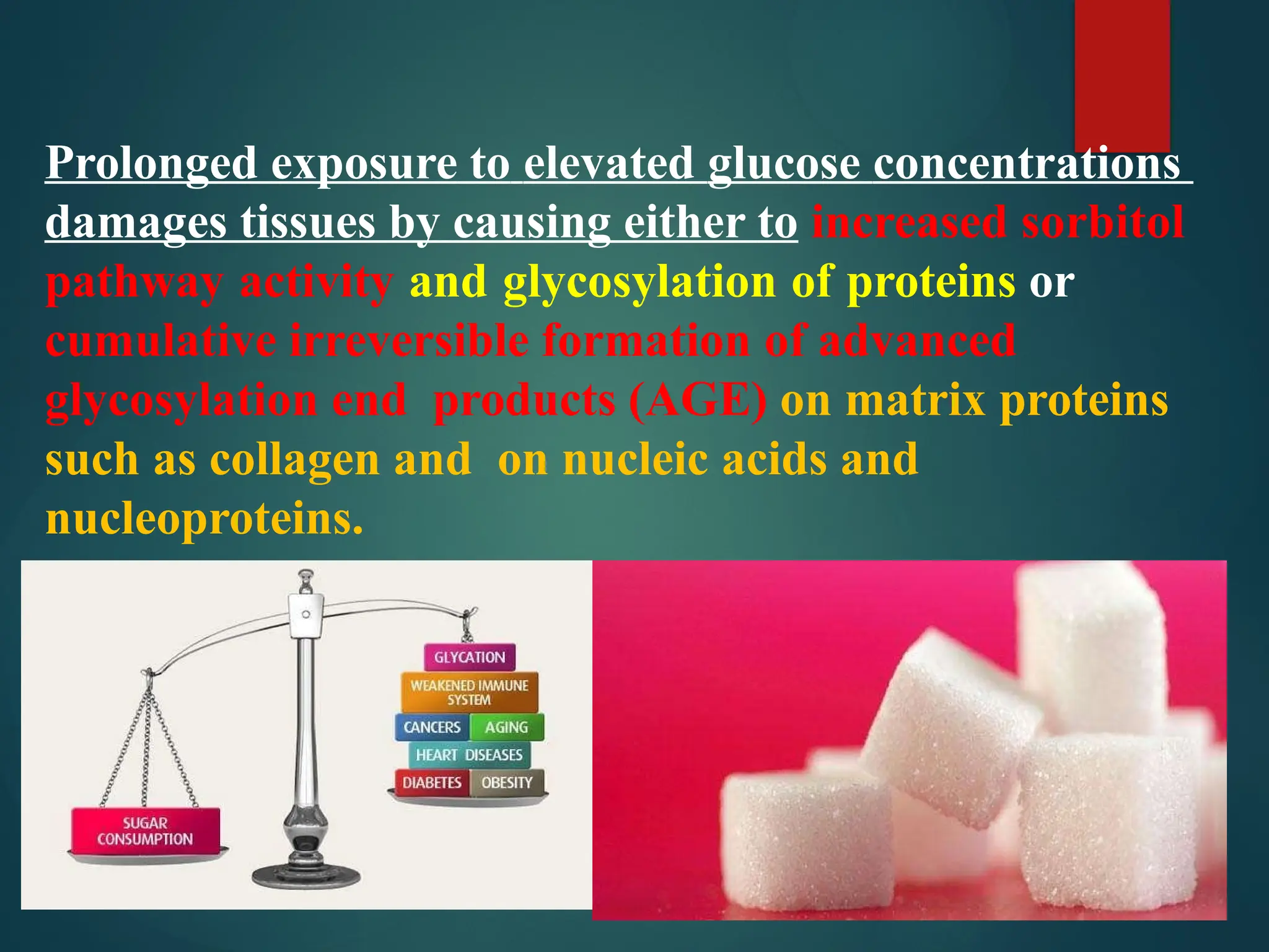
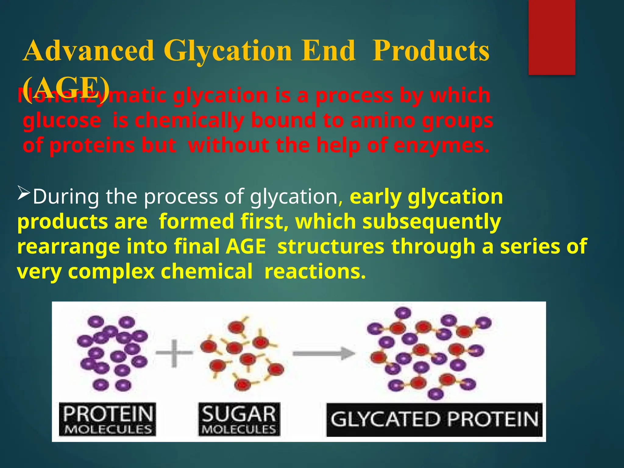
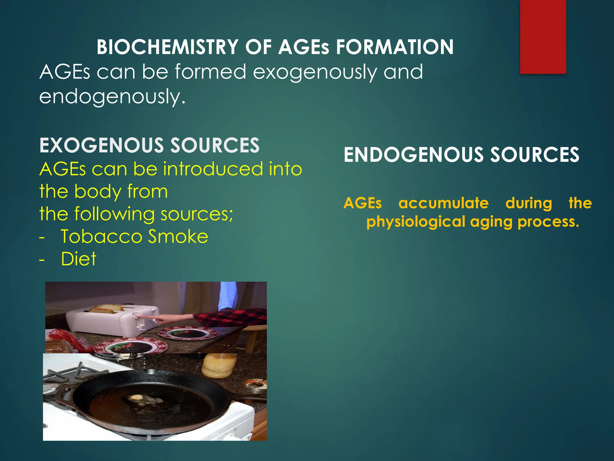
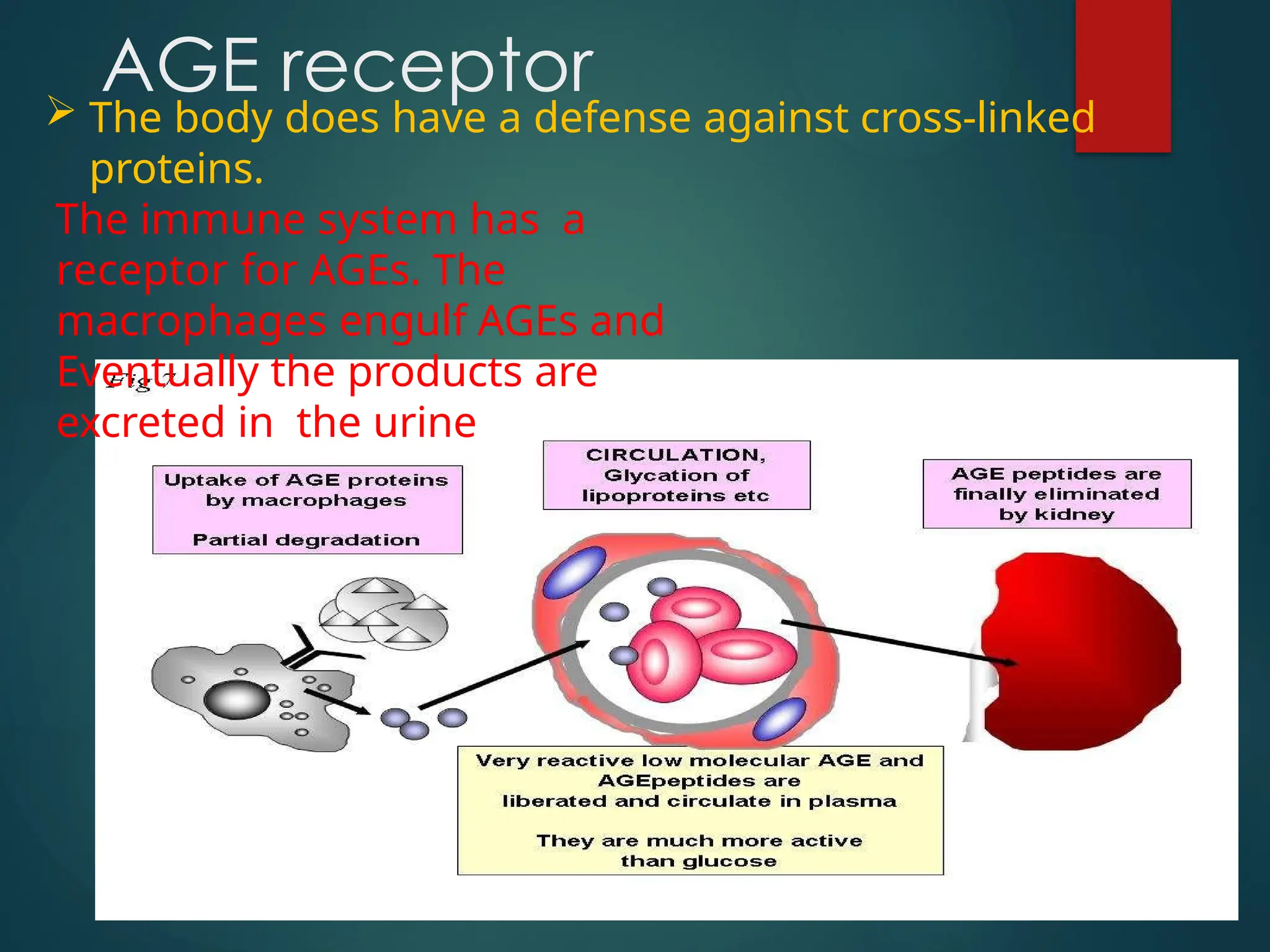
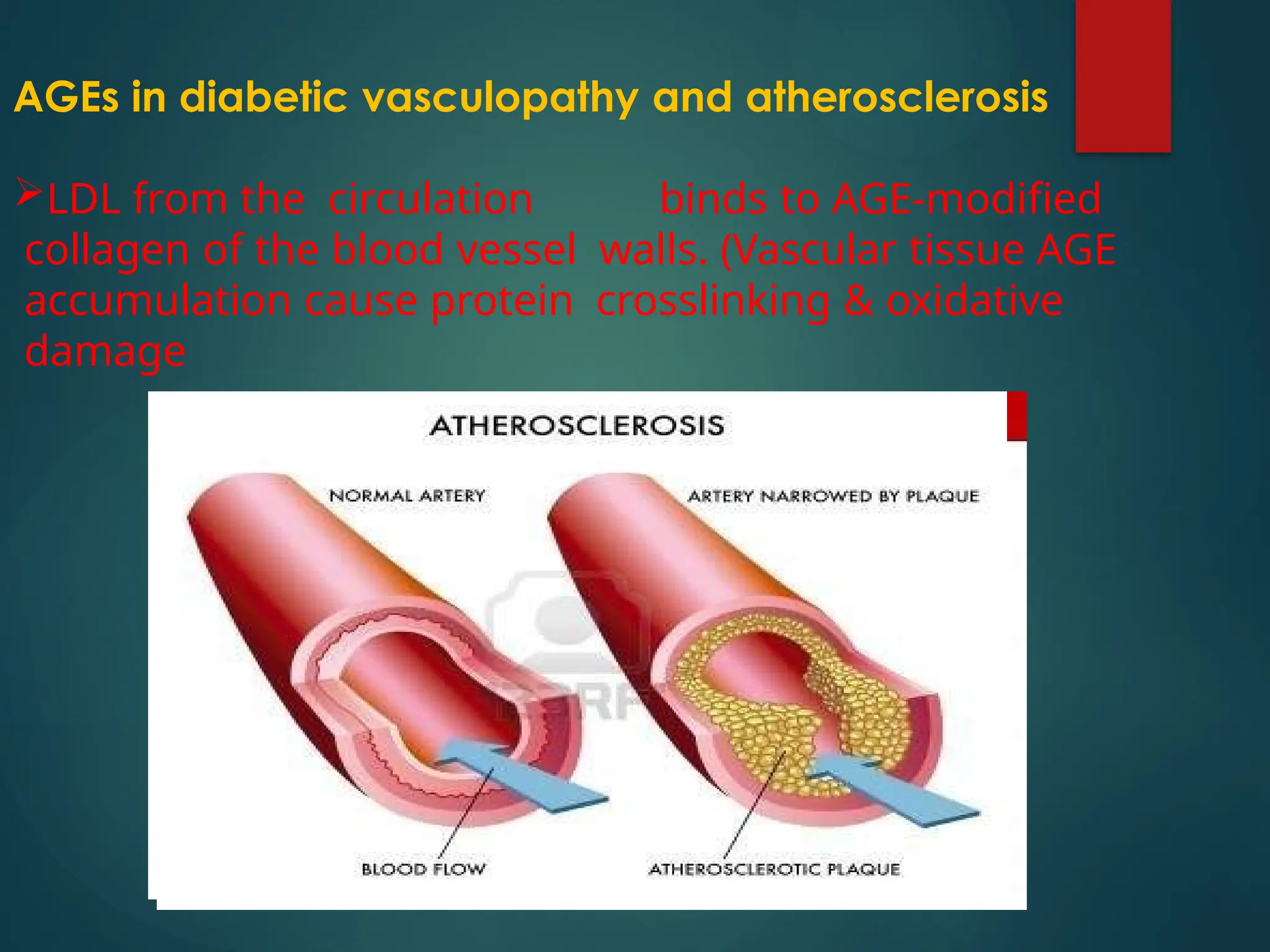
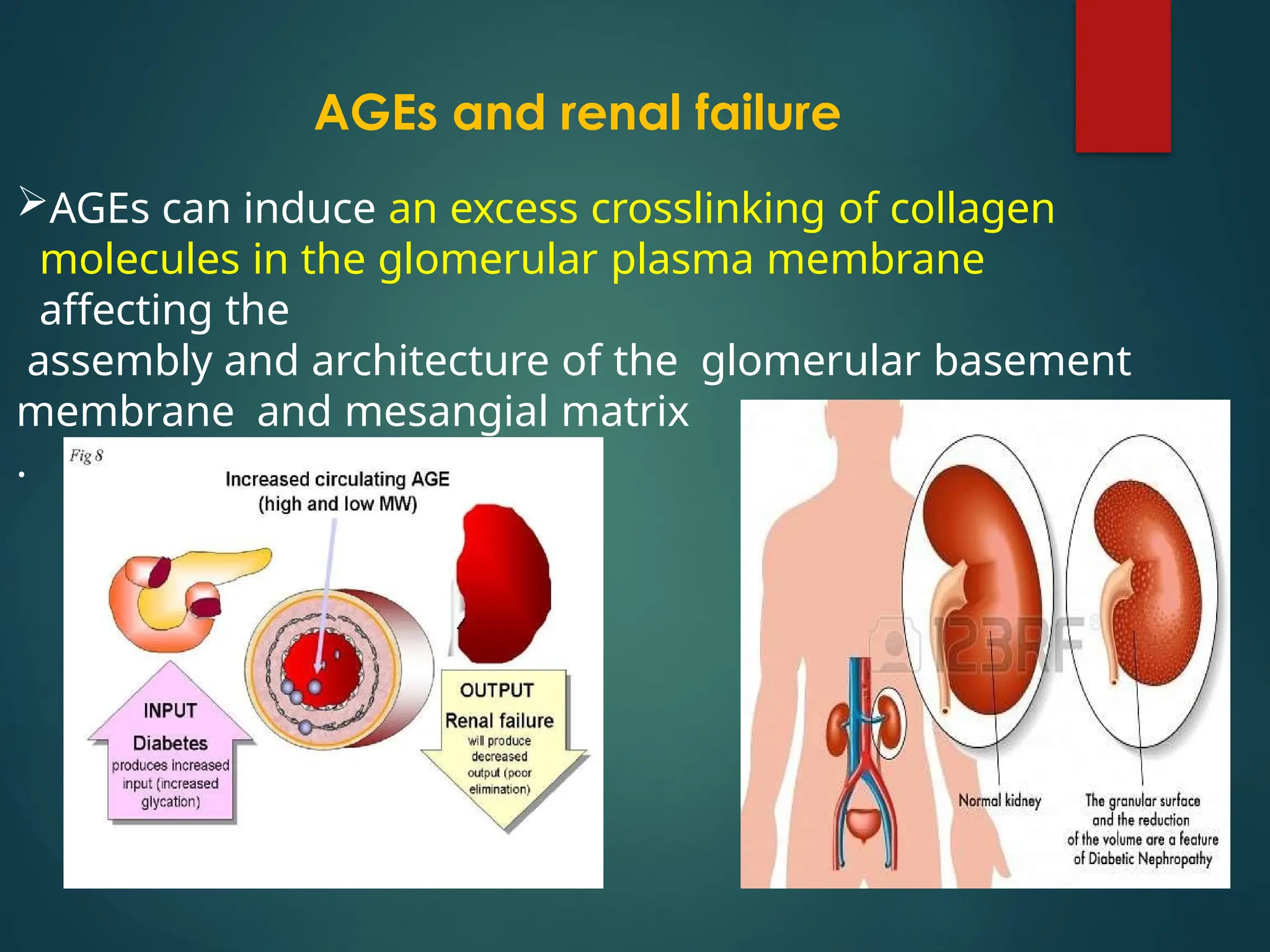
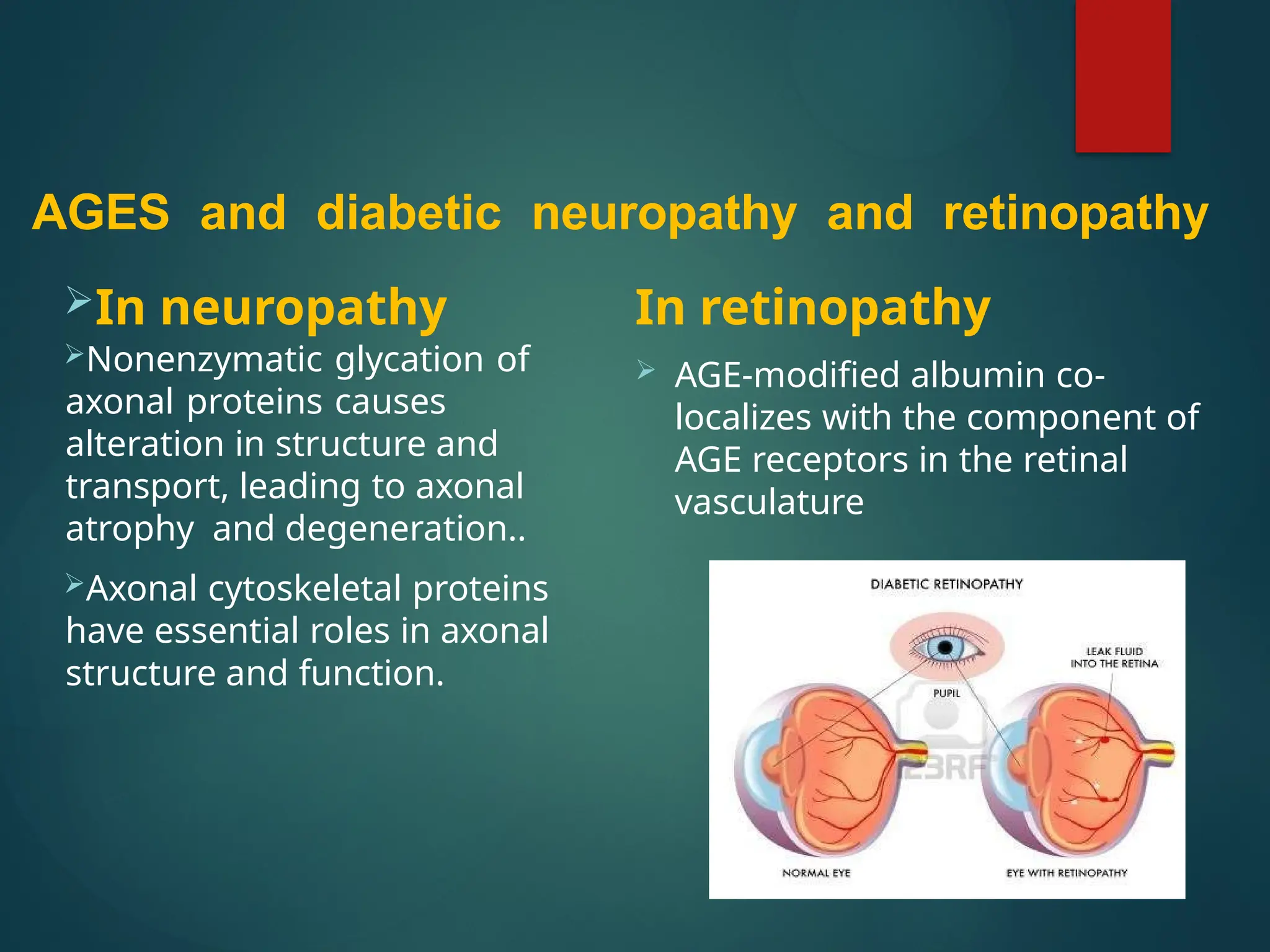
Advanced Glycation End
Products
Prolonged exposure to elevated glucose concentrations
damages tissues by causing either to increased sorbitol
pathway activity and glycosylation of proteins or
cumulative irreversible formation of advanced
glycosylation end products (AGE) on matrix proteins
such as collagen and on nucleic acids and
nucleoproteins.
Nonenzymatic glycation is a process by which
glucose is chemically bound to amino groups
of proteins but without the help of enzymes.
During the process of glycation, early glycation
products are formed first, which subsequently
rearrange into final AGE structures through a series of
very complex chemical reactions.
Advanced Glycation End Products
(AGE)
BIOCHEMISTRY OF AGEs FORMATION
AGEs can be formed exogenously and
endogenously.
EXOGENOUS SOURCES
AGEs can be introduced into
the body from
the following sources;
- Tobacco Smoke
- Diet
ENDOGENOUS SOURCES
AGEs accumulate during the
physiological aging process.
 The body does have a defense against cross-linked
proteins.
The immune system has a
receptor for AGEs. The
macrophages engulf AGEs and
Eventually the products are
excreted in the urine
AGE receptor
AGEs in diabetic vasculopathy and atherosclerosis
LDL from the circulation binds to AGE-modified
collagen of the blood vessel walls. (Vascular tissue AGE
accumulation cause protein crosslinking & oxidative
damage
AGEs and renal failure
AGEs can induce an excess crosslinking of collagen
molecules in the glomerular plasma membrane
affecting the
assembly and architecture of the glomerular basement
membrane and mesangial matrix
.
AGES and diabetic neuropathy and retinopathy
In neuropathy
Nonenzymatic glycation of
axonal proteins causes
alteration in structure and
transport, leading to axonal
atrophy and degeneration..
Axonal cytoskeletal proteins
have essential roles in axonal
structure and function.
In retinopathy
 AGE-modified albumin co-
localizes with the component of
AGE receptors in the retinal
vasculature
advanced glycation end product 222 .pptx

advanced glycation end product 222 .pptx

  • 1.
  • 2.
    Prolonged exposure toelevated glucose concentrations damages tissues by causing either to increased sorbitol pathway activity and glycosylation of proteins or cumulative irreversible formation of advanced glycosylation end products (AGE) on matrix proteins such as collagen and on nucleic acids and nucleoproteins.
  • 3.
    Nonenzymatic glycation isa process by which glucose is chemically bound to amino groups of proteins but without the help of enzymes. During the process of glycation, early glycation products are formed first, which subsequently rearrange into final AGE structures through a series of very complex chemical reactions. Advanced Glycation End Products (AGE)
  • 4.
    BIOCHEMISTRY OF AGEsFORMATION AGEs can be formed exogenously and endogenously. EXOGENOUS SOURCES AGEs can be introduced into the body from the following sources; - Tobacco Smoke - Diet ENDOGENOUS SOURCES AGEs accumulate during the physiological aging process.
  • 5.
     The bodydoes have a defense against cross-linked proteins. The immune system has a receptor for AGEs. The macrophages engulf AGEs and Eventually the products are excreted in the urine AGE receptor
  • 6.
    AGEs in diabeticvasculopathy and atherosclerosis LDL from the circulation binds to AGE-modified collagen of the blood vessel walls. (Vascular tissue AGE accumulation cause protein crosslinking & oxidative damage
  • 7.
    AGEs and renalfailure AGEs can induce an excess crosslinking of collagen molecules in the glomerular plasma membrane affecting the assembly and architecture of the glomerular basement membrane and mesangial matrix .
  • 8.
    AGES and diabeticneuropathy and retinopathy In neuropathy Nonenzymatic glycation of axonal proteins causes alteration in structure and transport, leading to axonal atrophy and degeneration.. Axonal cytoskeletal proteins have essential roles in axonal structure and function. In retinopathy  AGE-modified albumin co- localizes with the component of AGE receptors in the retinal vasculature