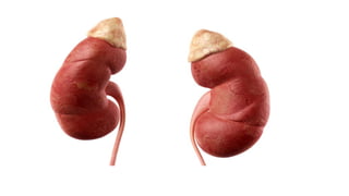
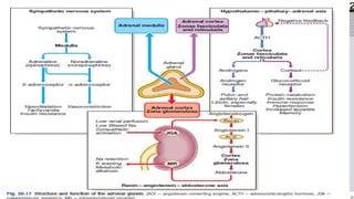
THE ADRENAL GLANDSback ground
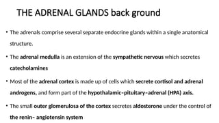
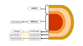
• The adrenals comprise several separate endocrine glands within a single anatomical
structure.
• The adrenal medulla is an extension of the sympathetic nervous which secretes
catecholamines
• Most of the adrenal cortex is made up of cells which secrete cortisol and adrenal
androgens, and form part of the hypothalamic–pituitary–adrenal (HPA) axis.
• The small outer glomerulosa of the cortex secretes aldosterone under the control of
the renin– angiotensin system
7.
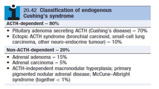
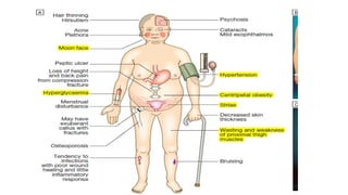
Cushing’s syndrome
• Cushing’ssyndrome is caused by excessive activation of glucocorticoid
receptors.
• It is most commonly iatrogenic, due to prolonged administration of synthetic
glucocorticoids such as prednisolone.
• Endogenous Cushing’s syndrome is uncommon but is due to chronic over-
production of cortisol by the adrenal glands.
• either as the result of an adrenal tumour or because of excessive production of
ACTH by a pituitary tumour or ectopic ACTH production by other tumours.
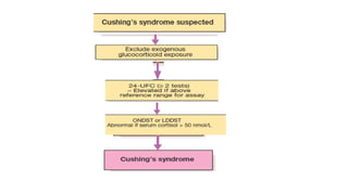
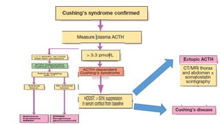
10.
The diagnosis ofCushing’s is a two step process:
1. to establish whether the patient has Cushing’s
syndrome
2. to define its cause
14.
Management
• Untreated severeCushing’s syndrome has a 50% 5-year mortality. Most patients are
treated surgically, but medical therapy may be given in severe cases for a few weeks prior
to operation to improve the clinical state.
• A number of drugs are used to inhibit corticosteroid biosynthesis, including metyrapone
and ketoconazole.
• The dose of these agents is best titrated against serum cortisol levels or 24-hour urine
free cortisol.
15.
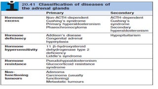
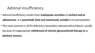
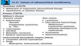
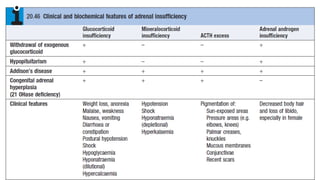
Adrenal insufficiency
• Adrenalinsufficiency results from inadequate secretion of cortisol and/or
aldosterone. It is potentially fatal and notoriously variable in its presentation.
• The most common is ACTH deficiency (secondary adrenocortical failure), usually
because of inappropriate withdrawal of chronic glucocorticoid therapy or a
pituitary tumour.
18.
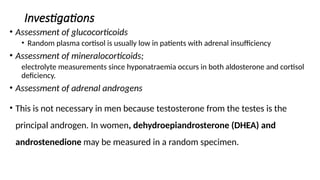
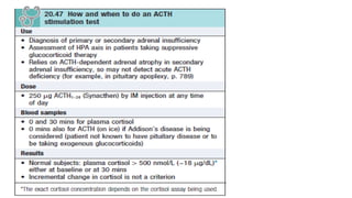
Investigations
• Assessment ofglucocorticoids
• Random plasma cortisol is usually low in patients with adrenal insufficiency
• Assessment of mineralocorticoids;
electrolyte measurements since hyponatraemia occurs in both aldosterone and cortisol
deficiency.
• Assessment of adrenal androgens
• This is not necessary in men because testosterone from the testes is the
principal androgen. In women, dehydroepiandrosterone (DHEA) and
androstenedione may be measured in a random specimen.
20.
Other tests toestablish the cause
in patients with elevated ACTH, further tests are required to establish the cause
of Addison’s disease.
• Adrenal autoantibodies are frequently positive in autoimmune adrenal failure.
• Tuberculosis causes adrenal calcification, visible on plain X-ray or ultrasound
scan.
• An HIV test should be performed if risk factors for infection are
present.
• Adrenal metastases are a rare cause of adrenal insufficiency.
21.
• Patients withevidence of autoimmune adrenal failure should be
screened for other organ-specific autoimmune diseases, such as
thyroid disease, pernicious anaemia and type 1 diabetes
22.
Management
• Patients withadrenocortical insufficiency always need glucocorticoid
replacement therapy and usually, but not always, mineralocorticoid therapy.
• There is some evidence that adrenal androgen replacement may also be
beneficial in women.
• Other treatments depend on the underlying cause.
23.
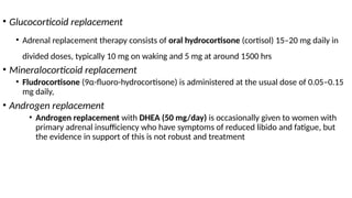
• Glucocorticoid replacement
•Adrenal replacement therapy consists of oral hydrocortisone (cortisol) 15–20 mg daily in
divided doses, typically 10 mg on waking and 5 mg at around 1500 hrs
• Mineralocorticoid replacement
• Fludrocortisone (9α-fluoro-hydrocortisone) is administered at the usual dose of 0.05–0.15
mg daily,
• Androgen replacement
• Androgen replacement with DHEA (50 mg/day) is occasionally given to women with
primary adrenal insufficiency who have symptoms of reduced libido and fatigue, but
the evidence in support of this is not robust and treatment
25.
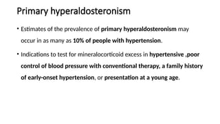
Primary hyperaldosteronism
• Estimatesof the prevalence of primary hyperaldosteronism may
occur in as many as 10% of people with hypertension.
• Indications to test for mineralocorticoid excess in hypertensive ,poor
control of blood pressure with conventional therapy, a family history
of early-onset hypertension, or presentation at a young age.
26.
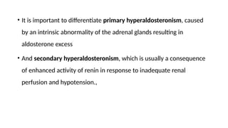
• It isimportant to differentiate primary hyperaldosteronism, caused
by an intrinsic abnormality of the adrenal glands resulting in
aldosterone excess
• And secondary hyperaldosteronism, which is usually a consequence
of enhanced activity of renin in response to inadequate renal
perfusion and hypotension.,
27.
Clinical features
• Individualswith primary hyperaldosteronism are usually asymptomatic but may
have features of sodium retention or potassium loss.
• Sodium retention may cause oedema, while hypokalaemia may cause muscle
weakness (or even paralysis, especially in Chinese), polyuria (secondary to renal
tubular damage, which produces nephrogenic diabetes insipidus) and occasionally
tetany (because of associated metabolic alkalosis and low ionised calcium).
• Blood pressure is elevated but accelerated phase hypertension is rare.
28.
Investigations; Biochemical
• Routineblood tests may show a hypokalaemic alkalosis.
• Sodium is usually at the upper end of the reference range in primary
hyperaldosteronism.
29.
• The keymeasurements are plasma renin and aldosterone and in
many centres, the aldosterone : renin ratio (ARR) is employed as a
screening test for primary hyperaldosteronism in hypertensive
patients.
30.
imaging and localization
•Imaging with CT or MRI will identify most APAs but it is important to recognize the
risk of false-positives (non-functioning adrenal adenomas are common) and false-
negatives (imaging may have insufficient resolution to identify adenomas with
diameter of less than 0.5 cm).
• If the imaging is inconclusive and there is an intention to proceed with surgery on
the basis of strong biochemical evidence of an APA, then adrenal vein
catheterisation with measurement of aldosterone (and cortisol to confirm
positioning of the catheters) is required.
31.
Management
Mineralocorticoid receptor antagonists(spironolactone and eplerenone) are
valuable in treating both hypokalaemia and hypertension in all forms of
mineralocorticoid excess.
Up to 20% of males develop gynaecomastia on spironolactone.
Amiloride (10–40 mg/day), which blocks the epithelial sodium channel regulated
by aldosterone, is an alternative.
32.
Cont.,…..
• In patientswith an APA, medical therapy is usually given for a few weeks to
normalise whole-body electrolyte balance before unilateral adrenalectomy.
• Laparoscopic surgery cures the biochemical abnormality but, depending on the
pre-operative duration, hypertension remains in as many as 70% of cases, probably
because of irreversible damage to the systemic microcirculation
33.
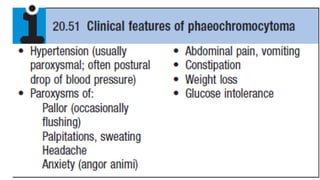
Phaeochromocytoma and paraganglioma
•These are rare neuro-endocrine tumors that may secrete catecholamines
(adrenaline/epinephrine, noradrenaline/norepinephrine).
• Approximately 80% of these tumours occur in the adrenal medulla
(phaeochromocytomas), while 20% arise elsewhere in the body in sympathetic
ganglia (paragangliomas)
34.
• Most arebenign but approximately 15% show malignant features.
• Around 30% are associated with inherited disorders, including
neurofibromatosis, von Hippel–Lindau syndromeand MEN 2 .
• Paragangliomas are particularly associated with mutations in the
succinate dehydrogenase B, C and D genes.
36.
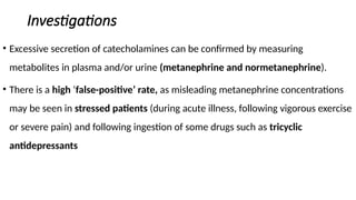
Investigations
• Excessive secretionof catecholamines can be confirmed by measuring
metabolites in plasma and/or urine (metanephrine and normetanephrine).
• There is a high ‘false-positive’ rate, as misleading metanephrine concentrations
may be seen in stressed patients (during acute illness, following vigorous exercise
or severe pain) and following ingestion of some drugs such as tricyclic
antidepressants
37.
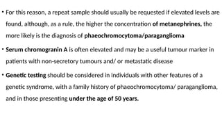
• For thisreason, a repeat sample should usually be requested if elevated levels are
found, although, as a rule, the higher the concentration of metanephrines, the
more likely is the diagnosis of phaeochromocytoma/paraganglioma
• Serum chromogranin A is often elevated and may be a useful tumour marker in
patients with non-secretory tumours and/ or metastatic disease
• Genetic testing should be considered in individuals with other features of a
genetic syndrome, with a family history of phaeochromocytoma/ paraganglioma,
and in those presenting under the age of 50 years.
38.
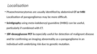
Localisation
• Phaeochromocytomas areusually identified by abdominal CT or MRI
Localisation of paragangliomas may be more difficult.
• Scintigraphy using meta-iodobenzyl guanidine (MIBG) can be useful,
particularly if combined with CT.
• 18F-deoxyglucose PET is especially useful for detection of malignant disease
and for confirming an imaging abnormality as a paraganglioma in an
individual with underlying risk due to genetic mutation.
39.
Management
• Medical therapyis required to prepare the patient for surgery, preferably for a
minimum of 6 weeks to allow restoration of normal plasma volume.
• The most useful drug in the face of very high circulating catecholamines is the
α-blocker phenoxybenzamine (10–20 mg orally 3–4 times daily) because it is a
non-competitive antagonist, unlike prazosin or doxazosin.
40.
• If α-blockadeproduces a marked tachycardia, then a β-blocker such as
propranolol can be added.
• On no account should a β-blocker be given before an α-blocker, as this may
cause a paradoxical rise in blood pressure due to unopposed α-mediated
vasoconstriction.


![Acute and chronic liver disease [Autosaved].pptx](https://cdn.slidesharecdn.com/ss_thumbnails/acuteandchronicliverdiseaseautosaved-250921172441-4babef2c-thumbnail.jpg?width=640&height=640&fit=bounds)


![ONFH[AVN HIP] -TRIPLE REGIME -A NOVAL SURGICAL CONCEPT .pptx](https://cdn.slidesharecdn.com/ss_thumbnails/onfhavnhip2026koaconcalicutdrgokuldevdrmashraf-260210064517-213ec005-thumbnail.jpg?width=640&height=640&fit=bounds)


![CTEV [ clubfoot] DR ARUN LAL ,DR MOHAMED ASHRAF travancore medical college k...](https://cdn.slidesharecdn.com/ss_thumbnails/ctevclubfootdrarunlaldrmohamedashraftravancoremedicalcollegekollamkeralaindia-260208063247-18fc466c-thumbnail.jpg?width=640&height=640&fit=bounds)


