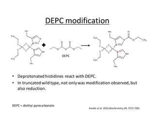
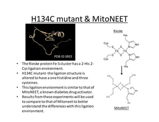
Spectroscopic	Analysis	and	
Chemical	Modification	of	the	
H134C	Mutant	of	the	Thermus
thermophius Rieske Protein
Victor	Rodríguez	and	Laura	M.	Hunsicker-Wang
Trinity	University,	San	Antonio,	TX
Abstract
The	Rieske protein	is	found	in	the	bc1	complex	(complex	III)	
and	plays	an	important	role	in	transporting	electrons	and	
protons	through	the	electron	transport	chain.	The	Rieske
protein	contains	a	[2Fe-2S]	cluster	ligated	by	a	2-cystines	and	
2- histidines.	The	reduction	potential	of	this	cluster	is	pH-
dependent	and	varies	across	species.			An	H134C	mutant	of	
the	Thermus thermophilus Rieskesubstitutes	one	of	the	
ligating	histidines for	a	cysteine,	changing	the	ligation	
structure	of	the	cluster	from	a	2Cys-2His	to	a	3Cys-1His	
environment.	To	study	the	effects	of	this	mutation,	the	protein	
is	subjected	to	modification	with	diethyl	pyrocarbonate
(DEPC)	and	to	pH	changes.	The	behavior	of	this	protein	is	
compared	to	the	wild	type	protein	as	observed	through	
circular	dichroism and	UV	visible	spectroscopy.	Because	of	the	
similarity	in	the	iron-sulfur	cluster	ligands,	H134C	will	also	be	
compared	to	another	mitochondrial	protein,	mitoNEET,	which	
contains	a	3Cys-1His	environment.
Rieske Proteins
• The	Rieske protein	is	found	in	complex	III	
(cytochrome	bc1 complex)	as	part	of	the	
electron	transport	chain.	
• This	complex	oxidizes	ubiquinol to	
ubiquinone	and	transports	the	electrons	
through	the	complex	into	the	Fe-S	cluster	in	
Reiske.
http://www.scienceprofonline.org/metabolism/electron-transport-chain-cellular-respiration.html
http://en.wikipedia.org/wiki/Coenzyme_Q_%E2%80%93_cytochrome_c_reductase
H134C	mutant	&	MitoNEET
• The	Rieske protein	Fe-S	cluster	has	a	2-His	2-
Cys	ligation	environment.	
• H134C	mutant-the	ligation	structure	is	
altered	to	have	a	one	histidine and	three	
cysteines.	
• This	ligation	environment	is	similar	to	that	of	
MitoNEET,	a	known	diabetes	drug	activator.	
• Results	from	these	experiments	will	be	used	
to	compare	to	that	of	Mitoneet to	better	
understand	the	differences	with	this	ligation	
environment.
Rieske
MitoNEET
PDB	ID	3REE
pH-Dependent	UV-Visible	spectra
0
0.1
0.2
0.3
0.4
0.5
0.6
0.7
0.8
300 400 500 600 700 800
Absorbance	(A)
Wavelength	(nm)
4.39
6.63
7.54
8.65
9.61
10.41
10.97
11.99
• Previous	studies	show	that	H134C	has	a	large	range	of	pH	
stability
• H134C	has	a	pKa of	9.86.
Data	collected	by	Abhishek Chhetri
H134C
H134C	Crystals
• Crystals	for	H134C	Rieske
mutant	were	isolated	under	
the	following	conditions:	
30%	PEG	4000,	0.2	M	
MgCl2,	0.1	M	Tris-HCl pH	
8.5	or	20%	PEG	2000	MME,	
0.01	M	NiCl2·	6H2O,	0.1	M	
Tris pH	8.5.	
• Best	data	collected	to	1.66	
Å	resolution!
• Space	group	P2221
Preliminary	electron	density	map	(2Fo-Fc)	
showing	3	Cys,	1	His	[2Fe2S]	cluster
His	154
Cys 134
Cys 132 Cys 151
DEPC	modification
• Deprotonated	histidines react	with	DEPC.	
• In	truncated	wild	type,	not	only	was	modification	observed,	but	
also	reduction.
DEPC
DEPC	=	diethyl	pyrocarbonate
Konkle	et	al.	2010	Biochemistry	49,	7272-7281
Reaction	with	DEPC- UV-Vis
pH	6
pH	7
pH	8
pH	6
pH	7
pH	8
Konkle	et	al.	2010	Biochemistry	49,	7272-7281
truncTtRp H120Q/H162Q
• truncTtRp	and	H120Q/H162Q	are	modified	by	DEPC
• Higher	pH	causes	faster	modification	and	more	modification	in	truncTtRp
Effect	of	pH	on	DEPC	reaction	- CD
pH	=	6 pH	=	7
pH	=	8 pH	=	9
H120Q/H162Q
Konkle	et	al.	2010	Biochemistry	49,	7272-7281• More	spectral	changes	with	higher	pH
• Proteins	become	reduced	after	modification
H134C	Reaction	with	DEPC- UV
-0.05
0.05
0.15
0.25
0.35
0.45
0.55
200 300 400 500 600 700 800
Difference
Wavelength	(nm)
pH	6.0
-0.05
0.05
0.15
0.25
0.35
0.45
0.55
200 300 400 500 600 700 800
Difference
Wavelength	(nm)
pH	7.6
• H134C		was	reacted	with	DEPC	
• difference	UV-Vis	spectra	over	40	minutes	at	different	pH	values.	
-0.05
0.05
0.15
0.25
0.35
0.45
0.55
200 300 400 500 600 700 800
Difference
Wavelelngth	(nm)
pH	8.2
H134C	is	modified	by	DEPC,	but	the	LMCT	bands	are	minimally	affected
-0.1
0
0.1
0.2
0.3
0.4
0.5
200 300 400 500 600 700 800
Difference
Wavelength	(nm)
pH	9.0
pH-dependence	of	modification
• Reaction	rate	increases	with	pH,	but	the	extent	of	modification	decreases	
above	pH	7.6
• The	profile	differs	from	truncTtRp (see	previous	slide)
ΔAbs.	240	nm
Time	(min)
0
0.05
0.1
0.15
0.2
0.25
0.3
0.35
0.4
0.45
0.5
0 5 10 15 20 25
pH	6.0
pH	7.6
pH	8.2
pH	9.0
• H134C	reacted	with	DEPC	
over	90	minutes	(upper	
figure).	
• A	change	in	signal	is	
observed	over	time	and	
follow	a	similar	patter	to	
what	was	observed	for	
reduction	in	wild	type	
Rieske.	
• To	fully	understand	the	
changes	of	the	signals,	the	
mutant	protein	was	reduced	
and	oxidize	(lower	figure)
CD	spectra	change	but	the	protein	does	not	appear	to	become	reduced
H134C	Reaction	with	DEPC- CD
24	hour	reaction	with	DEPC
• The	CD	signal	at	450	nm	increases	until	3	hours,	and	then	
returns	to	the	original	level.		
• One	interpretation	is	that	the	DEPC	modification	reverses	
over	time,	indicating	a	labile	adduct.
-13
-11
-9
-7
-5
-3
-1
1
3
5
230 280 330 380 430 480 530 580
mdeg
Wavelength	(nm)
H134C	at	pH	8.2	reacetd	with	DEPC	for	24	hours
unreacted
0	hours
3	hours
10	hours
23	hours
27	hours
0
2
4
0 3 6 9 12 15 18 21 24 27
Absorbtion Time	(hours)
H134C	reacted	with	
DEPC	at	450	nm
Conclusions
• H134C	pKa of	protein	is	9.86.
• Crystal	structure	confirms	the	3Cys	1	His	
Structure
• Protein	is	modified	by	DEPC	and	not	reduced	
within	a	90	minute	reaction.
• DEPC	modification	may	reverses	over	longer	
times,	indicating	a	labile	adduct	in	this	3	Cys,	1	
His	ligation	environment.
Acknowledgements
• McNair	Scholar	Program
• National	Science	Foundation
• P.	John	Hart	Lab	at	the	University	of	Texas	
Health	Science	Center	San	Antonio	for	crystals

ACS poster final