PUERPERAL PYREXIA
• “A rise of temperature reaching
100.4 degree F or more (Measured
orally) on two separate occasions
24 hours apart (excluding first 24
hours) within the first 10 days
following delivery is called
Puerperal pyrexia”
• In some countries postabortal fever
is also included.
PUERPERAL SEPSIS
• “Aninfection of the genital tract
which occurs as a complication of
delivery is termed puerperal
sepsis.”
• Puerperal pyrexia is considered to
be due to genital tract infection
unless proved otherwise.
6.
INCEDENCE
• There isa marked decline in puerperal
sepsis during the past few years due
to:-
• Improved obstetric care
• Availability of wider range of
antibiotics
PREDISPOSING FACTORS
• Damageof Cervicovaginal mucous
membrane
• Large placental wound surface area
• Blood clots presents at placental site
ANTEPARTUM FACTORS:
• Malnutrition and anaemia
• Preterm labour
• PROM
• Chronic illness
• Prolonged rupture of membrane >18
hours
9.
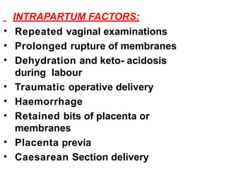
INTRAPARTUM FACTORS:
• Repeatedvaginal examinations
• Prolonged rupture of membranes
• Dehydration and keto- acidosis
during labour
• Traumatic operative delivery
• Haemorrhage
• Retained bits of placenta or
membranes
• Placenta previa
• Caesarean Section delivery
10.
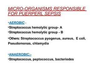
MICRO-ORGANISMS RESPONSIBLE
FOR PUERPERLSEPSIS
•AEROBIC:-
•Streptococcus hemolytic group- A
•Streptococcus hemolytic group - B
•Others: Streptococcus pyogenus, aureus, E coli,
Pseudomonas, chlamydia
•ANAEROBIC:-
•Streptococcus, peptococcus, bacteriodes
11.
MODE OF INFECTION
•Puerperal sepsis is essentially a wound
infection
• Placental site, lacerations of the genital
tract or cesarean section wounds
• It may get infected by ENDOGENOUS or
EXOGENOUS organisms.
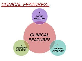
1. LOCAL INFECTION
•Slight temperature rise
• Generalized malaise
• Headache
• Redness and swelling to local
wound
• Pus formation
14.
2. UTERINE INFECTION
MILD:-
•Rise in temperature and pulse rate
• Offensive and copious lochial
discharge
• Subinvoluted and tender uterus
• SEVERE:-
• Acute onset with high grade
temperature with chills and rigor
• Rapid pulse rate
• Scanty and orderless lochia
15.
3. SPREADING INFECTION
•Parametritis
• Pelvic pritonitis
• General
peritonitis
• Thrombophlebit
is
• Septicemia
PROPHYLAXIS
ANTENATAL:
• Improvement ofnutritional status
• Eradication of any septic status
• INTRANATAL:
• Full surgical asepsis during labour
• Prophylactic antibiotics: Cefriaxone 1g
IV immediate after cord clamping and
second dose: after 8 hour is
recommended
18.
POSTNATAL:
• Aseptic precautionsatleast one
week following delivery
• Too many visitors are restricted
• Sterilized senitory pads are to be
used
• Infected babies and mothers should
be in isolated room
19.
GENERAL CARE:-
• Isolationof the patient.
• Adequate fluid and calorie (IV).
• Corrected anaemia.
• Progress chart should be
maintained.
TREATMENT
20.
ANTIBIOTICS
• Gentamicin, 2mg/kg IV loading
dose followed by 1.5 mg/kg IV
every 8 hours
• Ampicillin, 1g IV every 6 hours
• Clindamycin 900 mg, IV every 8
hours
• Cefotaxime 1 g, 8 hourly IV is an
alternative
• Metronidazole 0.5 g IV, 8 hourly
21.
PERINEAL WOUND:-
• Stichesof perineal wound may have
to be removed to facilitate drainage
of pus and relieve pain.
• Wound has to be cleaned with sitz
bath several times per day and
dressed with antiseptic ointment or
powder.
• After the infection is controlled,
secondary wound closure may be
done on later date.
SURGICAL TREATMENT
22.
RETAINED UTERINE PRODUCTS:-
•With diameter of 3 cm or less may be
disregarded or left alone
• Other wise surgical evacuation after
antibiotic coverage for 24 hours should be
done to avoid risk of septicemia
• SEPTIC THROMBOPHLEBITIS:-
• IV Heparin for 7-10 days
23.
PELVIC ABCESS:-
• Drainageby colpotomy under
ultrasound guidance
WOUND DEHISCENCE:
• Dehiscence of episiotomy or abdominal
wound following caesarean section:-
• Scrubbing the wound
• Debridement of all necrotic tissues
• Secondary suture
24.
• LAPAROTOMY:
• Hasgot limited indications
• IV fluids and antibiotics usually
controls the peritonitis
• When the peritonitis is unresponsible
to antibiotics laprotomy is indicated
• HYSTERECTOMY:
• In case of uterine rupture or
perforation
• Multiple abcess, gangrenous uterus
• Ruptured tubo-ovarian abcess
25.
NECROTYSING FASCITIS:
• Woundscrubbing
• Debridement of all necrotic tissues
• Use of effective antimicrobial agents
• BACTEREMIC OR SEPTIC SHOCK:
• Fluid and electrolyte balance
• Respiratory supports
• Circulatory support (dopamine/
dobutamine)
• Infection control
SYMPTOMS
• May beasymptomatic sometimes
• Abnormal lochial discharge : excessive or
prolonged
• Irregular at times excessive uterine
bleeding
• Irregular cramp like pain
• Rise of temperature in case of sepsis
MANAGEMENT
• Antibiotics incase of infection
• Exploration of uterus for
retained products
• Pessary in prolapse or
retroversion
• Methergine to enhance
involution process
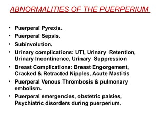
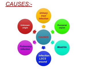
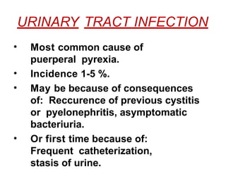
URINARY TRACT INFECTION
•Most common cause of
puerperal pyrexia.
• Incidence 1-5 %.
• May be because of consequences
of: Reccurence of previous cystitis
or pyelonephritis, asymptomatic
bacteriuria.
• Or first time because of:
Frequent catheterization,
stasis of urine.
RETENTION OF URINE
Commoncomplication in early
puerperium.
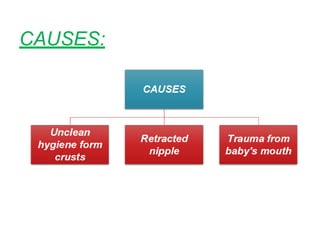
CAUSES:
• Bruising
• Edema of bladder neck
• Reflex from the perineal
injury
• An accustomised position
39.
TREATMENT
• Indwelling catheterfor 48 hours
• Following removal of a catheter
recidual urine is to be measured
• If it is more than 100 ml
drainage is resumed
• Appropriate urinary antiseptics up to
5-7 days
40.
INCONTENENCE OF URINE
•Not a common symptom following
birth
• It may be:-
• Stress incontenence (late
puerperium)
• Overflow incontenence
( following retention of urine)
• True incontenence (soon
41.
SUPRESSION OF URINE
•“If the 24 hours urine excretion is
less than 400 ml or less, supression
of urine is diagnosed.”
• The cause is to be sought for and
appropriate management is
instituted.
BREAST ENGORGEMENT
• Breastengorgement is due to
exaggerated normal venous and
lymphatic engorgement of the breasts
which precedes lactation.
• This in turn prevents escape of milk
from the lacteal system
45.
• The primiparouspatient and the
patient with inelastic breasts are
more likely develop breast
engorgement
• Engorgement is an indication that
the baby is not in step with stage
of lactation
ONSET:
• It usually manifests after the milk
secretion starts ( 3r
dand 4t
hday
postpartm)
PREVENTION:
• Avoid prelectealfeeds
• Initiate early breast feeding
• Exclusive breast feeding on
demand
• Feeding in correct position
48.
TREATMENT:
• Support withthe binders
• Mannual expression of milk
• Administer analgesics for pain
• Frequently and regular feeding
the baby
• In severe cases gentle use of
breast
• Warm compression
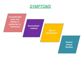
SYMPTOMS
• Condition mayremain
asymptomatic
• Sometimes painful when feeding
the baby
• When infected, the infection may
spread to the deeper tissue
proceding mastitis
52.
PROPHYLAXIS
• Local cleanlinessduring
pregnancy and puerperium
• Clean the crusts before and
after feeding
• Application of lotion to soothen
the epithelium
53.
TREATMENT
• Correct attachementduring
feeding
• Purified lanonin with mother's
milk applied 3 or 4 times a day for
healing
• In severe cases expression of
milk by breast pump
54.
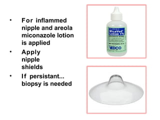
• For inflammed
nippleand areola
miconazole lotion
is applied
• Apply
nipple
shields
• If persistant...
biopsy is needed
55.
RETRACTED AND FLATNIPPLE
• Commonly seen in primiparous
mother
• Manual expression of milk is initiated
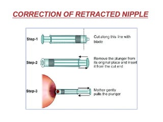
• Correction of retracted nipple
57.
ACUTE MASTITIS
• Incidenceof mastitis is 2-5 %
in lactating women
• Less than 1% in nonlactating.
• Organisms involved are...
• Streptococcus aureus,
• S. epidermidis and
• Streptococci viridans
59.
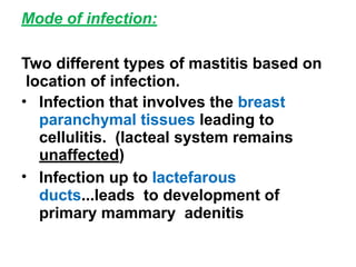
Mode of infection:
Twodifferent types of mastitis based on
location of infection.
• Infection that involves the breast
paranchymal tissues leading to
cellulitis. (lacteal system remains
unaffected)
• Infection up to lactefarous
ducts...leads to development of
primary mammary adenitis
60.
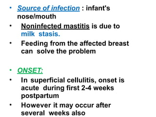
• Source ofinfection : infant's
nose/mouth
• Noninfected mastitis is due to
milk stasis.
• Feeding from the affected breast
can solve the problem
• ONSET:
• In superficial cellulitis, onset is
acute during first 2-4 weeks
postpartum
• However it may occur after
several weeks also
MANAGEMENT
• Support bybinders
• Plenty of oral fluids
• Good attachment when feeding the
baby
• Initiate feeding from uninfected breast
first to establish let down
• The infected site is emptied manually
with each feed
• Penicillins like flucloxacillin are is the
drugs of choice. 500 mg 6 hourly.
erythromycin is alternative
66.
• Antibiotic therapyis to continue up to 7
days
• Analgesics
• Milk flow is maintained by feeding the
baby
• It will prevent proliferation of
staphylococcus in the stagnant milk
• The ingested staphylococcus will
be digested without any harm
67.
BREAST ABCESS
FEATURES ARE:
•Flushed breasts not responding
to antibiotics
• Browny edema on the overlying
skin
• Marked tenderness with
fluctuation
• Swinging temperature
68.
MANAGEMENT
• Incision anddrainage under
general anaesthesia
• Deep radial incision extending
from near the areolar margin to
prevent injury of the lacteferous
ducts
• Incision perpendicular to the
lactiferous duct can increase the risk
of fistula formation and ductal
occlusion
69.
• Finger explorationhas to be done
to break the walls of loculi.
• The cavilty is loosely packed with
gause which should be replaced
after 24 hours by a smaller pack
• Continue till it heals up
• Abscess can also be drained by
serial percutaneous niddle
aspiration under ultrasound
guidance
• Surgical draiange is commonly
done
70.
• Breast feedingis continued
at uninvolved side
• The infected side is mechanically
expressed by pump every two
hours and with every let down
• Recurrence risk is about 10 %
• Once cellulitis has resolved breast
feeding from the involved side may
be resumed
71.
BREAST PAIN
May bedue to....
• Engorgement
• Infection ( candida albicans)
• Nipple trauma
• Mastitis
• Occasionally on latching-on or
let down reflex
72.
MANAGEMENT
• Appropriate nursingtechnique
• Positioning
• Breast care
• Use of myconazole oral lotion or gel
on the nipples and in infant's mouth
thrice daily for two weeks are helpful
73.
LACTATION FAILURE
CAUSES ARE:
•Infrequent suckling
• Depression or anxiety state in
puerperium
• Unwilling to nursing
• Ill development of nipples
• Endogenous supression of prolactin
• Prolactin inhibition
74.
MANAGEMENT
ANTENATAL:
• Counsell motherregading benefits
of nursing her baby
• To take care of any breast
abnormality.. breast engorgement
• Maintaining adequate breast
hygiene especially in the last two
months of pregnancy
75.
PUERPERIUM:
• Encourage adequatefluid intake
• To nurse the baby regularly
• Treat the painfull local lesions to
prevent nursing phobia
• Metoclopramide 10 mg thrice daily,
intranasal oxytocin and sulpiride
( selective dopamine intagonist) has
been found to increase milk production.
• They act by stimulating prolactin
secretion
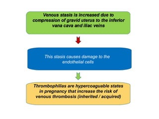
This stasis causesdamage to the
endothelial cells
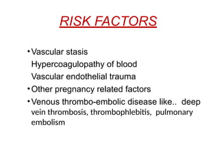
Thrombophilias are hypercoaguable states
in pregnancy that increase the risk of
venous thrombosis (inherited / acquired)
80.
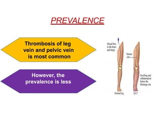
DEEP VEIN THROMBOSIS
•Clinical diagnosis is unreliable.
• In majority it remains
asymptomatic
• SYMPTOMS INCLUDE:
• Pain in the caff muscles
• On examination asymmentric
leg edema
• A positive Homan's sign
PELVIC THROMBOPHLEBITIS
• Originatesin the thrombosed veins
at placental site by organisms such
as an anaerobic streptococci or
bacteriosides
• When localised in the pelvis its
called pelvic thrombophlebitis.
• There is specific features but it
is suspected when there is
constatnt fever inspite of
antibiotics administration.
83.
EXTRA PELVIC SPREAD
•Through the right ovarian vein to
inferior vana cava and hence to the
lungs
• Through left ovarian vein to left renal
vein and hence to the left kidney
• Retrograde extension to iliofemoral
veins to produce the clinical pathological
entity called “phlegmasia alba dolens”
( adjacent cellulitis in femoral vein)
84.
CLINICAL FEATURES:
• Usuallydevelops in second week of
puerperium
• Mild pyrexia
• High grade fever with chills and rigor
• Constitutional disturbances like...
headache, malaise, rising pulse rate
• Swelling, pain, white , cold over
affected leg
85.
PROPHYLAXIS
• PREVENTIVE MEASURES:
•Prevention of trauma, sepsis,
anemia, dehydration
• Use of elastic compression
stocking
• Leg exercise, Early ambulation
86.
MANAGEMENT
• Bed restwith foot end kept higher
to heart level
• Pain management
• Antibiotics
• Anticoagulants- Heparin- 15000 units
IV followed by 10,000 units 6-8 hourly
for 4 to 6 injections. up to 7 to 10
days
• Administration of fibrinolytic agents
• Venous thrombectomy
87.
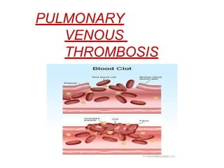
PULMONARY EMBOLISM
• Classicalsymptoms of massive
pulmonary embolism are...
• Sudden collapse
• Acute chest pain
• Air hunger
• Death usually occurs within short time
from shock and vagal inhibition
DIAGNOSIS
• ECG
• Arterialblood gas
• D-Dimer: value (More than 500 ng/ mL)
• Doppler utrasound
• Lung scans
• Pulmonary CT angiography
• MRA: Magnetic resonance angiography
90.
MANAGEMENT
• Prophylactic measures
•Active treatment:
• Resuscitation: cardiac massage, oxygen
therapy, heparin bolus IV of 5000 units and
morphine 15 mg
• IV fluids
• Incase of recurrent.. embolectomy,
placement of caval filters, ligation of inferior
vana cava and ovarian veins
91.
OBSTETRIC PALSIES
(Syn.POSTPARTUM TRAUMATICNEURITIS)
• The commonest form of obstetric
palsy encountered in puerperium
is...
“FOOT DROP”
• Usually unilateral
• Appears shortly after delivery/ first
day postpartum
92.
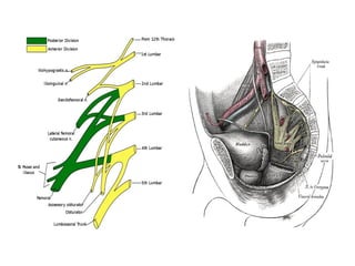
• It isdue to stretching of the
lumbosacral trunk by the
prolapsed intervertebral disc
between L5 and S1
• Backward rotation of the
sacrum during labour may
also be a contributory factor
• Direct pressure either by fetal head
or forcep blade on the lumbosacral
cord or sacral plexus
94.
• Condition isusually mild
• May pass unnoticed
• Neurological examination reveals
lower motor neurone type of lesions
with placcidity and wasting of
muscles in areas supplied by femoral
nerve or lumbosacral plexus
• Secondary loss is always present
95.
• Management ofdamaged
lumbosacral nerve roots is same as
that of the proplapsed
intervertebral disc in consultation
with an orthopaedic surgeon.
• Paraplegia due to epidural
haematoma or abcess is rare.
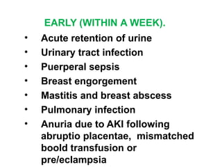
EARLY (WITHIN AWEEK).
• Acute retention of urine
• Urinary tract infection
• Puerperal sepsis
• Breast engorgement
• Mastitis and breast abscess
• Pulmonary infection
• Anuria due to AKI following
abruptio placentae, mismatched
boold transfusion or
pre/eclampsia
INTRODUCTION
• In thefirst 3 months after delivery,
the incidence of mental illness is
high.
• Overall incidence is about 15-20%.
• Sleep deprivation, hormone
elevation near the end of gestation
and massive postpartum
withdrawal contribute to the high
risk
103.
HIGH RISK FACTORS
PASTHISTORY:
• Psychiatric illness
• Puerperal psychiatric
illness
FAMILY HISTORY:
• Major psychiatric illness
• Marital conflicts
• Lack of social support
PUERPERAL BLUES
• Itis transient state of mental
illness observed 4-5 days after
delivery
• Lasts for few days
• Incidence is 50 %
106.
MANIFESTATIONS ARE:
• Depression
•Anxiety
• Tearfulness
• Insomnia
• Helplessness
• Negative feelings towards the infant
• No specific metabolic or endocrine
abnormalities detected
• But lowered troptophan (neurotransmitor
serotonin) level is observed. it indicats
altered neurotransmitter function
POSTPARTUM DEPRESSION
• Observedin 10-20 % of
mothers
• More gradual in onset over the
first 4-6 months following
delivery or abortion
• Changes in the hypothalamo-
pitutary- adrenal axis may be
a cause
109.
MANIFESTED BY:
• Lossof energy
• Loss of appetite
• Insomnia
• Social withdrawal
• Irritability
• Suicidal ideation
• Risk of reccurence is 50-100% in
subsequence pregnancies
110.
TREATMENT:
• Is startedearly
• Fluoxetine or paroxetine
(Selective serotonin reuptake
inhibitors)
• General supportive measures
111.
POSTPARTUM PSYCHOSIS
• Observedin 0.14-0.26 % of
mothers
• Commonly seen in women with
past history and family history
• Onset is relatively sudden
• Lasts for 4 days
112.
MANIFESTED BY:
• Fear
•Restlessness
• Confusion followed by
hallucinations, delusions and
disorientation
• Suicidal, infanticidal ideation
• Temporary seperation and
clinical supervision is needed
• Risk for reccurence 20-25%
113.
MANAGEMENT
• A psychiatristmust be consulted urgently
• Hospitalization is needed
• Chlopramazine 150 mg stat and 50-150
mg three time /day is started
• Sublingual estradiol 1 mg TDS
causes significant improvement
• Electro convulsive therapy if remains
unresponsive or in depressive psychosis
• Lithium in manic depressive psychosis
• Breast feeding is restricted in case of
lithium administration
114.
PSYCHOLOGICAL RESPONSES TOTHE
PERINATAL DEATHS AND MANAGEMENT
• Most perinatal events are joyful
• But when a fetal /neonatal death
occurs, social attention must be given
to grieving parents and family
• It may also be because of
unexcpected hysterectomy, birth of
malformed or chronically ill infant
• Prolonged seperation from chronically
ill infant can also cause grief
115.
Physician, nurse andattending staff
must understand patient's reaction
The common maternal somatic
symptoms are;
• Insomnia
• Fatigue
• Sighing respiration
• Feeling of guilt
• Anger
• Hostility ( feeling of
opposition)
116.
MANAGEMENT OF PERINATAL
GRIEVING
•Facilitating grieving process with
consolation (comfort), support,
sympathy
• Others are:
• Supporting the couple in seeing/ holding/
taking photographs of infant
• Autopsy requests
• Planning investigations
• Follow up visits
• Plan for subsequent pregnancy