Bangalore Call Girl Whatsapp Number 100% Complete Your Sexual Needs
A novel microfluidic device for rapid melanoma diagnosis
1. Final Project Paper Bioengineering 121
A Novel Microfluidic Device for Rapid Melanoma Diagnosis
Danielle Beeve, Luke Cassereau, Regine Labog, and Tomoya Saito
University of California, Berkeley, Department of Bioengineering
Over 3 million Americans a year are diagnosed with skin cancer and 1 in 5 will be
diagnosed in their lifetime making skin cancer the most common cancer. While usually benign, a
small percentage of people are diagnosed with a potentially lethal metastatic melanoma.
Metastatic melanoma causes over 70% of skin cancer related deaths. The threat from melanoma
can be eliminated if diagnosed early enough allowing treatment to start prior to the development
of aggressive metastatic tumors. We are proposing a microfluidic device that can generate fast
bedside quantitative results while also reducing costs and need for invasive biopsies. Successful
design and application of this device would not only improve melanoma diagnosis but also serve
as a proof of concept for similar devices for other cancer types.
Introduction the skin cancer-related deaths, and it requires
Skin cancer is the most common type of the most immediate attention. Fortunately,
cancer, with 1 in 5 American diagnosed with only 5% of patients have melanoma.
skin cancer in their lifetime. There are 3.5 However, due to its rarity and the fact that
million cases of skin cancer per year in the few people regularly visit dermatologists,
United States alone, and skin-cancer related potential melanomas are often overlooked.
deaths are as high as 200,000 per year Current diagnostics are not sufficient, as they
worldwide. are too slow, qualitative, and expensive. It is
All cancer is based on aberrant cell usually a 4-step process and there are
behavior leading to uncontrolled growth. associated issues with each step:
Treatment effectiveness decreases with tumor 1. Patient visual examination: depends on
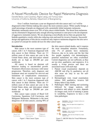
progression. Therefore, early diagnosis and ABCDE rule (Fig 2), qualitative, not all
treatment onset are essential for survival and skin areas are easy to see for self-
limitations of complications (metastases). examination
Skin cancer is the most common type of 2. Dermatologist visit, visual examination
cancer, with 1 in 5 American diagnosed with redone: Still qualitative, invasive full
skin cancer in their lifetime. There are 3.5 biopsy taken if any doubt.
million cases of skin cancer per year in the 3. Pathologist: H&E staining and sectioning
United States alone, and skin-cancer related followed by image analysis, still
deaths are as high as 200,000 per year qualitative and subject to error, depends
worldwide. on images used, level of staining is prone
Skin cancer types consist of Basal Cell to variability, Expensive to have a full
Carcinoma, Squamous Cell Carcinoma, and time pathologist.
Malignant Melanoma. Melanoma is the most
dangerous of all skin cancers, causing 90% of
Basal Cell Squamous Cell Malignant
Carcinoma Carcinoma Melanoma
Figure 1: Skin Cancer Types Figure 2: ABCDE Rule for Visual Melanoma
Diagnosis
2. Final Project Paper Bioengineering 121
4. Full-body imaging: only occurs if isolated 5 key genes, which in a DNA chip
pathologist determines growth is format were able to distinguish melanoma
potentially a melanoma, used to search for from benign skin growths.
other metastases, repeated regularly to A microfluidic device would allow
insure no reoccurrence. Also prone to mRNA measurements in a faster format. Only
error as it is a qualitative analysis. a small sample size is needed and no
At present, more advance medical amplification is required. There is also an
facilities have transitioned to additional added benefit of no full biopsy, which is
biochemical testing. Nevertheless, the better for patients. There is less risk of mRNA
development of new approaches to improve degradation: having an enclosed device means
existing cancer diagnostics and therapeutics there is no RNAase exposure besides that on
has proven to be insufficient. the outside of the sample. For further details
on biochemistry, refer to the supplementary
Potential of Biochemistry paper by Luke Cassereau.
Promising diagnostic designs have arisen
using biochemical analysis. Properly applied Device Design
biochemical assays could provide faster Our proposed device involves direct
results in a quantitative manner. It would mRNA measurements of five relevant genes
allow more accurate diagnosis in less time to melanoma: TRYP1, Melan-A, KIT,
which would allow treatment to begin MYO5A, and ENDRB. This method provides
immediately thus increasing change of much faster results than traditional methods,
survival. Many different targets can be while still being able to accurately predict
selected including proteins, DNA methylation melanoma/skin growth severity. For more
patterns, and mRNA, but the best choice of information on the five genes, refer to the
biomarker is mRNA. mRNA is indicative of supplementary paper by Luke Cassereau.
future behavior of potential tumor cells, The overview of our device design can be
which is a prognosis/potential risk of seen in Figure 3. The device design is based
melanoma or metastases. Previous work has on PDMS-based fluid flow physics. It
Figure
3:
Device
design
overview
3. Final Project Paper Bioengineering 121
SDS
NaC12H25SO4
http://upload.wikimedia.org/wikipedia/commons/thumb/4/4c/
Sodium_dodecyl_sulfate.svg/800px-Sodium_dodecyl_sulfate.svg.png
Figure
5:
SDS
Structure
http://www.molecularstation.com/cell/cell-lysis/
chosen to lyse the skin cells for our device.
Figure 4: Cell lysis overview SDS is often used in DNA extraction and
involves
two major steps: 1) Cell Lysis and 2) protein unraveling for polyacrylamide gel
Detection. Each will be discussed in detail. electrophoresis (SDS-PAGE). SDS acts as a
detergent and begins to break apart the cell
Cell Lysis membrane on contact. While there are many
In order to extract mRNA from the skin detergents that can accomplish cell lysis, SDS
sample, cell lysis is clearly necessary. There has additional advantages. Not only does it
are countless ways to perform cell lysis, require the least amount of time among
which include mechanical, electrical, detergents (30 seconds) to complete cell lysis,
chemical, and thermal techniques.1 We but as a strong anionic detergent, it also has
determined that a chemical technique would the ability to immediately denature enzymes
be the most practical for our application due such as DNAse and RNAse.3 SDS is
to its simplicity and relatively low cost. The purposely used in this manner to inhibit
chemical technique utilizes a detergent RNAse and prevent mRNA deterioration in
solution, which is used to agitate the cell our device. The original device design was
membrane (Fig 4). The detergent has going to require an RNAse inhibitor solution
hydrophobic long, linear alkyl chains that separately mixed with the skin sample
disorganize and break the membrane’s lipid solution prior to cell lysis, but SDS made that
bilayer. step unnecessary.
There is unfortunately no standard The structure of SDS (Fig 5) shows a tail
protocol for selecting a detergent to use for of 12 carbon atoms, attached to a sulfate
membrane lysis. In general, nonionic and group, giving the molecule the amphiphilic
zwitterionic detergents are milder and less properties required of a detergent. The
denaturing than ionic detergents and are used structure also provides it with a binding that is
to solubilize membrane proteins where it is cooperative, which means that the binding of
critical to maintain protein function and/or one molecule of SDS increases the likelihood
retain native protein:protein interactions for that another molecule of SDS will bind to that
enzyme assays or immunoassays. CHAPS, a protein. This alters most proteins into rigid
zwitterionic detergent, and the Triton-X series rods whose length is proportional to
of nonionic detergents are commonly used for molecular weight.4 The amount of detergent
these purposes. In contrast, ionic detergents needed for optimal protein extraction depends
are strong solubilizing agents and tend to on the critical micelle concentration (CMC),
denature proteins, thereby destroying protein aggregation number, temperature and nature
activity and function.2 of the membrane and the detergent.4 CMC for
Sodium dodecyl sulfate (SDS, SDS in pure water at 25°C is 0.0082 M,5 and
NaC12H25SO4), an anionic surfactant, was
4. Final Project Paper Bioengineering 121
the aggregation number at this concentration method uses a fluorophore and quencher that
is usually considered to be about 62.6 are attached to each end of the stem structure
The SDS is diluted to a 0.2% of the molecular beacons. In the absence of
concentration solution for our application.7 target mRNA, the quencher is located right
The channel length required to mix SDS and next to the fluorophore and prevents any
sample completely is approximately 8 cm, but fluorescent signal. Once hybridization occurs
our device has a 15.2 cm length to ensure however the fluorophore is released from the
lysis.8 SDS and cell sample flow rate is vicinity of the quencher and you are left with
roughly ~0.2 µl/min.8 This process may take a strong fluoresecent signal indicating the
anywhere from 30-190 seconds.1,3 presence of target mRNA of interest. For our
particular device design we have decided to
Detection adhere a set of molecular beacons down to the
To detect our particular genes of interest bottom of each of the 5 wells that correspond
we have chosen to use currently existing to our 5 different genes of interest by using
Molecular Beacon technology. This technique avidin-biotin surface adhesion to glass. Biotin
involves the use of a DNA (or RNA in our is attached to the quencher side of the stem of
case) stem-loop structure where the loop each molecular beacon while avidin is
contains a complementary probe sequence for adsorbed onto a glass slide that will be used
one of our 5 individual target genes of as the substrate to bind our PDMS to. Once a
interest. The stem contains complementary solution of these biotin-enhanced molecular
base pairs that keep the structure together in beacons comes into contact with the avidin
the absence of target mRNA, and it is absorbed onto the glass slide the biotin fits
designed in such a way that in the presence of into the avidin like a lock-and-key mechanism
target mRNA exactly complimentary to the and the molecular beacons are anchored into
probe sequence the stem-loop will place. Each of the 5 wells will have a set of
spontaneously unfold and hybridize to the molecular beacons containing a different
target. If there is even one base pair mismatch complementary probe sequence and as the
between the target mRNA and probe sample solution from the patient’s cells flows
sequences this spontaneous hybridization will along the device, target mRNA of interest (if
not occur, making this and extraordinarily it is present in the sample) will hybridize to
specific genetic detection method. the molecular beacon probes causing
The quantitative aspect of this detection fluorescence. This fluorescent signal can then
be detected using a plate reader that has been
!"#$%&'(#)"#$&&(#!"#$%&'(#*+,+(#!"#(#,-../,0,1"
specialized to fit our device design, and the
presence or absence of fluorescent signal for
Figure
6:
Molecular
Beacon
Figure
7:
Molecular
Beacon
Binding
in
device
5.
Final Project Paper Bioengineering 121
Mixing
Channel
Cell Sample
YES YES YES YES YES NO
TRYPI MELAN-A KIT MYO5A ENDRB Control Outlet
SDS
Figure 8: Device design with red showing fluid flow. Green wells indicate a positive reading and red wells indicate a
negative reading.
each of the 5 genes will indicate whether refer to the supplementary paper by Regine
melanoma is present in the patient sample. Labog.
For more specifics (including pictorial
diagrams of the molecular mechanisms and Expected Impact
corresponding references) on this entire We have designed a point-of-care
detection process see Danielle Beeve’s paper. diagnostic that highlight key factors to
significantly improve upon current
Fabrication technologies:
The device is fabricated using soft 1. Reduces cost, wait time, and invasiveness
lithography techniques. A silicon wafer is 2. Provides quantitative results and accurate
lithographically patterned with a mask design, prognosis
using SU-8 negative photoresist. The 3. Can be used to quickly decide best
resulting wafer mold is then patterned onto approach for each individual patient in
PDMS. 1mm holes punched at the two inlets one doctor’s visit
and the outlet on the PDMS device. The glass With these potential improvements, we hope
slide is coated with molecular beacons in each that this design will be implemented into
well with the sequence of DNA that we are standard diagnostic methodology in the near
looking for. The PDMS is then bonded on a future.
glass slide. For further details on the
fabrication method, refer to the Future Work
supplementary paper by Regine Labog. Many improvements can be made to our
current proposed device. These include
Overall design alternative skin sample acquisition methods,
The full movement of liquid through our alternative cell lysis methods, and alternative
device can be seen in Figure 8. The SDS inlet genetic detection methods. Alternative
is split into two channels that later meet to detection methods include Quartz Crystal
flank the sample solution. Once combined, Microbalance (QCM), Surface Acoustic
they flow together in a mixing channel Waves (SAW), and DNA microchip
composed of a series of S-curves. As the technologies. RNA isolation and addition of
lysate flows to the detection line, pressure PBS buffer to SDS are also considerations in
valves stops the flow for a certain period of order to possibly improve detection signal.
time at each well to ensure proper mixing and One long-term goal is to apply this device to
detection. After the fifth well containing the other cancers and diseases. This will require a
relevant gene, the flow goes onto a sixth well clear understanding of which relevant genes
that contains a control, and then into an outlet. for each disease can be used with the
For more information on the overall design, molecular beacon technology.
6. Final Project Paper Bioengineering 121
Discussion 4. "Detergents for Cell Lysis." Protein
Melanoma is a deadly disease and it is Purification, Modification and
clear that current diagnostics are far from Detection: Pierce Protein Research.
ideal. Biochemistry and microfluidics provide Thermo Fisher Scientific. Web. 16 Dec.
a potential solution to this problem. Possible 2010.
benefits include reduced cost, shorter wait <http://www.piercenet.com/browse.cfm?
times, less invasiveness, quantitative results, fldID=5558F7E4-5056-8A76-4E55-
and higher accuracy. The potential for 4F3977738B63>.
extension to other cancers and genetic 5. P. Mukerjee and K. J. Mysels, "Critical
diseases makes this novel diagnostic device a Micelle Concentration of Aqueous
viable choice for future research. Surfactant Systems", NSRDS-NBS 36,
US. Government Printing Office,
References Washington,.D.C., 197 1.
1. J. Kim, M. Johnson, P. Hill and B. K. 6. N.J. Turro. A. Yekta, J. Am. Chem. Soc.,
Gale, Microfluidic sample preparation: 1978, 100, 5951
cell lysis and nucleic acid purification, 7. Yu, L., Huang, H., Dong, X., Wu, D.,
Integr. Biol., 2009, 1(10), 574–586. Qin, J., Lin, B., Electrophoresis 2008, 29,
2. "Cell Lysis Solutions." Protein 5055–5060.
Purification, Modification and 8. X. Chen, D. Cui, C. Liu and H. Cai, Chin.
Detection: Pierce Protein Research. J. Anal. Chem., 2006, 34,1656–1660.
Thermo Fisher Scientific. Web. 16 Dec.
2010. Note: References from the papers of group
<http://www.piercenet.com/browse.cfm? members shall be coupled to this list.
fldID=5559C287-5056-8A76-4E25-
8975D8025374>.
3. Pang, Z., Al Mahrouki, A., Berezovski,
M., Krylov, S. N., Electrophoresis 2006,
27, 1489–1494.