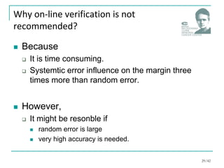
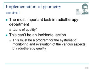
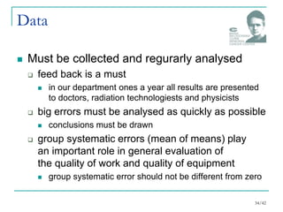
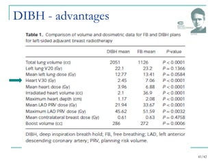
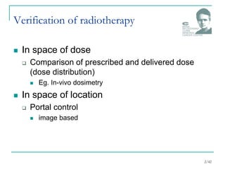
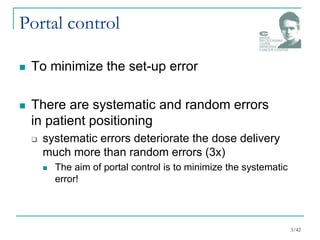
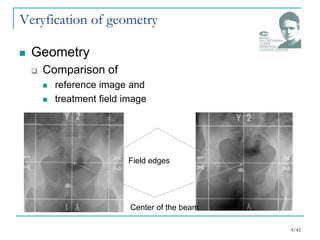
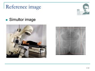
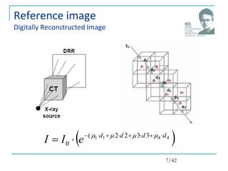
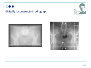
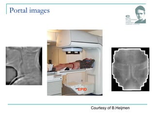
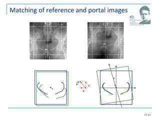
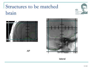
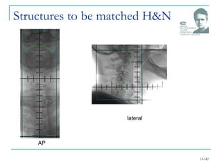
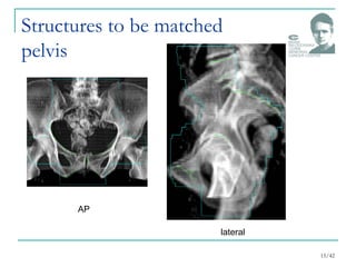
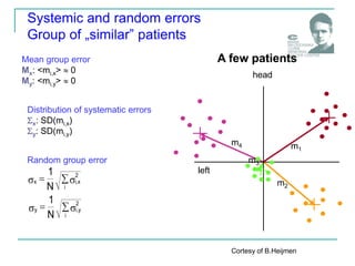
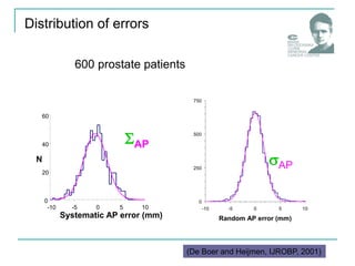
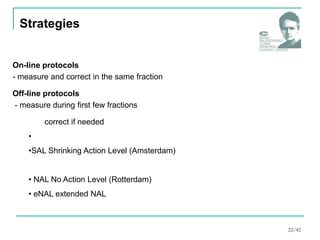
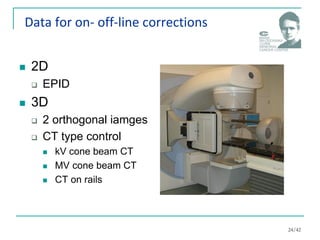
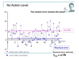
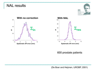
IGRT2
How to use image information?
Paweł Kukołowicz
Verification of radiotherapy
 In space of dose
 Comparison of prescribed and delivered dose
(dose distribution)
 Eg. In-vivo dosimetry
 In space of location
 Portal control
 image based
2/42
Portal control
 To minimize the set-up error
 There are systematic and random errors
in patient positioning
 systematic errors deteriorate the dose delivery
much more than random errors (3x)
 The aim of portal control is to minimize the systematic
error!
3/42
Veryfication of geometry
 Geometry
 Comparison of
 reference image and
 treatment field image
Field edges
Center of the beam
4/42
Reference image
 Simultor image
5/42
Reference image
 Simultor image
6/42
Reference image
Digitally Reconstructed Image
 
4
4
1
1 3
3
2
2
(
0
d
d
d
d
e
I
I 








 



7/42
DRR
digitally reconstructed radiograph
8/42
Quality of DRR
 Depends on
 slice separation
 it is recommended to use 3 mm slice separation
 but
 3 mm slice separation makes contouring very
tedious
 interpolation tools
 these tools must be checked !
9/42
Portal images
EPID
Courtesy of B.Heijmen
Edges
zero of the second derivative of intensity
11/42
Matching of reference and portal images
X
Y
P

12/42
Structures to be matched
brain
13/42
AP
lateral
Structures to be matched H&N
14/42
AP
lateral
Structures to be matched
pelvis
15/42
AP
lateral
Correction strategies
0
-10
-5
5
10
0 5 10 15 20 25
Fraction
Systematic and random errors
Single patient, one direction
Mean value = systematic error
Standard deviation = random error
AP direction
Cortesy of B.Heijmen
-10
-5
0
5
10
0 5 10 15 20 25
Fraction
patient 2
patient 1
patient 3
Systematic and random errors
For a few patients
mm
Cortesy of B.Heijmen
Systematic and random errors – 2D
head
left
Cortesy of B.Heijmen
Mean group error
Mx: <mi,x>  0
My: <mi,y>  0
Distribution of systematic errors
x: SD(mi,x)
y: SD(mi,y)
Random group error



i
2
x
,
i
x
N
1



i
2
y
,
i
y
N
1
m1
m3
m2
m4
A few patients
head
left
Systemic and random errors
Group of „similar” patients
Cortesy of B.Heijmen
600 prostate patients
Distribution of errors
AP
0
250
500
750
-10 -5 0 5 10
Random AP error (mm)
N
AP
0
20
40
60
-10 -5 0 5 10
Systematic AP error (mm)
N
Cortesy of B.Heijmen
(De Boer and Heijmen, IJROBP, 2001)
On-line protocols
- measure and correct in the same fraction
Off-line protocols
- measure during first few fractions
correct if needed
•
•SAL Shrinking Action Level (Amsterdam)
• NAL No Action Level (Rotterdam)
• eNAL extended NAL
Strategies
22/42
10
-5
0
5
10
15
0 5 15 20 25 30 35
AP displacements (mm)
Prostate cancer patient
Fraction
On-line correction
A few MU
image
Remainder MU
most dose delivered with very small errors:
correction of both systematic
and random errors
23/42
Data for on- off-line corrections
 2D
 EPID
 3D
 2 orthogonal iamges
 CT type control
 kV cone beam CT
 MV cone beam CT
 CT on rails
24/42
NAL (de Boer and Heijmen, IJROBP, 2001)
 Fraction 1,2, and 3
 set-up a patient according to protocol
 portal control, (mix,miy,miz), i =1 ,2,3
 before 4th fraction
 calculate the systematic error
(mx,mean,my,mean,mz,mean)
 from 4th fraction on
 set-up a patient according to prtocol
 shift couch with –(mx,mean,my,mean,mz,mean)
 irradiate
de Boer and Heijmen, Med. Phys. 2002
10
-5
0
5
10
15
0 5 15 20 25 30 35
Fraction
: initial error after set-up
Residual error
No NAL
No Action Level
The random error remains the same!
: error after correction
Residual error estimate
res  /N
(De Boer and Heijmen, IJROBP, 2001)
600 prostate patients
0
20
40
60
-10 -5 0 5 10
Systematic AP error (mm)
N
With no correction
in
0
20
40
60
80
100
-10 -5 0 5 10
Systematic AP error (mm)
N
With NAL
res
NAL results
(1) De Boer et al. 2002
(2) Kaatee et al. 2002
(3) De Boer et al. 2003
(4) De Boer et al. 2004
How precise may be radiotherapy?
Residual (after NAL) bony anatomy
displacements [mm]:
Prostate
(1)
Cervix
(2)
LR CC AP
 1.7 1.5 1.6
res 1.1 1.1 1.1
 2.6 2.9 2.7
res 1.2 1.7 1.6
Lung
(3)  2.0 2.4 2.4
res 1.3 0.6 1.2
head & neck
(4)  1.6 1.4
res 1.1 1.2
1.6
1.0
28/42
Why on-line verification is not
recommended?
 Because
 It is time consuming.
 Systemtic error influence on the margin three
times more than random error.
 However,
 It might be resonble if
 random error is large
 very high accuracy is needed.
29/42
Set Up Margin
Internal Margin
Margins
 Set-up margin
 to compensate set-up errors
 errors measured with
respect to external
coordinate system (laser
system)
 Internal margin
 to compensate movement of
the target caused by
physiology (eg. breathing)
 errors measured with
respect to internal anatomy
coordinate system
( eg. pubis symphisis)
30/42
   2
int
2
ernal
up
set
tot 



 
   2
int
2
ernal
up
set
tot 

 
 
How to add margins?
 If set-up and internal errors may be treated
as not correlated, than we add errors in
quadrature
systematic
random
31/42
tot
tot
M 




 7
,
0
2
Margins
 Two formulas
tot
tot
M 




 7
,
0
5
,
2
To cover the CTV for 90% of the
patients with the 95%
isodose (analytical solution).
Herk Red, 47: 1121 - 1135, 2000
Margin size which ensures at least
95% dose is delivered to (on
average) 99% of the CTV.
Stroom, Red, 43: 905-919,1999
32/42
Implementation of geometry
control
 The most important task in radiotherapy
department
 „Lens of quality”
 This can’t be an incidental action
 This must be a program for the systematic
monitoring and evaluation of the various aspects
of radiotherapy quality
33/42
Data
 Must be collected and regurarly analysed
 feed back is a must
 in our department ones a year all results are presented
to doctors, radiation technologiests and physicists
 big errors must be analysed as quickly as possible
 conclusions must be drawn
 group systematic errors (mean of means) play
an important role in general evaluation of
the quality of work and quality of equipment
 group systematic error should not be different from zero
34/42
Breathing and related problems
35/42
CT for planning
 Artefacts
36/42
without breathing control with breathing control
Changes of GTV position
37/42
In relations to bronchial tree
CT for planning
 With breathing control
38/42
RPM system
Pattern of breathing
39/42
Patient A
Patient B
Deep-inspiration breath hold technique
 DIBH
 for patients with left breast cancer
 for some of tchem (they have to inhale and keep inhale
for some time 10 – 15 sec)
40/42
DIBH - advantages
41/42
Thank you for your attention!
p.kukolowicz@zfm.coi.pl

6. image guided radiation therapy 12.pdf

  • 1.
    IGRT2 How to useimage information? Paweł Kukołowicz
  • 2.
    Verification of radiotherapy In space of dose  Comparison of prescribed and delivered dose (dose distribution)  Eg. In-vivo dosimetry  In space of location  Portal control  image based 2/42
  • 3.
    Portal control  Tominimize the set-up error  There are systematic and random errors in patient positioning  systematic errors deteriorate the dose delivery much more than random errors (3x)  The aim of portal control is to minimize the systematic error! 3/42
  • 4.
    Veryfication of geometry Geometry  Comparison of  reference image and  treatment field image Field edges Center of the beam 4/42
  • 5.
  • 6.
  • 7.
    Reference image Digitally ReconstructedImage   4 4 1 1 3 3 2 2 ( 0 d d d d e I I               7/42
  • 8.
  • 9.
    Quality of DRR Depends on  slice separation  it is recommended to use 3 mm slice separation  but  3 mm slice separation makes contouring very tedious  interpolation tools  these tools must be checked ! 9/42
  • 10.
  • 11.
    Edges zero of thesecond derivative of intensity 11/42
  • 12.
    Matching of referenceand portal images X Y P  12/42
  • 13.
    Structures to bematched brain 13/42 AP lateral
  • 14.
    Structures to bematched H&N 14/42 AP lateral
  • 15.
    Structures to bematched pelvis 15/42 AP lateral
  • 16.
  • 17.
    0 -10 -5 5 10 0 5 1015 20 25 Fraction Systematic and random errors Single patient, one direction Mean value = systematic error Standard deviation = random error AP direction Cortesy of B.Heijmen
  • 18.
    -10 -5 0 5 10 0 5 1015 20 25 Fraction patient 2 patient 1 patient 3 Systematic and random errors For a few patients mm Cortesy of B.Heijmen
  • 19.
    Systematic and randomerrors – 2D head left Cortesy of B.Heijmen
  • 20.
    Mean group error Mx:<mi,x>  0 My: <mi,y>  0 Distribution of systematic errors x: SD(mi,x) y: SD(mi,y) Random group error    i 2 x , i x N 1    i 2 y , i y N 1 m1 m3 m2 m4 A few patients head left Systemic and random errors Group of „similar” patients Cortesy of B.Heijmen
  • 21.
    600 prostate patients Distributionof errors AP 0 250 500 750 -10 -5 0 5 10 Random AP error (mm) N AP 0 20 40 60 -10 -5 0 5 10 Systematic AP error (mm) N Cortesy of B.Heijmen (De Boer and Heijmen, IJROBP, 2001)
  • 22.
    On-line protocols - measureand correct in the same fraction Off-line protocols - measure during first few fractions correct if needed • •SAL Shrinking Action Level (Amsterdam) • NAL No Action Level (Rotterdam) • eNAL extended NAL Strategies 22/42
  • 23.
    10 -5 0 5 10 15 0 5 1520 25 30 35 AP displacements (mm) Prostate cancer patient Fraction On-line correction A few MU image Remainder MU most dose delivered with very small errors: correction of both systematic and random errors 23/42
  • 24.
    Data for on-off-line corrections  2D  EPID  3D  2 orthogonal iamges  CT type control  kV cone beam CT  MV cone beam CT  CT on rails 24/42
  • 25.
    NAL (de Boerand Heijmen, IJROBP, 2001)  Fraction 1,2, and 3  set-up a patient according to protocol  portal control, (mix,miy,miz), i =1 ,2,3  before 4th fraction  calculate the systematic error (mx,mean,my,mean,mz,mean)  from 4th fraction on  set-up a patient according to prtocol  shift couch with –(mx,mean,my,mean,mz,mean)  irradiate de Boer and Heijmen, Med. Phys. 2002
  • 26.
    10 -5 0 5 10 15 0 5 1520 25 30 35 Fraction : initial error after set-up Residual error No NAL No Action Level The random error remains the same! : error after correction Residual error estimate res  /N
  • 27.
    (De Boer andHeijmen, IJROBP, 2001) 600 prostate patients 0 20 40 60 -10 -5 0 5 10 Systematic AP error (mm) N With no correction in 0 20 40 60 80 100 -10 -5 0 5 10 Systematic AP error (mm) N With NAL res NAL results
  • 28.
    (1) De Boeret al. 2002 (2) Kaatee et al. 2002 (3) De Boer et al. 2003 (4) De Boer et al. 2004 How precise may be radiotherapy? Residual (after NAL) bony anatomy displacements [mm]: Prostate (1) Cervix (2) LR CC AP  1.7 1.5 1.6 res 1.1 1.1 1.1  2.6 2.9 2.7 res 1.2 1.7 1.6 Lung (3)  2.0 2.4 2.4 res 1.3 0.6 1.2 head & neck (4)  1.6 1.4 res 1.1 1.2 1.6 1.0 28/42
  • 29.
    Why on-line verificationis not recommended?  Because  It is time consuming.  Systemtic error influence on the margin three times more than random error.  However,  It might be resonble if  random error is large  very high accuracy is needed. 29/42
  • 30.
    Set Up Margin InternalMargin Margins  Set-up margin  to compensate set-up errors  errors measured with respect to external coordinate system (laser system)  Internal margin  to compensate movement of the target caused by physiology (eg. breathing)  errors measured with respect to internal anatomy coordinate system ( eg. pubis symphisis) 30/42
  • 31.
      2 int 2 ernal up set tot          2 int 2 ernal up set tot       How to add margins?  If set-up and internal errors may be treated as not correlated, than we add errors in quadrature systematic random 31/42
  • 32.
    tot tot M       7 , 0 2 Margins Two formulas tot tot M       7 , 0 5 , 2 To cover the CTV for 90% of the patients with the 95% isodose (analytical solution). Herk Red, 47: 1121 - 1135, 2000 Margin size which ensures at least 95% dose is delivered to (on average) 99% of the CTV. Stroom, Red, 43: 905-919,1999 32/42
  • 33.
    Implementation of geometry control The most important task in radiotherapy department  „Lens of quality”  This can’t be an incidental action  This must be a program for the systematic monitoring and evaluation of the various aspects of radiotherapy quality 33/42
  • 34.
    Data  Must becollected and regurarly analysed  feed back is a must  in our department ones a year all results are presented to doctors, radiation technologiests and physicists  big errors must be analysed as quickly as possible  conclusions must be drawn  group systematic errors (mean of means) play an important role in general evaluation of the quality of work and quality of equipment  group systematic error should not be different from zero 34/42
  • 35.
    Breathing and relatedproblems 35/42
  • 36.
    CT for planning Artefacts 36/42 without breathing control with breathing control
  • 37.
    Changes of GTVposition 37/42 In relations to bronchial tree
  • 38.
    CT for planning With breathing control 38/42 RPM system
  • 39.
  • 40.
    Deep-inspiration breath holdtechnique  DIBH  for patients with left breast cancer  for some of tchem (they have to inhale and keep inhale for some time 10 – 15 sec) 40/42
  • 41.
  • 42.
    Thank you foryour attention! p.kukolowicz@zfm.coi.pl