
Recommended
More Related Content
Similar to 4.ANTIBODY-DEPENDENT RESPONSES.ppt
Similar to 4.ANTIBODY-DEPENDENT RESPONSES.ppt (20)
More from AnguaniVictor
Recently uploaded
Recently uploaded (20)
4.ANTIBODY-DEPENDENT RESPONSES.ppt
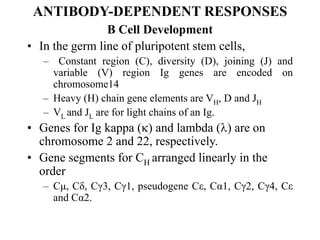
- 1. ANTIBODY-DEPENDENT RESPONSES B Cell Development • In the germ line of pluripotent stem cells, – Constant region (C), diversity (D), joining (J) and variable (V) region Ig genes are encoded on chromosome14 – Heavy (H) chain gene elements are VH, D and JH – VL and JL are for light chains of an Ig. • Genes for Ig kappa () and lambda () are on chromosome 2 and 22, respectively. • Gene segments for CH arranged linearly in the order – Cμ, Cδ, Cγ3, Cγ1, pseudogene Cε, Cα1, Cγ2, Cγ4, Cε and Cα2.
- 2. Immunoglobulins (Igs) • Igs are members of the immunoglobulin gene superfamily which includes – T cell receptor; MHC class II and I molecules; β2 – microglobulin; CD2; CD3; CD4; CD8 and C-reactive proteins. • Somatic recombination occurs before contact with antigen exposure during B cell development in the bone marrow.
- 3. Igs cont • One D and one J rearrange with DJ segment and similarly one V and J rearrange with VJ. – V(D) J recombination is expressed in developing B cells and recognize recombination signal sequences. • Lymphoid progenitor cells receive signals – Cytokines and recombinase from bone marrow stromal cells and – Undergo D-J joining on H-chain to become early pro-B cells and express CD45 (B220) and class II MHC molecules.
- 4. VDJ Rearrangement • A functional VDJH rearrangement essential for – Normal pro-B cell differentiation into pre-B cells. • Those that fail to make a functional VDJH rearrangement – Undergo apoptosis, phagocytized by bone marrow macrophages. • Pro-B cells become pre-B cells when they express membrane H chains with surrogate light chains in the pre-B cell receptor.
- 5. VDJ cont • Following proliferation, – Synthesized and µ-chain expressed on cell membrane and – Develops into an immature B cell that is very sensitive to an antigen. • Different stages in B cell development distinguished by – Expression of mu (µ) heavy chain, surface Ig (SmIg) and CD antigens
- 6. TABLE 10: Stages in B Cell Development Stem cell Early pro-B cell Late pro-B cell Large pre-B cell Small pre-B cell Immat ure B cell Mature B cell H chain genes Germ line D-J joining V-DJ joining VDJ rearran ged VDJ rearran ged VDJ rearran ged VDJ rearran ged L chain genes Germ line Germ line Germ line Germ line V-J joining VJ rearran ged VJ rearran ged Surfac e Ig none none none µ chain in pre- B recepto r µ chain in cytopla sm and on surface Membr ane IgM Membr ane IgM and IgD
- 7. Clomplementarity DR(CDR) • Each B cell generates a set of functional antibody genes to produce – One VDJ gene for the heavy chain and one VJ gene for either kappa () or lambda () of light chain. • Transcription of these genes permits formation of antibody molecules with a single combining specificity – Able to interact with antigenic determinant in a very specific manner through BCR. • Variable domains contain – Three hypervariable (complementarity determining regions, CDRs) comprising N-terminal domains of both heavy and light chains.
- 8. Allelic Exclusion • Mature and activated B cells may co-express surface IgM and surface IgD. – Express only one heavy chain allotype and one light chain isotype, despite the genetic capacity to encode two heavy chains and four light chains. – A reproductive rearrangement of one allele blocks the rearrangement of the other. – This phenomenon is referred to as allelic or isotypic exclusion, a cornerstone of clonal selection of B- lymphocytes.
- 9. Ig Regulation cont Regulation of B cell Development • Interleukin – 7 (IL –7) secreted by stroma cell and stem cell factor (SCF) initiate – Cytoplasmic cascades resulting in expression of proteins required for B cell development.
- 10. Light Chain Selection Light chain V-J joining occurs first for kappa () chain and productive rearrangement results into immature B cell expressing IgM () BCR and then IgM (). • When neither nor is productively rearranged, the cell undergoes apoptosis. • B cells usually rearrange DH and DL segments of both chromosomes simultaneously. • During pre-B cell stage, light chain V-J joining usually occurs first for (kappa) chain. • Developing cells are positively selected when the pre-B cell receptor binds its ligand. • Presence of a rearranged VH or VL gene signals B cells to suppress further recombination of genes.
- 11. Membrane Receptors • Membrane Ig (SmIg) comprises IgM and IgD co-expressed on surface of naïve virgin mature B cells. – Antigenic specific receptors are mainly expressed on B cells (BCR) and T cells (TCR). • Variable domains contain – Three hypervariable (complementarity determining region, CDR) and constant heavy region responsible for biological functions of the Ig. • TCR not secreted from the T cell – Expressed on the T cell membrane with a – Signal transduction complex, CD3 (invariant TCR) formed from identical subunits.
- 12. Ig Diversity • The body is capable of recognizing 10 to 7 antigenic deteminants • Diversity is achieved through several mechanisms including – Expression of germ line multiple variable (V) region genes; – VJ and VDJ combinations, – Random assortment of H and L chains; – Imprecise DNA recombination; – Various mutations which may occur in the V genes of H or L chains in the life time of a B cell and receptor editing.
- 13. Ig Diversity • Antibody diversity due to a – Combination process of immunoglobulin genes assembled from D and J regions. • Different V genes evolve by gene duplication and mutation giving rise to – 500- 1000 V-heavy chain different genes and about 12 diversity (D) and 4 joining (J) genes.
- 14. Ig Diversity cont • For both kappa (κ) and lambda (λ) glycoproteins, – There are about 200 different V-light chain genes and 6J genes. • Junctional diversity results from – Addition of L imprecise joining of gene segments : – Addition of nucleotides to the DNA sequences. – tdT adds up to 15 nucleotides to the DNA sequence of VH and JH regions. • B cells produce IgM and IgD receptors simultaneously thru alternative mRNA splicing. – One mRNA transcribed that encodes VDJH - Cμ – Cδ. – Ig diversity my be due to somatic hypermutation.
- 15. Isotype Switching • Isotype switching increases functiona diversiy thru stimulation and Th2 cytokine production. – Rearranged VDJH is always expressed first with membrane Cμ in the developing B cell with both membrane Cμ and Cδ in the mature B cell and secreted Cμ as the plasma cell. – B cell undergoes isotype switching from IgM to IgG. • Sequence of appearance of immunoglobulins – Pre-B cells appear at 13-16 weeks of gestation; – B lymphocytes with IgM, IgG and IgD in foetal liver and 12 weeks and thereafter in peripheral blood; – IgM first, then IgG and IgA.
- 16. Adult Ig Levels • Neonate respond to antigen exposure with IgM then IgG – Adults produce IgG 5-15 days post-infection and – Infant elaborate only IgM for 20-30 days or up to 6 months of age. • Adult levels are achieved in 2 yrs (IgM), 5-6 yrs (IgG) and 10 yrs (IgA). Clonal Selection Theory • Clonal selection theory (CST) is illustrated below – B cells are selected by an antigen from a library or clones of the lymphocytes.
- 17. Effector B cells (Plasma cells) Memory B ells (Residual lgs) Antigen Effector T cells (CTL) Memory T cells Peptide from macrophages (APCs) Fig. 12A Fig. 12B Fig 12. Clonal Selection Theory: An antigen complementary with the specific BCR gives rise to effector plasma cells which secrete antibodies and memory B cells. Then antigen processed and presented by macrophages is recognized by TCR on the T cell clone which is triggered to differentiate into effector T cells (CTL) and memory T cells. Refer to the text for specific details.
- 18. B Cell Proliferaion • Each cell carries an antigenic receptor specific for epitopes of foreign antigens. • Complementarity – Between the epitopes and B cell antigenic receptors together with provision of other signals – Cell proliferates and differentiates into – Antibody forming cells (AFC) or plasma cells (effector cells) and memory cells.
- 19. Burnet Clonal Selection Theory • According to Burnet’s clonal selection theory, – Subsets of virgin immunocytes (termed clones) in circulation acquire immunity through selection by a specific antigen. – Recruitment of individual B cells through their interaction with antigenic determinants proliferate and eventually differentiate into • Plasma cells that secrete antibodies (effector cells) and B cells (memory cells) capable of responding to the inducing antigen.
- 20. Clonal Selection cont • Similarly in clonal selection of T cells – Recruitment of T cells leads to differentiation into • Effector cytotoxic lymphocytes (CTL) that eliminate virus, tumour cells or allografts and • Memory T cells capable of responding to the re- exposed antigen.
- 21. B Cell Activation • Antigen specific signals are derived – From interaction between B cells (APCs) and T cells – Provision of cytokines (T cell dependent activation) leading to production of specific antibodies. • In non-specific B cell activation, – No involvement of T cells and polyclonal antibodies are produced. • Natural antibodies arise independently of any known antigenic stimulation and – Can react with exogenous antigens.
- 22. Antigen Specific B Cell Actvation • Require processing and presentation of antigen in an immunogenic form by the accessory APCs. – B cells appear to ingest only soluble antigens by the process of pinocytosis. – Antigenic determinants generated are re-expressed on the surface of B cells in association with class II MHC antigens. • Leads to the activation of CD4 T cells liberating IL-4 and IL–5 cytokines
- 23. Ag Specific B Cell Activation cont • IL-4 stimulates B cell proliferation and mediates IgM+ B cell switching to IgG and other 1gs and – Development of Fc- receptors on B cells. – IL – 5 induces B cell proliferation and differentiation into IgM secreting cells. • A model of T cell dependent B cell activation is illustrated below
- 24. T Cell Dependent B Cell Activation Fig. 13: T cell Dependent Activation of B cells. Endocytosis of antigen digestion and interaction with MHC molecules and CD4 T cells leads to differentiation of B cells into plasma cells that secrete antibodies. Refer to text for details. Source: users.rcn.com/ikimball.ma.ultranet/BiologyPages/B/B_and T-cells.html
- 25. Non Specific B Cell Activation • Polyclonal activation occurs due to – B cell expression of membrane receptors for C3d and Epstein-Barr virus (EBV); – Protein A – bearing staphylococci and gram-negative bacterial lipolysaccharides. • Other polyclonal B cell activators include – Mitogens eg plant lectins (phytohaemagglutinin, pokeweed mitogen); – Anti-Igs; antibodies to certain B cell differentiation antigens; – Bacterial and parasite derived products; tumour promoting agents.
- 26. Ig Levels • Levels of immunoglobulins produced depend on – Ethnic background, age and sex; – History of allergy; – Recurrent infections or endemicity of parasites in a particular population. Kinetics of Primary and Secondary Responses • Primary response characterized by – Frst measurable immune response – A longer lag phase and – IgM predominant antibody.
- 27. Secondary IR • Secondary response is associated with – A second exposure to same antigen, – Shorter lag phase, – Quicker onset and a higher magnitude, – Long lived memory and anamnestic response – IgG as the predominant antibody.
- 28. CD40- CD40L Interaction • Interaction between CD40 L provide important signals in – B cell central functions like proliferation, – Up regulation of membrane markers, – Isotype switching and memory B cell formation. • CD40 ligand (CD40L) – Member of the tumour necrosis factor superfamily expressed on the surface of • Activated T cells, dendritic cells, macrophages and epithelial cells.
- 29. CD40-CD40L Interaction cont • Interaction of CD40L with CD40 on B cells leads – B cell activation, Ig secretion and Ig class switching. • Ig class switch involves DNA recombination in which – A substitution of one Ig heavy chain constant region (CH) gene – Effected for another one with subsequent change in the CH region and biological activities of the Ig.
- 30. Kinetics of Pri and Sec IR Fig. (Image) 14. Kinetics of Primary (10) and Secondary (20) Immune Responses: Initial administration of antigen at day O leads to a lag phase before IgM antibodies are generated 10-14 days post-immunization period. When the antigen is again given at 28 days, as a booster, the immune response detected is more pronounced and durable and the lag period is shorter. Primary response is characterized by IgM antibody production while a secondary response is predominantly of IgG and other classes later Source: www.gla.ac.uk/- jmb17n/Teaching/L2taching/memvacc/pictures/secab.jpg.
- 31. Immunological Tolerance • Route of antigen administration determines nature of response induced. – Presentation of antigen via the gastrointestinal tract favours tolerance. – Intravenous injection also favours tolerance through induction of CD8 T cell suppressor circuits. • High zone tolerance designates the antigen dose above immunizing level – One below immunizing range termed low zone tolerance.
- 32. Tolerance Induction • Tolerance induced to a single epitope and no other antigenic determinant is termed split-tolerance – Manifests as depression of specific immune response involving either single or all isotypes and – Delayed type hypersensitivity or may be partial. • Immunological tolerance established – Early in ontogeny and in adult life partly through clonal abortion; clonal deletion; clonal anergy or silencing.
- 33. Clonal Abortion • Lymphocytes mature through a vulnerable stage – In which contact with a specific antigen results into inevitable death of the clones • When elimination involves functionally mature lymphocytes lacking T cell help the process is referred to as clonal deletion.
- 34. Clonal Anergy • When B cells encounter autoantigens, – There is down-regulatory signaling process (silencing) resulting into induction of a refractory state of hyporeactivity designated clonal anergy. • B cells are also rendered unresponsive through – Antigen dependent modulation of SmIg. – Iinteraction of antigen with IgM and IgD on neonatal B cells leads to Ig redistribution, capping and endocytosis of immune complexes formed.
- 35. Clonal Anergy cont • Mature B cells expressing IgM and IgD or other isotypes – Receptor require very high concentrations of antigen for tolerization. – Multivalent antigen immobilizes the antigen receptors and freezes the membrane in overwhelming high concentrations – Leading to a situation referred to as immunological paralysis
- 36. Basic Ig Structure • Antibodies are glycoproteins consisting of – Subunits containing two identical light chains (L chains) and two identical heavy chain (H chains) – With variable (V) regions at the N-terminal of both H and L chains. • Hypervariable regions in the V region – Construct the antigen binding site designated complementarity determining regions (CDRs) • The C-terminus of H and L chains form – Constant (C) regions consist of two kinds of kappa () 60% and lamda (λ), 40% for the L chains.
- 37. Basic Ig Formula • The basic formula of Ig subunit is – H2 L2, a four chain structure (some Igs composed of polymers of the basic monomeric form). • Five different kinds of C regions for H chains define isotypes (classes) – Mu (u) chains (H chain of IgM); – Gamma () chains (IgG); – Alpha () chains (IgA); – Delta () chains (IgD), and epsilon () chains (IgE).
- 38. Ig Structure cont • Light chain composed of 220 amino acid residues • Heavy chains composed of 440-550 amino acids held together by covalent disulfide bonds. • Variable regions – Contained within the (NH2) terminal and of the polypeptide chain (1-110 amino acids) and – Constant region comprising 111-220 or 440-550 amino acids residues.
- 39. Regulatory Structures on V and C • Isotypes are constant region determinants of Ig heavy chains – Distinguishes various classes and subclasses of Igs. – Determinants interact with Fc receptors, Ig binding factors and rheumatoid factors (RF) or anti-isotype antibodies. • Five classes of heavy chain isotypes exist – IgG1, IgG2, IgG3, IgG4; IgA1, IgA2; IgM; IgE and IgD.
- 40. Regulatory Structures • Idiotypes are antigenic determinants located in the V region of various isotypes. • Allotypes represent genetic variants (alleles) – Minly in the constant (C) region of the IgG and IgA and also in the V region. – Those associated with IgG are designated Gm – Am refer to alpha-chain IgA specific allotypes.
- 41. Gm and Am markers • Immunoglobulin heavy chain (Gm) and light chain (Km) allotypes are – Markers of susceptibility to a wide range of immune-mediated diseases (malignancies, infectious diseases and autoimmune disorders) Idiotypic network interaction • Idiotypes are antigenic determinants located in the V region of an Ig – Gives rise to production of anti-idiotypic antibodies and T cells. • Recognition structures involved are – B cell idiotypes (V-regions of Ig) and T cell idiotypes (V-regions of the T cell receptor).
- 42. TABLE 11: Immunoglobulin Profiles And Properties Isotypes IgM Ig D IgG1 IgG2 IgG3 IgG4 IgA1 IgA2 sIgA IgE Heavy chain domains Sedimentation coefficient (S) Carbohydrate content (%) Molecular weight (Mw x 1000) Serum concentrations (mgdL-1) Classical complement activation Maternal placental transmission Immediate hypersensitivity mediator Agglutinating potency Anti-toxin potency Neutralizing capacity Opsonic capacity 5 19 12 970 120 ++++ - - +++ + + - 4 7-8 14 184 3 - - - - - - - 4 6 3 146 900 ++ + + + +++ + +++ 4 6 3 146 300 + ± + + +++ + +++ 4 7 3 170 100 ++ + + + +++ + +++ 4 6 12 146 50 - + + + +++ + +++ 4 7 11 160 300 - - - + + + + 4 7 11 160 50 - - - + + + + 4 11 11 385 5 - - - + ++ +++ - 5 8 12 195 0.0 5 - - ++ + - - - - Mean normal immunoglobulin levels in serum, colostrums, milk and saliva are ( mg /ml): IgG 6-13; IgA 0.6 – 3; IgM 05 – 3; IgD <0.14 and IgE < 0. 0004
- 43. Ag.Ab Reactions • Antigens react with antibodies to form soluble or precipitating immune complexes. • Antibodies possess antigen-combining sites – Formed by at least 4 regions of extreme variability in the V regions of both H and L chains (hypervariable regions) – Antigen makes contact with about 10 – 12 amino acids in this region referred to as a cleft. • When an antibody binds strongly to only the immunizing antigen and to a lesser extent to other similar determinants it is termed specific. • A substance eg hapten with a single antigenic determinant (monovalent) combines with one paratope on the antibody.
- 44. Law of Mass Action • According to the Law of Mass Action, [Ag] + [Ab] = [Ag. Ab] (Antigen) (Antibody) (Immune complex, IC) Equilibrium constant, K = [Ag. Ab] [Ag] + [Ab] • Antibody affinity refers to the strength of a single antigen-antibody bond generated by the summation of positive and negative forces. • Antibody avidity is the sum total of the binding forces where both the antigen and antibody are multivalent – Relative strength of the antigen-antibody binding is dependent on the valency of the reacting substances.
- 45. Antibody Cross-Reactivity • Some antigens display shared structural similarity and the produced antIbody – Possess determinants demonstrate polyspecific reactions referred to as cross-reactivity. • The most common heterophile substance is – Forssman antigen found in red blood cells and pneumococci and salmonella. – Other shared antigens are between Treponema pallidum bacterium and cardiolipin (mammalian heart tissue component).
- 46. IgG Molecule • IgG molecule exists as – A monomer in membrane-bound form on the surface of B- lymphocytes and in secreted form. • IgG levels attained by 5-6 years of age – Is about 150 kDa in Mw consisting of 4 classes (IgG1, gG2, IgG3, and IgG4) • The only Ig able to cross the placenta to protect infant. – Selective transfer of IgG prevents maternal allergy producing IgE antibodies and ABO isoagglutinins of IgM accessing the child. – Maternally acquired measles IgG interfere with active immunization in children under 12 months of age.
- 47. IgG Molecule cont • IgG is produced by plasma cells in secondary lymphoid organs (lymph nodes and spleen) – Constitutes about 70 – 75% of total immunoglobulins produced daily in the body. – Structure of IgG displays a basic structure of an Ig as depicted below consisting of • Two light (L) chains (Mw 25kDa) and two heavy (H) chains (Mw 55 – 77kDa) held together by inter-and intra-chain disulphide (S-S) bonds. • Each chain is divided into – Variable (V) antigen binding fragment (Fab) and a constant (C) or crystallizable fragment (Fc). • Light chains are further divided into two subtypes, – Kappa (κ) and lamda (λ) on the basis of differences in antigenic determinants.
- 48. IgG cont • Hinge region is between the papain and pepsin cleavage sites rich in prolines – Separates the two combining sites – Contribute to molecular mobility of the Ig – Cysteines contribute to interchain disulphide bonds. • Domains are conserved units of molecular structures of Ig with a protein conferring a unique shape. – Different monomers and different levels of glycosylation contributes to variations in Ig molecular weights. • The polypeptide chains fold into – Three dimensional globular structures designated domains schematically presented below
- 49. IgG Cleavage Sites • Ig hydrolysis of the polypeptides at specific sites in the hinge region results in – IgG cleavage by pepsin digestion yields F(ab)2 and one Fc fragment – Papain digestion produces two Fab and one Fc fragment as shown in Fig. 18. • IgA molecule resistant to proteinase activity – Hinge region made up of proline residues reinforced by galactose – containing oligosaccharides
- 50. IgG Hinge Region • Hinge region is between the papain and pepsin cleavage sites rich in prolines – Separates the two combining sites – Contribute to molecular mobility of the Ig – Cysteines contribute to interchain disulphide bonds. • Domains are conseved units of molecular structures of Ig with a protein conferring a unique shape. – Different monomers and different levels of glycosylation contributes to variations in Ig molecular weights. • The polypeptide chains fold into – Three dimensional globular structures designated domains schematically presented below.
- 51. IgG Subclasses • IgG subclasses consist of four polypeptides – γ1, γ2, γ3 and γ4, designated IgG1, IgG2, IgG3 and IgG4, respectively. • Average normal serum concentrations are – 60 – 70% (IgG1); 14 – 20% (IgG2); 4 – 8% (IgG3); 2 – 6% (IgG4) of normal IgG levels.
- 52. IgG Subclasses • Distribution of the IgG subclasses based on – Number and arrangement of inter-heavy chain disulphide bonds. – In IgG1 the disulphide bond located between CH1 and CH2 in the constant region and – Between VH and CH1 in IgG2, IgG3 and IgG4.
- 53. IgG Subclass Activities • With respect to the number of disulphide chain bonds – IgG1 and IgG4 possess two, IgG2 four and fifteen exist in IgG3. • IgG associated biological properties differ at the subclass level. – Complement activation is in the order of IgG3 > IgG1 > IgG2 > IgG4.
- 54. IgG Subclass Activities cont • Transmission across the placenta rates – IgG1> IgG3 and IgG4 while IgG2 transmission is either minimal or non-existent. • All the four IgG subclasses are potent anti- toxin and opsonic antibodies.
- 55. IgM Molecule cont IgM molecule • IgM molecule exists as a monomer on surface of B cells – All mature B-lymphocytes possess both IgM and IgD in the membrane bound form as an antigenic receptor. • Exists as a receptor pentamer with Mw of about 970 kDa (each monomer about 180 kDa) with – Ten antigen-binding sites. – Monomers are joined together through disulphide bridges with J- chain about 15 kDa. • IgM produced in the lymph nodes and spleen (about 10% total serum Ig produced during the primary immune response)
- 56. IgM cont • IgM rich in carbohydrates constituting about 12% of its Mw and its heavy chain has a 4th CH domain. • IgM rises rapidly (3-4 weeks) of life then gradually – Adult levels achieved at 2 years and intrauterine infection in infants leads to increased IgM levels and sometimes IgA •
- 57. IgM cont • Circulating IgM molecule – Pentamer of five four-chain polypeptides – Held together by disulphide linkages and J- chain polypeptides. • Pentameric structure endows – IgM with potent agglutinating properties and efficient complement activation
- 58. IgA Molecule Serum IgA exists as – A monomer of about 160 kDa Mw (80%) – Or dimmer of about 415 kDa (20%) held together by J-chain (15 kDa). Secretory IgA (sIgA) • sIgA exists primarily as – A dimer of Mw 390 kDa held together by a J- chain (15 kDa ) and a secretory component (polypeptide of 70 kDa in Mw)
- 59. sIgA cont • A secretory component synthesized by mucosal epithelial cells and delivered from the poly – Ig-receptor. • IgA is then released as secretory IgA (sIgA) in the lumen of mucosa and – The secretory piece covalently linked to Fc of IgA and – Wrapped around the Fc-portion of the IgA dimmer. • The basic structure of sIgA illustrated in a planet form below • Combined serum and sIgA – The most abundant Ig in the body fluids
- 60. Fig. 21B: A planet form of sIgA shows the secretory component (protection protein) wrapped around the constant regions of the heavy chains. Source: www.planetbiotchnology.com/technology.htm.
- 61. Synthesis and Functions of sIgA • Primed T cells regulate IgA committed B cells and the production of IgA immunoglobulins. • Peyer’s patches consist of – Cuboidal epithelium cells(unique APCs called M cells, actively pinocytic) – Take up the antigen and deliver it to underlying mucosal lymphoid tissue. • T cells are sensitized and precursor IgA committed B cells are stimulated, – Leave the Peyer’s patches and migrate into mucosal tissue and secretory glands.
- 62. sIgA cont • B cells then differentiate into plasma cells, – Secrete IgA specific for antigen first encountered in MALT. • A dimeric form is produced in external secretions (saliva, acromial, bronchial and GIT secretions) • Secretory IgA – Confers protection in the sub-epithelial mucosal areas(upper respiratory, urogenital and gastrointestinal tracts) – Present in secretions like saliva, nasal fluid, sweat and colostrum and – Prevents antigen uptake derived from infectious and non-infectious agents.
- 63. sIgA Functions • sIgA provides – Protection against respiratory tract infections, allergic reactions and autoimmune diseases. – Immune exclusion of bacterial and viral pathogens, bacterial toxins and noxious agents. – Transport of pathogen immune-IgA complexes into the bile and potentiates mucous viscosity in the blocking and removal of infectious agents. – Mediates antibody-dependent T cell – mediated cytotoxicity (ADCC) – Interferes with utilization of growth factors like iron by bacterial pathogens.
- 64. sIgA Functions cont • sIgA potentiates the activation of immune response by – Increasing antigen uptake by M cells into Peyer’s patches and – Lymphocyte activation through anti-idiotypic antibody stimulation. IgE molecule • IgE is a reagenic or homocytotropic Ig responsible for Type I hypersensitivity reaction. – Exists as monomer (Mw 188 kDa) in membrane-bound and secreted form (about 0.001%, the lowest concentrated fraction of all Igs in serum. – Mediates ADCC through binding to Fc-R on eosinophils against helminths.
- 65. IgE cont • IgE, like IgM, has a 4th chain in the constant region. • IgE structure similar to IgG monomer with five domains like IgM, devoid of the hinge region. • Monomeric IgE bound onto APC by CD23 – Leading to enhanced antigen presentation and activation of IgE - committed B cells. • Several mechanisms operate in the control of IgE responses. – IgE levels is more susceptible to both anti-idiotype and T cell regulation. – T cell derived IL – 4 increases IgE production while IFN – γ decreases its production.
- 66. IgD Molecule • IgD monomer about Mw 185 kDa with 14% carbohydrate and – Constitutes only 1% of circulating antibodies. • The structure of IgD corresponds to a standard Ig with – A very long hinge region of about 64 amono acid residues. • Secreted IgD is extremely labile – Present in very low levels in serum without any known effector function. • IgD appears to play an important role in the initiation of a primary antibody response.
- 67. Benched Jone proteins • Multiple myeloma has massive quantities of one type of Ig in the serum designated (myeloma proteins). • Patients also secrete intact light chains in the urine. • The secreted light chains are termed Bence Jones proteins.
- 68. Monoclonal Antibodies • Köhler and Milstein devised a technique of – Combining unlimited growth potential of myeloma cells and – Predetermined antibody specificity of normal immune spleen cells in process referred to as somatic cell hybridization. • Cells immunized with a desired antigen are fused with myeloma cells in presence of an agent
- 69. Monoclonal Ab cont • Myeloma cells lose the ability to synthesize hypoxanthine – guanine phosphoribosyltransferase (HGPRT) that – Enables cells to synthesize purines and extracellular source of hypoxanthine as a precursor. • When cells are exposed to aminopterin (a folic acid analog) they – Become fully dependent on HGPRT for survival and cannot synthesize antibodies of their own.
- 70. Mn Ab cont • The cell fusion mixture is transferred to a culture medium containing hypoxanthine, aminopterin and pyrimidine thymidine (HAT) medium – Unfused myeloma cells cannot grow because they lack HGPRT and unfused normal cells have limited life span. – Hypbridoma cells then grown indefinitely as the spleen cell partner supplies HGPRT and the myeloma partner is immortal.
- 71. Uses of Monoclonal Abs • Uses of monoclonal antibodies include diagnosis, research and human therapy. – Diagnosis fluorescent molecule used to aid in imaging the target and a strongly – radioactive atom (Iodine-13) used to aid in killing the target. • In human medicine monoclonal antibodies are used in immunosuppression of immune system such as :the employment of muromonals – CD3 (KT3) and anti-CD3 monoclonal – Infliximab binds to TNFD-; – Omalizumab binds to IgE preventing it from attaching on mast cells (allergy); – Daclizumab binds to IL-2 receptor of activated