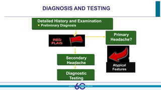
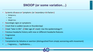
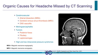
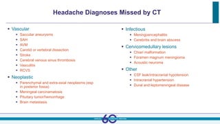
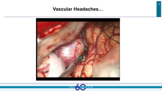
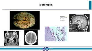
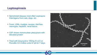
Noah Rosen MD FAHS
Zucker Hofstra Northwell Health
noheadaches@gmail.com
Secondary Headaches
Disclosures
Reference Arial 10pt left aligned Footer Arial 10pt right aligned
3
 Allergan:
 Advisory Board, Research Support, Speaker’s Panel
 Amgen:
 Advisory Board
 Promius Pharma:
 Advisory Board
 Supernus:
 Advisory Board
 Teva:
 Advisory Board
 Eli Lilly:
 Advisory Board, Research Support
 Curelator:
 Consultant paid in options
 1. To recognize “red flags” for headaches with a discernable cause
 Is it time to rename “secondary headaches”?
 2. To reinforce understanding on how to diagnose, manage and treat dangerous
and rapidly progressive headaches due to specific causes.
 3. To review headaches that we may not encounter on a regular basis.
 How much of our practice is primary headache disorders due to access reasons?
 Who is actually taking care of these patients?
Reference Arial 10pt left aligned Footer Arial 10pt right aligned
4
Learning Objectives
DIAGNOSIS AND TESTING
Detailed History and Examination
 Preliminary Diagnosis
Primary
Headache?
NO
Secondary
Headache
Diagnostic
Testing
Atypical
Features
YES
The Best 2 Tools We Have
 History
 Headache onset
 Duration
 Location
 Severity and Quality
 Associated Features
 Aggravating Factors
 Exam
 General exam!
 Neurologic exam
HX
PE
SNOOP (or some variation…)
7
• Systemic disease or Symptoms (or Secondary risk factors)
– Malignancy
– Fever
– Weight loss
• Neurologic signs or symptoms
• Onset that is sudden (acute or thunderclap)
• Onset “later in life”. (Older age of onset) Use the epidemiology!!!
• Previous headache history with new or different headache features
• Progressive
• Postural
• Precipitation by Valsalva or exertion (distinguished from simply worsening with movement)
• …. Pregnancy…. Papilledema….
Organic Causes for Headache Missed by CT Scanning
 Cerebrovascular
 Arterial dissection (MRA)
 Cerebral venous sinus thrombosis (MRV)
 CNS vasculitis
 Meningoencephalitis
 Tumors
 Posterior fossa
 Pituitary
 Leptomeninges
 High and low intracranial pressure syndromes
MRA = Magnetic resonance angiography
MRV = Magnetic resonance venography
Bousser MG, et al. In: Wolff’s Headache and Other Head Pain. 2001.
Dodick DW. Adv Stud Med. 2003;3:S550-S555.
Headache Diagnoses Missed by CT
 Vascular
 Saccular aneurysms
 SAH
 AVM
 Carotid or vertebral dissection
 Stroke
 Cerebral venous sinus thrombosis
 Vasculitis
 RCVS
 Neoplastic
 Parenchymal and extra-axial neoplasms (esp
in posterior fossa)
 Meningeal carcinamatosis
 Pituitary tumor/hemorrhage
 Brain metastasis
 Infectious
 Meningoencephalitis
 Cerebritis and brain abscess
 Cervicomedullary lesions
 Chiari malformation
 Foramen magnum meningioma
 Acoustic neuroma
 Other
 CSF leak/intracranial hypotension
 Intracranial hypertension
 Dural and leptomeningeal disease
10
What to discuss…
Too Many Headaches to Cover!!!
Reference Arial 10pt left aligned Footer Arial 10pt right aligned
11
Will focus on selected disorders:
1. Headaches secondary to Vascular Disorders
2. Headaches associated with Spinal Fluid Disorders
3. Headaches associated with CNS Infections
4. Potpourri
Vascular Headaches…
12
Secondary Headache: Subarhachnoid Hemorrhage
 First or worst Headache
 Often missed
 Maximum headache in
<60sec
 LOC, focal findings,
seizures, cardiac death
 Early CT +in > 90%, LP +
early in 100%
Subarachnoid Hemorrhage: Beware of Blood in the PITS
Parenchymal
Intraventricular
Truncal
Sulcal
Subarachnoid Hemorrhage
 Risks: rebleed and
vasospasm
 Early surgery,
then increase perfusion
 10% have multiple
aneurysms
 Best evidence: control blood
pressure!
Intracerebral Hemorrhage
 Focal deficits plus headache
 Progresses quickly
 Difficult to arouse
 If hemosiderin staining
And young
Think cavernoma
If older
Think Amyloidosis
Reversible Cerebral Vasoconstriction Syndrome (RCVS)
 RCVS
 Most commonly precipitated post-partum
or with the use of vasoactive substances
 Cannabis
 Cocaine
 SSRIs
 Binge drinking
 Nasal decongestants
 May result in:
 SAH
 ICH
 Stroke
 PRES
 Treated with IV/PO calcium channel
blockers
Ducros et al. Brain 2007; 130:3091-3101
Temporal/ Giant Cell Arteritis
 Age >60 with subacute onset of headaches
 High risk to involve ophthalmic artery, posterior ciliary arteries, or
branch of external carotid
 Less common: cerebral and coronary arteries
 Could be considered subset of primary CNS vasculitis
 Symptoms:
 visual loss (arteritic anterior ischemic optic neuropathy)
 -Also
 Temporal artery tenderness, weight loss, malaise, fever, chills, and jaw claudication
 Polymyalgia rheumatica common
Giant Cell Arteritis
 Lab: ↑↑ ESR
 Also, Anemia, leukocytosis and
elevated liver enzymes
 Diagnosis: Need temporal
artery bx although increasing
evidence for ultrasound
 Treatment: High dose
steroids (sometimes other
immune suppressants
 CD4 Lymphocytes and
macrophages
Pressure Disorders:
From blood vessels to spinal fluid…
20
Pseudotumor
• Headache: Unremarkable daily headache
• May have coocurrence with migraine
• Pulsatile tinnitus, TVOs
• Papilledema
• Enlargement of blind spot
• Loss of inferonasal field
• other
• 6th Nerve palsy in 10 – 20%
• Empty Sella
• Lateral Sinus Abnormality
Pseudotumor
• Risk factors include female gender and obesity
• Not all cases are idiopathic
• Increased risk of thrombosis with cigarette smoking and with
the use of oral contraception.
• NORDIC IIHT trial demonstrated safety and efficacy
of acetazolamide for pseudotumour
• 38/86 tolerated the 4 grams per day in study
• There is mixed evidence for the use of stenting
• Shunts are possible, but complications common
• Optic Nerve Fenestration is option to preserve vision
22
MRI Findings in
Pseudotumor
7.2 Headache
attributed to
low
cerebrospinal
fluid pressure
7.2.1 Post-dural puncture headache
 A. Headache worsens within 15 minutes after sitting or
standing and improves within 15 minutes of lying and has
at least 1 of the following
 Neck stiffness, tinnitus, hypacusia, photophobia, nausea
 B. Dural puncture has been performed
 C. Headache develops within 5 days after dural puncture
 D. Headache resolves either:
 Spontaneously within 1 week
 Within 48 hours after effective treatment of the spinal fluid leak
(usually by blood patch)
 (True in 95% of cases)
7.2.2 CSF Fistula Headache
 A. Headache worsens within 15 minutes after sitting or
standing with at least one of the following:
 Neck stiffness, tinnitus, hypacusis, photophobia, nausea
 B. A known procedure or trauma has caused persistent CSF
leakage with at least one of the following:
 Evidence of low CSF pressure on MRI
 Evidence of CSF leakage on myelography, CT myelography or
cisternography
 CSF pressure <60mm H2O in sitting position
 C. Headache develops in close temporal relation to CSF
leakage
 D. Headache resolves within 7 days of sealing the CSF leak
7.2.3 Headache Attributed to Spontaneous (Idiopathic) CSF Leak
 A. Diffuse or dull headache that worsens within 15
minutes after sitting or standing with at least one of the
following:
 Neck stiffness, tinnitus, hypacusia, photophobia, nausea
 B. At least one of the following:
 Evidence of low CSF pressure on MRI
 Evidence of CSF leakage on myelography, CT myelography or
cisternography
 CSF pressure <60mm H2O in sitting position
 C. No history of dural puncture or other causes of CSF
Fistula
 Headache resolves within 72 hours after epidural blood
patching
Diffuse pachymeningeal (dural) enhancement
Bilateral subdural effusion/hematomas
Downward displacement of brain
Enlargement of pituitary gland
Engorgement of dural venous sinuses
Prominence of spinal epidural venous plexus
Venous sinus thrombosis & isolated cortical vein thrombosis
AJNR 2008.; 29:1164-70
MRI Signs of Intracranial Hypotension
JAMA 2006.;295(19):2286-96
AJNR 2008.; 29:853-56
Diffuse, uniform thickness
Located at convexity, along falx cerebri, tentorium & posterior fossa dura
Disappears after successful treatment
Diffuse Pachymeningeal, (Dural) Enhancement
Bilateral Subdural Effusion/Hematomas
Incidence: 10-50%
Tend to be thin (2-7 mm),
typically occur over
supratentorial convexity
Have variable MR signal,
depending on protein conc. &
presence of blood
Disappear after successful
treatment
Downward Displacement of The
Brain
Low lying cerebellar tonsils
Effacement of prepontine
cistern, flattening of pons
against clivus
Effacement of perichiasmatic
cistern with bowing of optic
chiasm over pituitary fossa
Engorgement of
Dural Venous Sinuses
On T1W the middle 1/3 of
dominant transverse sinus,
shows convex borders
All venous sinuses become
engorged
The falx & tentorium show marked
enhancement
AJNR 2007 ; 28:1489-93
Spinal Extradural Fluid Collections
From: AJNR 2009.; 30:147-51
Prominent of
Epidural Venous Plexus
AJNR 2009.; 30:147-51
Patients may even present with a compressive myelopathy
due to a prominent venous epidural plexus.
Complications
Patient with known intracranial hypotension who rapidly
deteriorated shows cerebellar, brainstem & cord infarctions.
AJNR 2009, doi:10.3174/ajnr.A1749
Intracranial hypotension due to Post op spinal CSF
leak
Patient had a tumor resection from the thoracic vertebrae & developed intracranial
hypotension found to be due to paraspinal thoracic pseudomeningocele.
CSF leak complicated by cortical vein thrombosis
Iatrogenic- post LP- intracranial hypotension with cortical vein thrombosis (arrow).
SEEPS
Subdural fluid collection
Enhancement of meninges
Engorgement of veins
Pituitary hyperemia
Sagging of brain Hypermobile joints sometimes
seen in those with SIH
Spinal Fluid Leaks
CNS Infections
39
Meningitis
 Headache, fever, stiff neck, confusion, decreased consciousness and cranial
neuropathies
 Bacterial: Rapidly declining… CT, LP, antibiotics ASAP
 Viral/Aseptic: Slower onset, “less sick”, lesson from Mollaret
 Chronic/Fungal/TB: Cranial neuropathies more common, stiff neck less
common. Often requires high volume tap to obtain positive cells.
41
Meningitis
Acute Meningitis
 Bacterial Meningitis:
 LP shows elevated PMN’s, elevated protein and reduced glucose.
 treat adults with Ampicillin and Ceftriaxone pending cultures.
 Viral Meningitis/ Encephalitis:
 LP shows mild increase of lymphocytes, increased protein and normal
glucose
 Can include enteroviruses, lymphocytic choriomeningitis virus, HIV, as well
as many others.
Chronic Meningitis
 Tuberculosis:
 LP shows increased lymphocytes, elevated protein
and low glucose. Large volume tap gives 50% yield of
acid fast mycobacteria.
 Treat with four drugs for 1st 2 months until sensitivity is
known.
 Neurosyphilis:
 Primary, Secondary, Tertiary
 Gummas, aortitis, chorioretinitis
 Three major syndromes: tabes dorsalis, Argyll
Robertson pupils, general paresis of the insane.
 LP for VDRL- may consider FTA-ABS in late syphilis.
 Treat with Penicillin 2.4 million units IM weekly for 3
weeks if non neurologic. Otherwise treat Penicillin G
2-4 million units IV q4 for 10 days.
Chronic Meningitis
 Lyme Disease:
 Mononuclear pleocytosis with increase protein. Can send PCR for Borrelia
or detect IgG antibodies via ELISA or Western Blot.
 Ceftriaxone 2 g IV qday or Penicillin G 4 million units IV q 4 for 2-4 weeks.
 Fungal Meningitis
 Can include Cryptococcus, Coccidioides, Candida, Histoplasma,
Blastomyces
 LP shows mononuclear increase, elevated protein and near normal glucose.
 Treat with Amphotericin B or some with fluconazole.
Leptospirosis
 Spirochetal disease most often Leptospira
Interrogans from cats, dogs, etc.
 Fever, chills, myalgia, nausea, diarrhea,
meningitis, hepatitis, renal failure.
 CSF shows mononuclear pleocytosis with
elevated protein
 Treat with doxycycline 100mg IV q12 or
Penicillin G 5 million units IV q6 for 7 days.
Brain Abscess
 Bacterial Abscess
 Subacute progressive headache (75%), altered mental
status(70%), focal neurological signs(50%), fever (50%)
 Ring enhancing lesion; pathogen by CSF 10%, by biopsy 80%
 Look for extradural cause
 Treat empirically with oxacillin 2g IV q4 (or penicillin G),
Metronidazole and Ceftriaxone. Consider Amphotericin B
 Subdural Empyema
 Cranial and Spinal Epidural Abscess
 High dose dexamethasone 60 to 100mg IV push followed by 10-
20 mg q6 (and call surgery!).
 Vancomycin and Ceftriaxone
 Toxoplasmosis
 Sulfadiazine and pyrimethamine, Clindamycin
 Cysticercosis
Viral Encephalitis
 Herpes Simplex Encephalitis
 Acyclovir 10-12.5 mg/ kg IV q8 for 2-3 weeks.
 Phenytoin
 Consider Steroids
 Mumps, Enterovirus, Arbovirus (Equine, St. Louis, California,
colorado tick virus, West Nile, Zika, Chikungunya, Dengue)
 Measles virus
 Rabies Virus
 Epstein Barr virus
 Cytomegalovirus
 Whipple’s Disease
 Elizabethkingia
Approach to the patient (The Basics)
 Think- Fever, Headache, Neurologic Sign
 History and Exam
 Acute or Chronic
 Predisposing Factors (diabetes, EtOH abuse, Malignancy, Steroids,
chemotherapy, AIDS)
 Systemic Infection (endocarditis, pneumonia, osteomyelitis, skull fracture,
otitis media, tick bite, animal bites)
 Exam should include: signs of fever, headache, change in mental status,
focal weakness and back pain, as well as: papilledema, meningismus, skin
rash, sinus tenderness, spine tenderness.
 Lumbar Puncture
Lumbar Puncture
Fever, Headache, Change in Mental Status
Consider CT imaging if: Papilledema, Focal Neurologic Deficit
(especially brainstem), Known Intracranial Mass Lesion, AIDS,
Lethargy, Stupor or Coma
Don’t Forget Your Opening Pressure!!
Cervicogenic Headache
 Headache caused by disorder of cervical spine
(bone, disc and soft tissue) demonstrated by
clinical, laboratory and/ or imaging evidence
 Need at least 2 of 4:
 Headache developed in temporal relation to onset
of disorder
 Headache significantly improved or resolved in
parallel with improvement of cervical disorder
 Cervical range of motion reduced and headache
worsened with manoeuvers
 Headache abolished following diagnostic blockade
of cervical structure or nerve supply
 Not better accounted by another diagnosis!
 HA attributed to cervical myofascial pain
 HA attributed to upper cervical radiculopathy
Temporomandibular Joint Disorder
 Temporomandibular joint dysfunction is
an umbrella term covering pain
and dysfunction of the muscles of
mastication (the muscles that move the jaw)
and the temporomandibular joints
 More common in young adults
 Pain in jaw and ear, restricted movement,
lateral motion, locking up, difficulty chewing
 Treated with exercises, bite plates,
injections, muscle relaxants, relaxation
 Causes: Trauma, Arthritis, Disk erosion,
Inflammatory conditions
Some Other Secondary Headaches
 Subdural hematoma
 Ischemic stroke
 Transient ischemic attack
 Cervicocephalic arterial dissection
 Cerebral venous thrombosis
 Unruptured arteriovenous
malformation
 -Postcarotid endarterectomy
 Bell’s palsy – associated with
retroauricular pain
 Brain tumors and abscesses
 Dental abscesses
 Sinusitis
 Trigeminal neuralgia
 Low-CSF pressure headache
 Acute glaucoma
 Arterial hypertension
 William Young and Stephen Silberstein
 Rashmi Halker Singh and all of our speakers
 My kids…
Thanks
54

2-secondary-headaches-all types ahs-2018.pptx

  • 2.
    Noah Rosen MDFAHS Zucker Hofstra Northwell Health noheadaches@gmail.com Secondary Headaches
  • 3.
    Disclosures Reference Arial 10ptleft aligned Footer Arial 10pt right aligned 3  Allergan:  Advisory Board, Research Support, Speaker’s Panel  Amgen:  Advisory Board  Promius Pharma:  Advisory Board  Supernus:  Advisory Board  Teva:  Advisory Board  Eli Lilly:  Advisory Board, Research Support  Curelator:  Consultant paid in options
  • 4.
     1. Torecognize “red flags” for headaches with a discernable cause  Is it time to rename “secondary headaches”?  2. To reinforce understanding on how to diagnose, manage and treat dangerous and rapidly progressive headaches due to specific causes.  3. To review headaches that we may not encounter on a regular basis.  How much of our practice is primary headache disorders due to access reasons?  Who is actually taking care of these patients? Reference Arial 10pt left aligned Footer Arial 10pt right aligned 4 Learning Objectives
  • 5.
    DIAGNOSIS AND TESTING DetailedHistory and Examination  Preliminary Diagnosis Primary Headache? NO Secondary Headache Diagnostic Testing Atypical Features YES
  • 6.
    The Best 2Tools We Have  History  Headache onset  Duration  Location  Severity and Quality  Associated Features  Aggravating Factors  Exam  General exam!  Neurologic exam HX PE
  • 7.
    SNOOP (or somevariation…) 7 • Systemic disease or Symptoms (or Secondary risk factors) – Malignancy – Fever – Weight loss • Neurologic signs or symptoms • Onset that is sudden (acute or thunderclap) • Onset “later in life”. (Older age of onset) Use the epidemiology!!! • Previous headache history with new or different headache features • Progressive • Postural • Precipitation by Valsalva or exertion (distinguished from simply worsening with movement) • …. Pregnancy…. Papilledema….
  • 8.
    Organic Causes forHeadache Missed by CT Scanning  Cerebrovascular  Arterial dissection (MRA)  Cerebral venous sinus thrombosis (MRV)  CNS vasculitis  Meningoencephalitis  Tumors  Posterior fossa  Pituitary  Leptomeninges  High and low intracranial pressure syndromes MRA = Magnetic resonance angiography MRV = Magnetic resonance venography Bousser MG, et al. In: Wolff’s Headache and Other Head Pain. 2001. Dodick DW. Adv Stud Med. 2003;3:S550-S555.
  • 9.
    Headache Diagnoses Missedby CT  Vascular  Saccular aneurysms  SAH  AVM  Carotid or vertebral dissection  Stroke  Cerebral venous sinus thrombosis  Vasculitis  RCVS  Neoplastic  Parenchymal and extra-axial neoplasms (esp in posterior fossa)  Meningeal carcinamatosis  Pituitary tumor/hemorrhage  Brain metastasis  Infectious  Meningoencephalitis  Cerebritis and brain abscess  Cervicomedullary lesions  Chiari malformation  Foramen magnum meningioma  Acoustic neuroma  Other  CSF leak/intracranial hypotension  Intracranial hypertension  Dural and leptomeningeal disease
  • 10.
  • 11.
    Too Many Headachesto Cover!!! Reference Arial 10pt left aligned Footer Arial 10pt right aligned 11 Will focus on selected disorders: 1. Headaches secondary to Vascular Disorders 2. Headaches associated with Spinal Fluid Disorders 3. Headaches associated with CNS Infections 4. Potpourri
  • 12.
  • 13.
    Secondary Headache: SubarhachnoidHemorrhage  First or worst Headache  Often missed  Maximum headache in <60sec  LOC, focal findings, seizures, cardiac death  Early CT +in > 90%, LP + early in 100%
  • 14.
    Subarachnoid Hemorrhage: Bewareof Blood in the PITS Parenchymal Intraventricular Truncal Sulcal
  • 15.
    Subarachnoid Hemorrhage  Risks:rebleed and vasospasm  Early surgery, then increase perfusion  10% have multiple aneurysms  Best evidence: control blood pressure!
  • 16.
    Intracerebral Hemorrhage  Focaldeficits plus headache  Progresses quickly  Difficult to arouse  If hemosiderin staining And young Think cavernoma If older Think Amyloidosis
  • 17.
    Reversible Cerebral VasoconstrictionSyndrome (RCVS)  RCVS  Most commonly precipitated post-partum or with the use of vasoactive substances  Cannabis  Cocaine  SSRIs  Binge drinking  Nasal decongestants  May result in:  SAH  ICH  Stroke  PRES  Treated with IV/PO calcium channel blockers Ducros et al. Brain 2007; 130:3091-3101
  • 18.
    Temporal/ Giant CellArteritis  Age >60 with subacute onset of headaches  High risk to involve ophthalmic artery, posterior ciliary arteries, or branch of external carotid  Less common: cerebral and coronary arteries  Could be considered subset of primary CNS vasculitis  Symptoms:  visual loss (arteritic anterior ischemic optic neuropathy)  -Also  Temporal artery tenderness, weight loss, malaise, fever, chills, and jaw claudication  Polymyalgia rheumatica common
  • 19.
    Giant Cell Arteritis Lab: ↑↑ ESR  Also, Anemia, leukocytosis and elevated liver enzymes  Diagnosis: Need temporal artery bx although increasing evidence for ultrasound  Treatment: High dose steroids (sometimes other immune suppressants  CD4 Lymphocytes and macrophages
  • 20.
    Pressure Disorders: From bloodvessels to spinal fluid… 20
  • 21.
    Pseudotumor • Headache: Unremarkabledaily headache • May have coocurrence with migraine • Pulsatile tinnitus, TVOs • Papilledema • Enlargement of blind spot • Loss of inferonasal field • other • 6th Nerve palsy in 10 – 20% • Empty Sella • Lateral Sinus Abnormality
  • 22.
    Pseudotumor • Risk factorsinclude female gender and obesity • Not all cases are idiopathic • Increased risk of thrombosis with cigarette smoking and with the use of oral contraception. • NORDIC IIHT trial demonstrated safety and efficacy of acetazolamide for pseudotumour • 38/86 tolerated the 4 grams per day in study • There is mixed evidence for the use of stenting • Shunts are possible, but complications common • Optic Nerve Fenestration is option to preserve vision 22
  • 23.
  • 24.
  • 25.
    7.2.1 Post-dural punctureheadache  A. Headache worsens within 15 minutes after sitting or standing and improves within 15 minutes of lying and has at least 1 of the following  Neck stiffness, tinnitus, hypacusia, photophobia, nausea  B. Dural puncture has been performed  C. Headache develops within 5 days after dural puncture  D. Headache resolves either:  Spontaneously within 1 week  Within 48 hours after effective treatment of the spinal fluid leak (usually by blood patch)  (True in 95% of cases)
  • 26.
    7.2.2 CSF FistulaHeadache  A. Headache worsens within 15 minutes after sitting or standing with at least one of the following:  Neck stiffness, tinnitus, hypacusis, photophobia, nausea  B. A known procedure or trauma has caused persistent CSF leakage with at least one of the following:  Evidence of low CSF pressure on MRI  Evidence of CSF leakage on myelography, CT myelography or cisternography  CSF pressure <60mm H2O in sitting position  C. Headache develops in close temporal relation to CSF leakage  D. Headache resolves within 7 days of sealing the CSF leak
  • 27.
    7.2.3 Headache Attributedto Spontaneous (Idiopathic) CSF Leak  A. Diffuse or dull headache that worsens within 15 minutes after sitting or standing with at least one of the following:  Neck stiffness, tinnitus, hypacusia, photophobia, nausea  B. At least one of the following:  Evidence of low CSF pressure on MRI  Evidence of CSF leakage on myelography, CT myelography or cisternography  CSF pressure <60mm H2O in sitting position  C. No history of dural puncture or other causes of CSF Fistula  Headache resolves within 72 hours after epidural blood patching
  • 28.
    Diffuse pachymeningeal (dural)enhancement Bilateral subdural effusion/hematomas Downward displacement of brain Enlargement of pituitary gland Engorgement of dural venous sinuses Prominence of spinal epidural venous plexus Venous sinus thrombosis & isolated cortical vein thrombosis AJNR 2008.; 29:1164-70 MRI Signs of Intracranial Hypotension
  • 29.
    JAMA 2006.;295(19):2286-96 AJNR 2008.;29:853-56 Diffuse, uniform thickness Located at convexity, along falx cerebri, tentorium & posterior fossa dura Disappears after successful treatment Diffuse Pachymeningeal, (Dural) Enhancement
  • 30.
    Bilateral Subdural Effusion/Hematomas Incidence:10-50% Tend to be thin (2-7 mm), typically occur over supratentorial convexity Have variable MR signal, depending on protein conc. & presence of blood Disappear after successful treatment
  • 31.
    Downward Displacement ofThe Brain Low lying cerebellar tonsils Effacement of prepontine cistern, flattening of pons against clivus Effacement of perichiasmatic cistern with bowing of optic chiasm over pituitary fossa
  • 32.
    Engorgement of Dural VenousSinuses On T1W the middle 1/3 of dominant transverse sinus, shows convex borders All venous sinuses become engorged The falx & tentorium show marked enhancement AJNR 2007 ; 28:1489-93
  • 33.
    Spinal Extradural FluidCollections From: AJNR 2009.; 30:147-51
  • 34.
    Prominent of Epidural VenousPlexus AJNR 2009.; 30:147-51 Patients may even present with a compressive myelopathy due to a prominent venous epidural plexus.
  • 35.
    Complications Patient with knownintracranial hypotension who rapidly deteriorated shows cerebellar, brainstem & cord infarctions. AJNR 2009, doi:10.3174/ajnr.A1749
  • 36.
    Intracranial hypotension dueto Post op spinal CSF leak Patient had a tumor resection from the thoracic vertebrae & developed intracranial hypotension found to be due to paraspinal thoracic pseudomeningocele.
  • 37.
    CSF leak complicatedby cortical vein thrombosis Iatrogenic- post LP- intracranial hypotension with cortical vein thrombosis (arrow).
  • 38.
    SEEPS Subdural fluid collection Enhancementof meninges Engorgement of veins Pituitary hyperemia Sagging of brain Hypermobile joints sometimes seen in those with SIH Spinal Fluid Leaks
  • 39.
  • 40.
    Meningitis  Headache, fever,stiff neck, confusion, decreased consciousness and cranial neuropathies  Bacterial: Rapidly declining… CT, LP, antibiotics ASAP  Viral/Aseptic: Slower onset, “less sick”, lesson from Mollaret  Chronic/Fungal/TB: Cranial neuropathies more common, stiff neck less common. Often requires high volume tap to obtain positive cells.
  • 41.
  • 42.
  • 43.
    Acute Meningitis  BacterialMeningitis:  LP shows elevated PMN’s, elevated protein and reduced glucose.  treat adults with Ampicillin and Ceftriaxone pending cultures.  Viral Meningitis/ Encephalitis:  LP shows mild increase of lymphocytes, increased protein and normal glucose  Can include enteroviruses, lymphocytic choriomeningitis virus, HIV, as well as many others.
  • 44.
    Chronic Meningitis  Tuberculosis: LP shows increased lymphocytes, elevated protein and low glucose. Large volume tap gives 50% yield of acid fast mycobacteria.  Treat with four drugs for 1st 2 months until sensitivity is known.  Neurosyphilis:  Primary, Secondary, Tertiary  Gummas, aortitis, chorioretinitis  Three major syndromes: tabes dorsalis, Argyll Robertson pupils, general paresis of the insane.  LP for VDRL- may consider FTA-ABS in late syphilis.  Treat with Penicillin 2.4 million units IM weekly for 3 weeks if non neurologic. Otherwise treat Penicillin G 2-4 million units IV q4 for 10 days.
  • 45.
    Chronic Meningitis  LymeDisease:  Mononuclear pleocytosis with increase protein. Can send PCR for Borrelia or detect IgG antibodies via ELISA or Western Blot.  Ceftriaxone 2 g IV qday or Penicillin G 4 million units IV q 4 for 2-4 weeks.  Fungal Meningitis  Can include Cryptococcus, Coccidioides, Candida, Histoplasma, Blastomyces  LP shows mononuclear increase, elevated protein and near normal glucose.  Treat with Amphotericin B or some with fluconazole.
  • 46.
    Leptospirosis  Spirochetal diseasemost often Leptospira Interrogans from cats, dogs, etc.  Fever, chills, myalgia, nausea, diarrhea, meningitis, hepatitis, renal failure.  CSF shows mononuclear pleocytosis with elevated protein  Treat with doxycycline 100mg IV q12 or Penicillin G 5 million units IV q6 for 7 days.
  • 47.
    Brain Abscess  BacterialAbscess  Subacute progressive headache (75%), altered mental status(70%), focal neurological signs(50%), fever (50%)  Ring enhancing lesion; pathogen by CSF 10%, by biopsy 80%  Look for extradural cause  Treat empirically with oxacillin 2g IV q4 (or penicillin G), Metronidazole and Ceftriaxone. Consider Amphotericin B  Subdural Empyema  Cranial and Spinal Epidural Abscess  High dose dexamethasone 60 to 100mg IV push followed by 10- 20 mg q6 (and call surgery!).  Vancomycin and Ceftriaxone  Toxoplasmosis  Sulfadiazine and pyrimethamine, Clindamycin  Cysticercosis
  • 48.
    Viral Encephalitis  HerpesSimplex Encephalitis  Acyclovir 10-12.5 mg/ kg IV q8 for 2-3 weeks.  Phenytoin  Consider Steroids  Mumps, Enterovirus, Arbovirus (Equine, St. Louis, California, colorado tick virus, West Nile, Zika, Chikungunya, Dengue)  Measles virus  Rabies Virus  Epstein Barr virus  Cytomegalovirus  Whipple’s Disease  Elizabethkingia
  • 49.
    Approach to thepatient (The Basics)  Think- Fever, Headache, Neurologic Sign  History and Exam  Acute or Chronic  Predisposing Factors (diabetes, EtOH abuse, Malignancy, Steroids, chemotherapy, AIDS)  Systemic Infection (endocarditis, pneumonia, osteomyelitis, skull fracture, otitis media, tick bite, animal bites)  Exam should include: signs of fever, headache, change in mental status, focal weakness and back pain, as well as: papilledema, meningismus, skin rash, sinus tenderness, spine tenderness.  Lumbar Puncture
  • 50.
    Lumbar Puncture Fever, Headache,Change in Mental Status Consider CT imaging if: Papilledema, Focal Neurologic Deficit (especially brainstem), Known Intracranial Mass Lesion, AIDS, Lethargy, Stupor or Coma Don’t Forget Your Opening Pressure!!
  • 51.
    Cervicogenic Headache  Headachecaused by disorder of cervical spine (bone, disc and soft tissue) demonstrated by clinical, laboratory and/ or imaging evidence  Need at least 2 of 4:  Headache developed in temporal relation to onset of disorder  Headache significantly improved or resolved in parallel with improvement of cervical disorder  Cervical range of motion reduced and headache worsened with manoeuvers  Headache abolished following diagnostic blockade of cervical structure or nerve supply  Not better accounted by another diagnosis!  HA attributed to cervical myofascial pain  HA attributed to upper cervical radiculopathy
  • 52.
    Temporomandibular Joint Disorder Temporomandibular joint dysfunction is an umbrella term covering pain and dysfunction of the muscles of mastication (the muscles that move the jaw) and the temporomandibular joints  More common in young adults  Pain in jaw and ear, restricted movement, lateral motion, locking up, difficulty chewing  Treated with exercises, bite plates, injections, muscle relaxants, relaxation  Causes: Trauma, Arthritis, Disk erosion, Inflammatory conditions
  • 53.
    Some Other SecondaryHeadaches  Subdural hematoma  Ischemic stroke  Transient ischemic attack  Cervicocephalic arterial dissection  Cerebral venous thrombosis  Unruptured arteriovenous malformation  -Postcarotid endarterectomy  Bell’s palsy – associated with retroauricular pain  Brain tumors and abscesses  Dental abscesses  Sinusitis  Trigeminal neuralgia  Low-CSF pressure headache  Acute glaucoma  Arterial hypertension
  • 54.
     William Youngand Stephen Silberstein  Rashmi Halker Singh and all of our speakers  My kids… Thanks 54