1. CNS infections can be caused by bacteria, viruses, fungi, parasites, or prions. Common bacterial infections include meningitis caused by streptococcus pneumoniae, neisseria meningitidis, and listeria monocytogenes.
2. Viruses that can cause CNS infections include poliovirus, rabies virus, herpes simplex virus, measles (subacute sclerosing panencephalitis), and JC virus (progressive multifocal leukoencephalopathy).
3. Fungal infections like cryptococcosis and parasitic infections including malaria, toxoplasmosis, and trypanosomiasis can also infect the CNS.

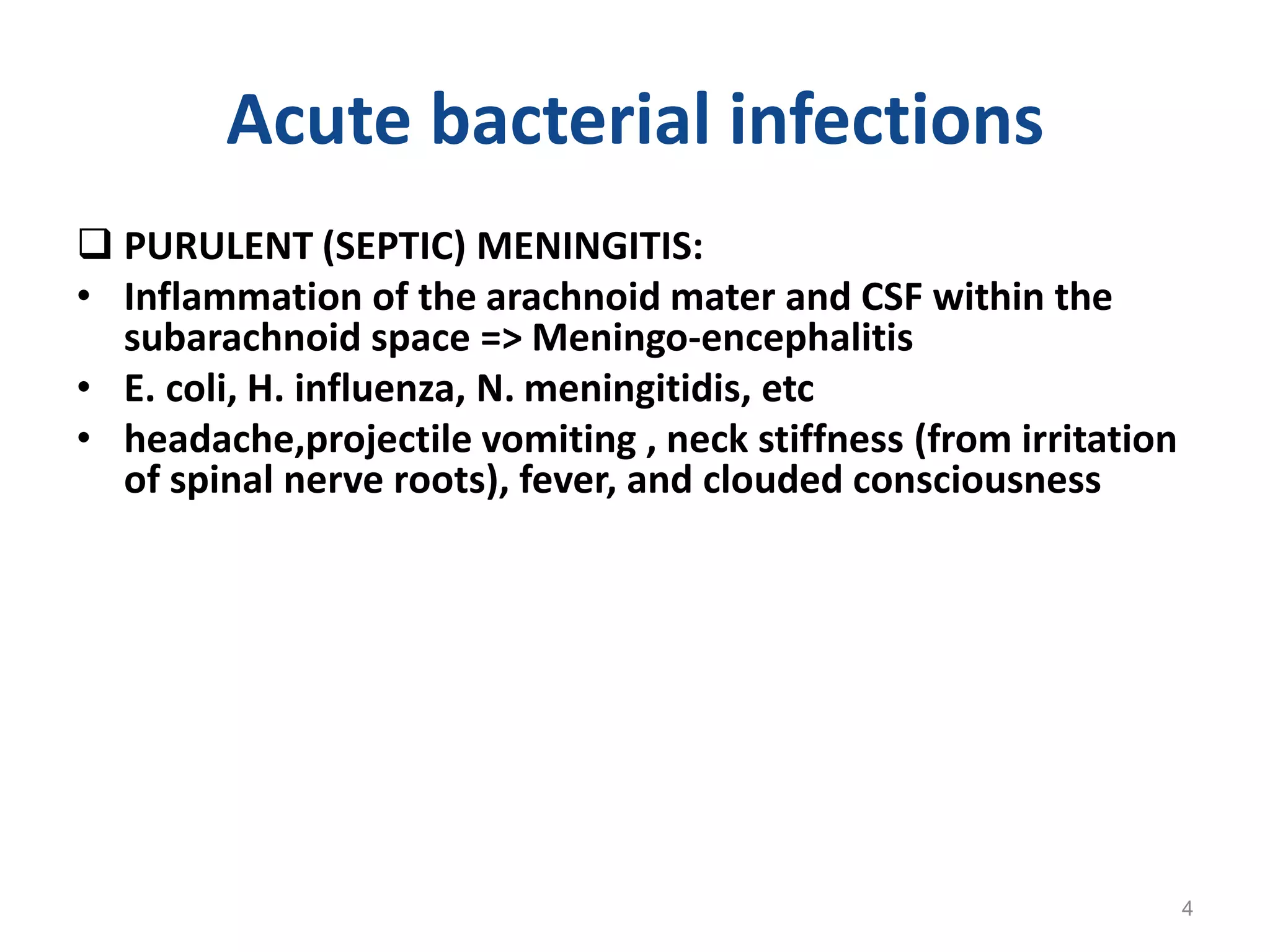


![Results of Cerebrospinal Fluid Testing.
Samuels MA et al. N Engl J Med 2007;357:1957-1965.
Substance CSF Plasma Ratio
CSF/plasma
Na+ [meq/kg
H2O]
147.0 150.0 0.89
K+ [meq/kg H2O] 2.9 4.6 0.62
Mg++ [meq/kg H2O] 2.2 1.6 1.39
Ca++ [meq/kg H2O] 2.3 4.7 0.49
Osmolality [mosm/kg H2O] 289.0 289.0 1.0
Protein [mg/dl] 20.0 6000.0 0.003
Glucose [mg/dl] 64.0 100.0 0.64
Cholesterol [mg/dl] 0.2 175.0 0.001
9](https://image.slidesharecdn.com/2-221130164036-8ae58b87/75/2-CNS-INFECTIONS-2015-pptx-9-2048.jpg)


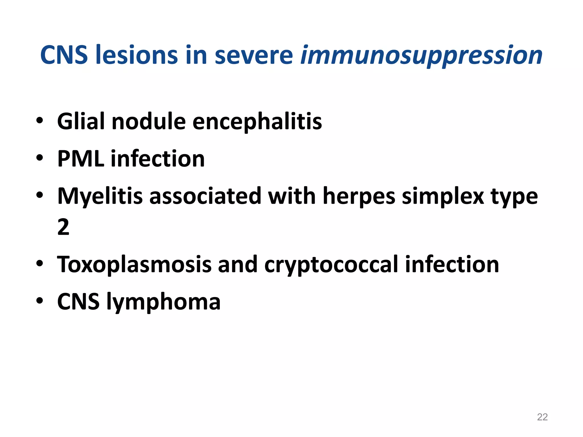
![HIV HIV-associated
[opportunistic]
CNS Lymphoma
CNS and HIV
Papovavirus (J C Virus)
Progressive multifocal
leukencephalopathy
Toxoplasma Gondii
Cryptococcus
neoformans
HIV meningoencephalitis
(subacute, giant cells)
Intravascular B-cell
Lymphoma
23](https://image.slidesharecdn.com/2-221130164036-8ae58b87/75/2-CNS-INFECTIONS-2015-pptx-23-2048.jpg)


