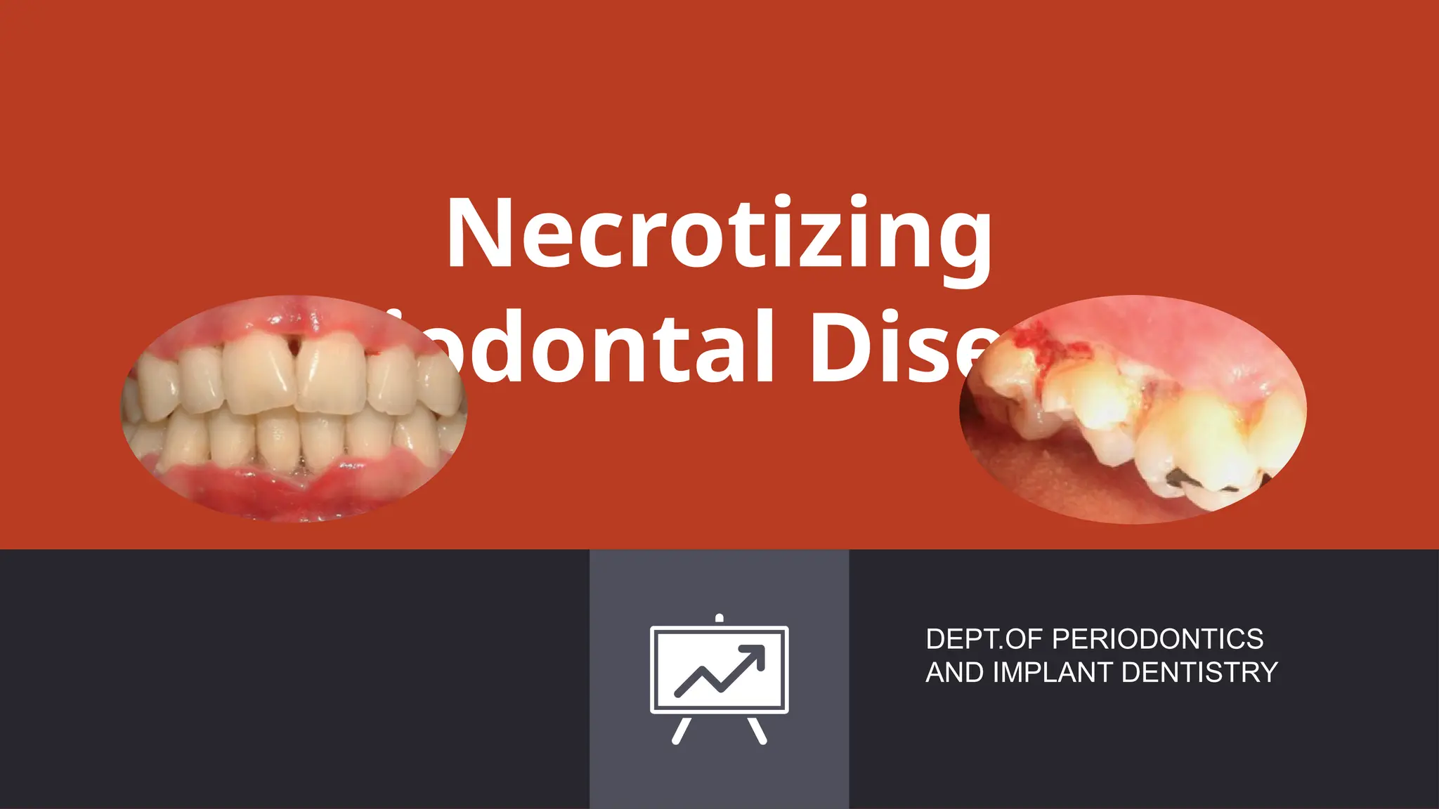
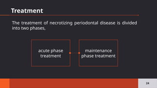
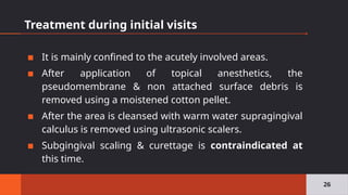
Necrotizing
Periodontal Diseases
DEPT.OF PERIODONTICS
AND IMPLANT DENTISTRY
Purpose statement
▪ The students must know the signs and
symptoms of the necrotizing periodontal
diseases.
▪ The students should be aware of the treatment
of the necrotizing periodontal diseases.
2
Learning Objective
3
Sr. No. LO Domain Level
1. Definition of necrotizing
periodontal diseases and its
signs and symptoms.
Cognitive Must know
2. Microbiology of necrotizing
periodontal diseases.
Cognitive Must know
3. Diagnosis of necrotizing
periodontal diseases.
Cognitive Must know
4. Treatment of necrotizing
periodontal diseases.
Cognitive Must know
▪ Necrotizing gingivitis: when only the gingival tissues
are affected.
▪ Necrotizing periodontitis: when the necrosis
progresses into the periodontal ligament and the
alveolar bone, leading to attachment loss.
▪ Necrotizing stomatitis: when the necrosis progresses
to deeper tissues beyond the mucogingival line,
including the lip or cheek mucosa, the tongue, etc.
4
5
NUP
NS
NOMA
NUG
Stages of oral necrotizing disease
▪ Stage 1- necrosis of the tip of the interdental papilla.
▪ Stage 2- necrosis of entire papilla
▪ Stage 3- necrosis extending to the gingival margin.
▪ Stage 4- necrosis extending to the attached gingiva.
▪ Stage 5– necrosis extending to labial & buccal mucosa.
▪ Stage 6- necrosis exposing alveolar bone.
▪ Stage 7– necrosis perforating skin of cheek.
6
Horning
&
Cohen,
1995
Characteristics of the lesion
▪ NPD is an inflammatory, destructive condition
characterized by ulcerated and necrotic papillae, giving
a punched‐out crater like appearance.
▪ The ulcers are covered by a yellowish–white or grayish
slough, which has been termed a “pseudomembrane”.
▪ The necrotizing lesions develop rapidly and are painful.
7
▪ Bleeding is readily provoked on
removal of the sloughed material
and exposure of the ulcerated
underlying connective tissue.
▪ In early phases of the disease,
lesions are typically confined to the
top of a few interdental papillae.
8
▪ The first lesions are often seen interproximally in the
mandibular anterior region.
▪ A characteristic and pronounced foetor ex ore is often
associated with NPD.
9
Necrotizing Gingivitis
▪ The lesions are seldom associated with deep pocket
formation, because extensive gingival necrosis often
coincides with loss of crestal alveolar bone.
▪ The gingival necrosis develops rapidly and within a few
days the involved papillae are often separated into one
facial and one lingual portion with an interposed necrotic
depression between them.
10
Necrotizing Periodontitis
11
▪ At this stage of the disease, the disease
process usually involves the
periodontal ligament and the alveolar
bone, and loss of attachment is now
established.
▪ Superficial necrotic lesions only rarely
cover a substantial part of the attached
gingiva, which becomes reduced in
width as the result of the disease
progression.
Sequestrum formation
▪ The disease progression may be
rapid and result in necrosis of
small or large parts of the alveolar
bone.
▪ Such a development is particularly
evident in severely
immunocompromised patients,
including HIV‐seropositive
individuals.
12
Involvement of alveolar mucosa
▪ When the necrotic process
progresses beyond the mucogingival
junction, the condition is denoted NS
(Williams et al. 1990)
▪ It may be life threatening. NS may
result in extensive denudation of
bone, resulting in major
sequestration with the development
of an oroantral fistula and osteitis
(SanGiacomo et al. 1990;).
13
Swelling of lymph nodes
▪ Swelling of the regional lymph nodes may occur in NPD,
but is particularly evident in advanced cases.
▪ Usually confined to the submandibular lymph nodes,
but the cervical lymph nodes may also be involved.
▪ In children with NPD, swelling of lymph nodes and
increased bleeding tendency are often the most
pronounced clinical findings (Jimenez & Baer 1975).
14
Fever and malaise
▪ Fever and malaise is not a consistent characteristic of
NPD.
▪ In mild & moderate stages of disease
Local lymphadenopathy & slight elevation in
temperature.
▪ In severe cases
High fever, increased pulse rate, leucocytois, loss of
appetite & general lassitude.
15
Oral hygiene
▪ The oral hygiene in patients with
NPD is usually poor. Moreover,
brushing of teeth and contact with
the acutely inflamed gingiva is
painful.
▪ Therefore, large amounts of
plaque on the teeth are common,
especially along the gingival
margin.
16
Acute and Chronic/recurrent forms
▪ In most instances, the
course of the diseases is
acute, as characterized
by the rapid destruction
of the periodontal
tissue.
17
▪ If inadequately treated or left untreated, the acute phase
may gradually subside.
▪ The symptoms then become less unpleasant for the
patient, but the destruction of the periodontal tissues
continues, although at a slower rate, and the necrotic
tissues do not heal completely.
▪ Such a condition has been termed chronic necrotizing
gingivitis, or periodontitis in the case of attachment loss
18
▪ In recurrent acute phases, subjective symptoms again
become more prominent and necrotic ulcers reappear.
▪ Several adjoining interdental craters may fuse, resulting
in total separation of facial and oral gingivae, which form
two distinct flaps.
▪ Recurrent forms of NG and NP may produce
considerable destruction of supporting tissues. The most
pronounced tissue loss usually occurs in relation to the
interproximal craters.
19
Microbiology
▪ The “constant flora” primarily contained Treponema spp.,
Selenomonas spp., Fusobacterium spp., and Bacteroides
melaninogenicus subsp. Intermedius (Prevotella intermedia).
▪ The “variable flora” consisted of a heterogeneous array of
bacterial types.
(Loesche et al. 1982; Ramos et al. 2012)
20
Pathogenic potential of microorganisms
▪ Among the bacteria isolated from necrotizing lesions,
spirochetes and fusiform bacteria can invade the
epithelium (Heylings 1967).
▪ The spirochetes can also invade the vital connective
tissue (Listgarten 1965).
▪ The pathogenic potential is further substantiated by the
fact that both fusobacteria and spirochetes can liberate
endotoxins (Mergenhagen et al. 1961; Kristoffersen & Hofstad 1970).
21
Differential diagnosis
23
Herpetic
gingivostomatiti
s
Gonococcal
gingivostomatiti
s
Tuberculous
gingival lesion
Agranulocytosis
Apthous
stomatitis
Candidiasis
Streptococcal
gingivostomatiti
s
Desquamative
gingivitis
Treatment
24
acute phase
treatment
maintenance
phase treatment
The treatment of necrotizing periodontal disease is divided
into two phases,
First visit
▪ The oral cavity is examined for the characteristic feature of
NUG, its distribution & possible involvement of
oropharyngeal region.
▪ Oral hygiene is evaluated with special attention to the
presence of pericoronal flaps, periodontal pockets & local
factors.
▪ History taking – H/o present illness, diet, socio-economic
background, smoking, chances of HIV infection, stress, and
profession.
25
Treatment during initial visits
▪ It is mainly confined to the acutely involved areas.
▪ After application of topical anesthetics, the
pseudomembrane & non attached surface debris is
removed using a moistened cotton pellet.
▪ After the area is cleansed with warm water supragingival
calculus is removed using ultrasonic scalers.
▪ Subgingival scaling & curettage is contraindicated at
this time.
26
Choice of drugs
▪ Metronidazole(250 mg) * TID (Loesche et al 1982)
▪ Other antibiotics such as Amoxycillin(500mg) in every 6
hours for 10 days or erythromycin (500mg every 6 hrs)
are used.
▪ The adjunctive use of metronidazole in HIV associated
NPD is reported to be extremely effective in reducing
acute pain & promoting rapid healing.(Scully et al 1991)
▪ Topical application of antibiotics is not indicated
27
▪ Hydrogen peroxide & other oxygen releasing agents also
have a long standing tradition in the treatment of NPD.
▪ Hydrogen peroxide (3%) is used for debridement in
necrotic areas & as a mouth rinse (equal portions 3%
H2O2 & warm water).
▪ Favorable effects of hydrogen peroxide may be due to
mechanical cleaning,& the influence on anaerobic
bacterial flora of the liberated oxygen. (Macphee & Cowley 1981).
28
▪ Appropriate treatment alleviates symptoms with in few
days (5 days).
▪ Physical rest advised.
▪ Suspected seropositive NPD patients, who are
unaware of their serostatus , should be referred to
the physicians.
29
Second visit
▪ Systematic subgingival scaling should be continued with
increasing intensity as the symptoms subside.
▪ Correction of restoration margins
▪ Polishing of restorations & root surfaces should be
completed after healing of ulcers.
▪ When ulcerated areas are healed local treatment is
supplemented with oral hygiene & patient motivation.
30
Third Visit
▪ Approximately 5 days after 2nd visit
▪ Patient counseling : Nutrition, Smoking cessation
▪ H2O2 rinse discontinued
▪ CHX for 2-3 weeks
▪ Maintenance Therapy
31
Maintenance phase treatment
▪ When the acute phase treatment has been completed,
necrosis & acute symptoms in NPD have disappeared.
▪ The formerly necrotic areas are healed & the gingival
craters are reduced in size, although some defects
usually persists.
▪ Bacterial plaque accumulates & therefore may
predispose to recurrences of NPD or to further
destruction because of a persisting chronic inflammatory
process or both.
▪ These sites therefore requires surgical correction. 32
Herrera D., et al. Acute periodontal lesions. Periodontol 2000, Vol. 65, 2014, 149–177.
▪ Shallow craters can be removed by simple
gingivectomy, while the elimination of deep defects may
require flap surgery.
▪ Treatment of NG is not completed until all gingival
defects have been eliminated & optimal conditions for
future plaque control have been established.
▪ Elimination of predisposing factors is also very important
to prevent recurrences.
33
Persistent or recurrent cases
▪ Adequate local therapy with optimal home care will
resolve most cases of NUG. If it persists despite therapy
or recurs , the patient should be revaluated with the
focus on the following factors,
▫ Reassessment of differential diagnosis to rule out the
disease that resembles NUG.
▫ Underlying systemic disease that cause immuno-
suppresion.
▫ Inadequate local therapy and/or compliance.
34
Summary
▪ Necrotizing periodontal disease includes necrotizing
gingivitis, necrotizing periodontitis and necrotizing
stomatitis and these may be considered as different
stages of the same pathologic process.
▪ This group of diseases always presents three typical
clinical features – papilla necrosis, bleeding and pain –
which makes them different from other periodontal
diseases.
35
▪ Although their prevalence is not high, their importance is
clear because they represent the most severe biofilm-
related periodontal conditions, leading to rapid tissue
destruction.
▪ Owing to their acute presentation, together with the
associated pain and tissue destruction, treatment should
be provided immediately upon diagnosis; this should
include superficial debridement, careful mechanical oral
hygiene. Surgical treatment of the sequelae should be
considered based on the needs of the individual case.
36
 Niklaus P Lang and Jan Lindhe. Clinical Periodontolgy and Implant
Dentistry, 6th
Ed.
 Newman and Carranza’s Clinical Periodontology, 13th
Ed.
 Herrera D., et al. Acute periodontal lesions. Periodontol 2000, Vol. 65,
2014, 149–177.
 Holmstrup, P. & Westergaard, J. (1994). Periodontal diseases in HIV‐
infected patients. Journal of Clinical Periodontology 21, 270–280.
 Horning, G.M. & Cohen, M.E. (1995). Necrotizing ulcerative gingivitis,
periodontitis, and stomatitis: Clinical staging and predisposing factors.
Journal of Periodontology 66, 990–998.
37
References
“
The two most powerful warriors are patience and time.
38

23 NUP.pptx Necrotizing Ulcerative Periodontitis Clinical features

  • 1.
  • 2.
    Purpose statement ▪ Thestudents must know the signs and symptoms of the necrotizing periodontal diseases. ▪ The students should be aware of the treatment of the necrotizing periodontal diseases. 2
  • 3.
    Learning Objective 3 Sr. No.LO Domain Level 1. Definition of necrotizing periodontal diseases and its signs and symptoms. Cognitive Must know 2. Microbiology of necrotizing periodontal diseases. Cognitive Must know 3. Diagnosis of necrotizing periodontal diseases. Cognitive Must know 4. Treatment of necrotizing periodontal diseases. Cognitive Must know
  • 4.
    ▪ Necrotizing gingivitis:when only the gingival tissues are affected. ▪ Necrotizing periodontitis: when the necrosis progresses into the periodontal ligament and the alveolar bone, leading to attachment loss. ▪ Necrotizing stomatitis: when the necrosis progresses to deeper tissues beyond the mucogingival line, including the lip or cheek mucosa, the tongue, etc. 4
  • 5.
  • 6.
    Stages of oralnecrotizing disease ▪ Stage 1- necrosis of the tip of the interdental papilla. ▪ Stage 2- necrosis of entire papilla ▪ Stage 3- necrosis extending to the gingival margin. ▪ Stage 4- necrosis extending to the attached gingiva. ▪ Stage 5– necrosis extending to labial & buccal mucosa. ▪ Stage 6- necrosis exposing alveolar bone. ▪ Stage 7– necrosis perforating skin of cheek. 6 Horning & Cohen, 1995
  • 7.
    Characteristics of thelesion ▪ NPD is an inflammatory, destructive condition characterized by ulcerated and necrotic papillae, giving a punched‐out crater like appearance. ▪ The ulcers are covered by a yellowish–white or grayish slough, which has been termed a “pseudomembrane”. ▪ The necrotizing lesions develop rapidly and are painful. 7
  • 8.
    ▪ Bleeding isreadily provoked on removal of the sloughed material and exposure of the ulcerated underlying connective tissue. ▪ In early phases of the disease, lesions are typically confined to the top of a few interdental papillae. 8
  • 9.
    ▪ The firstlesions are often seen interproximally in the mandibular anterior region. ▪ A characteristic and pronounced foetor ex ore is often associated with NPD. 9
  • 10.
    Necrotizing Gingivitis ▪ Thelesions are seldom associated with deep pocket formation, because extensive gingival necrosis often coincides with loss of crestal alveolar bone. ▪ The gingival necrosis develops rapidly and within a few days the involved papillae are often separated into one facial and one lingual portion with an interposed necrotic depression between them. 10
  • 11.
    Necrotizing Periodontitis 11 ▪ Atthis stage of the disease, the disease process usually involves the periodontal ligament and the alveolar bone, and loss of attachment is now established. ▪ Superficial necrotic lesions only rarely cover a substantial part of the attached gingiva, which becomes reduced in width as the result of the disease progression.
  • 12.
    Sequestrum formation ▪ Thedisease progression may be rapid and result in necrosis of small or large parts of the alveolar bone. ▪ Such a development is particularly evident in severely immunocompromised patients, including HIV‐seropositive individuals. 12
  • 13.
    Involvement of alveolarmucosa ▪ When the necrotic process progresses beyond the mucogingival junction, the condition is denoted NS (Williams et al. 1990) ▪ It may be life threatening. NS may result in extensive denudation of bone, resulting in major sequestration with the development of an oroantral fistula and osteitis (SanGiacomo et al. 1990;). 13
  • 14.
    Swelling of lymphnodes ▪ Swelling of the regional lymph nodes may occur in NPD, but is particularly evident in advanced cases. ▪ Usually confined to the submandibular lymph nodes, but the cervical lymph nodes may also be involved. ▪ In children with NPD, swelling of lymph nodes and increased bleeding tendency are often the most pronounced clinical findings (Jimenez & Baer 1975). 14
  • 15.
    Fever and malaise ▪Fever and malaise is not a consistent characteristic of NPD. ▪ In mild & moderate stages of disease Local lymphadenopathy & slight elevation in temperature. ▪ In severe cases High fever, increased pulse rate, leucocytois, loss of appetite & general lassitude. 15
  • 16.
    Oral hygiene ▪ Theoral hygiene in patients with NPD is usually poor. Moreover, brushing of teeth and contact with the acutely inflamed gingiva is painful. ▪ Therefore, large amounts of plaque on the teeth are common, especially along the gingival margin. 16
  • 17.
    Acute and Chronic/recurrentforms ▪ In most instances, the course of the diseases is acute, as characterized by the rapid destruction of the periodontal tissue. 17
  • 18.
    ▪ If inadequatelytreated or left untreated, the acute phase may gradually subside. ▪ The symptoms then become less unpleasant for the patient, but the destruction of the periodontal tissues continues, although at a slower rate, and the necrotic tissues do not heal completely. ▪ Such a condition has been termed chronic necrotizing gingivitis, or periodontitis in the case of attachment loss 18
  • 19.
    ▪ In recurrentacute phases, subjective symptoms again become more prominent and necrotic ulcers reappear. ▪ Several adjoining interdental craters may fuse, resulting in total separation of facial and oral gingivae, which form two distinct flaps. ▪ Recurrent forms of NG and NP may produce considerable destruction of supporting tissues. The most pronounced tissue loss usually occurs in relation to the interproximal craters. 19
  • 20.
    Microbiology ▪ The “constantflora” primarily contained Treponema spp., Selenomonas spp., Fusobacterium spp., and Bacteroides melaninogenicus subsp. Intermedius (Prevotella intermedia). ▪ The “variable flora” consisted of a heterogeneous array of bacterial types. (Loesche et al. 1982; Ramos et al. 2012) 20
  • 21.
    Pathogenic potential ofmicroorganisms ▪ Among the bacteria isolated from necrotizing lesions, spirochetes and fusiform bacteria can invade the epithelium (Heylings 1967). ▪ The spirochetes can also invade the vital connective tissue (Listgarten 1965). ▪ The pathogenic potential is further substantiated by the fact that both fusobacteria and spirochetes can liberate endotoxins (Mergenhagen et al. 1961; Kristoffersen & Hofstad 1970). 21
  • 23.
  • 24.
    Treatment 24 acute phase treatment maintenance phase treatment Thetreatment of necrotizing periodontal disease is divided into two phases,
  • 25.
    First visit ▪ Theoral cavity is examined for the characteristic feature of NUG, its distribution & possible involvement of oropharyngeal region. ▪ Oral hygiene is evaluated with special attention to the presence of pericoronal flaps, periodontal pockets & local factors. ▪ History taking – H/o present illness, diet, socio-economic background, smoking, chances of HIV infection, stress, and profession. 25
  • 26.
    Treatment during initialvisits ▪ It is mainly confined to the acutely involved areas. ▪ After application of topical anesthetics, the pseudomembrane & non attached surface debris is removed using a moistened cotton pellet. ▪ After the area is cleansed with warm water supragingival calculus is removed using ultrasonic scalers. ▪ Subgingival scaling & curettage is contraindicated at this time. 26
  • 27.
    Choice of drugs ▪Metronidazole(250 mg) * TID (Loesche et al 1982) ▪ Other antibiotics such as Amoxycillin(500mg) in every 6 hours for 10 days or erythromycin (500mg every 6 hrs) are used. ▪ The adjunctive use of metronidazole in HIV associated NPD is reported to be extremely effective in reducing acute pain & promoting rapid healing.(Scully et al 1991) ▪ Topical application of antibiotics is not indicated 27
  • 28.
    ▪ Hydrogen peroxide& other oxygen releasing agents also have a long standing tradition in the treatment of NPD. ▪ Hydrogen peroxide (3%) is used for debridement in necrotic areas & as a mouth rinse (equal portions 3% H2O2 & warm water). ▪ Favorable effects of hydrogen peroxide may be due to mechanical cleaning,& the influence on anaerobic bacterial flora of the liberated oxygen. (Macphee & Cowley 1981). 28
  • 29.
    ▪ Appropriate treatmentalleviates symptoms with in few days (5 days). ▪ Physical rest advised. ▪ Suspected seropositive NPD patients, who are unaware of their serostatus , should be referred to the physicians. 29
  • 30.
    Second visit ▪ Systematicsubgingival scaling should be continued with increasing intensity as the symptoms subside. ▪ Correction of restoration margins ▪ Polishing of restorations & root surfaces should be completed after healing of ulcers. ▪ When ulcerated areas are healed local treatment is supplemented with oral hygiene & patient motivation. 30
  • 31.
    Third Visit ▪ Approximately5 days after 2nd visit ▪ Patient counseling : Nutrition, Smoking cessation ▪ H2O2 rinse discontinued ▪ CHX for 2-3 weeks ▪ Maintenance Therapy 31
  • 32.
    Maintenance phase treatment ▪When the acute phase treatment has been completed, necrosis & acute symptoms in NPD have disappeared. ▪ The formerly necrotic areas are healed & the gingival craters are reduced in size, although some defects usually persists. ▪ Bacterial plaque accumulates & therefore may predispose to recurrences of NPD or to further destruction because of a persisting chronic inflammatory process or both. ▪ These sites therefore requires surgical correction. 32
  • 33.
    Herrera D., etal. Acute periodontal lesions. Periodontol 2000, Vol. 65, 2014, 149–177. ▪ Shallow craters can be removed by simple gingivectomy, while the elimination of deep defects may require flap surgery. ▪ Treatment of NG is not completed until all gingival defects have been eliminated & optimal conditions for future plaque control have been established. ▪ Elimination of predisposing factors is also very important to prevent recurrences. 33
  • 34.
    Persistent or recurrentcases ▪ Adequate local therapy with optimal home care will resolve most cases of NUG. If it persists despite therapy or recurs , the patient should be revaluated with the focus on the following factors, ▫ Reassessment of differential diagnosis to rule out the disease that resembles NUG. ▫ Underlying systemic disease that cause immuno- suppresion. ▫ Inadequate local therapy and/or compliance. 34
  • 35.
    Summary ▪ Necrotizing periodontaldisease includes necrotizing gingivitis, necrotizing periodontitis and necrotizing stomatitis and these may be considered as different stages of the same pathologic process. ▪ This group of diseases always presents three typical clinical features – papilla necrosis, bleeding and pain – which makes them different from other periodontal diseases. 35
  • 36.
    ▪ Although theirprevalence is not high, their importance is clear because they represent the most severe biofilm- related periodontal conditions, leading to rapid tissue destruction. ▪ Owing to their acute presentation, together with the associated pain and tissue destruction, treatment should be provided immediately upon diagnosis; this should include superficial debridement, careful mechanical oral hygiene. Surgical treatment of the sequelae should be considered based on the needs of the individual case. 36
  • 37.
     Niklaus PLang and Jan Lindhe. Clinical Periodontolgy and Implant Dentistry, 6th Ed.  Newman and Carranza’s Clinical Periodontology, 13th Ed.  Herrera D., et al. Acute periodontal lesions. Periodontol 2000, Vol. 65, 2014, 149–177.  Holmstrup, P. & Westergaard, J. (1994). Periodontal diseases in HIV‐ infected patients. Journal of Clinical Periodontology 21, 270–280.  Horning, G.M. & Cohen, M.E. (1995). Necrotizing ulcerative gingivitis, periodontitis, and stomatitis: Clinical staging and predisposing factors. Journal of Periodontology 66, 990–998. 37 References
  • 38.
    “ The two mostpowerful warriors are patience and time. 38

Editor's Notes

  • #11 The diagnosis of the disease process is consequently NP.
  • #12 The necrotic bone, denoted a sequestrum, initially is irremovable, but after some time becomes loose, whereafter it may be removed with forceps.
  • #21 An important aspect in the pathogenesis of periodontitis is the capacity of the microorganisms to invade the host tissues.