![Fetal Soft Markers in Obstetric Ultrasound
INTRODUCTION 3. Society of Obstetricians and Gynaecologists of Canada. Obstet-
ric/gynaecologic ultrasound [policy statement]. J Soc Obstet Gynaecol Can
1997;65:871–2.
roviding an obstetric ultrasound at 16 to 20 weeks’ ges-
P tation has become standard practice in Canada.1–3
Although there are many potential benefits, the pri-
4. Saari-Kemppainen A, Karjalainen O, Ylostalo P, Heinonen OP. Ultrasound
screening and perinatal mortality: controlled trial on systematic one-stage
screening in pregnancy. The Helsinki Ultrasound Trial. Lancet
1990;336(8712):387–91.
mary reason to routinely offer this scan is for the detection
5. Leivo T, Tuominen R, Saari-Kemppainen A, Ylostalo P, Karjalainen O,
of fetal abnormalities.4–6 Some obstetric ultrasound find- Heinonen OP. Cost-effectiveness of one-stage ultrasound screening in
ings are considered variants of normal but are noteworthy pregnancy: a report from the Helsinki ultrasound trial. Ultrasound Obstet
Gynecol 1996;7(5):309–14.
because they also increase the risk for underlying fetal
6. Long G, Sprigg A. A comparative study of routine versus selective fetal
aneuploidy. These findings are known as “soft markers” anomaly ultrasound scanning. J Med Screen 1998;5(1):6–10.
and should be considered distinct from fetal anatomic mal- 7. Nicolaides KH, Snijders RJ, Gosden CM, Berry C, Campbell S.
formations and (or) growth restriction that also increase Ultrasonographically detectable markers of fetal aneuploidy. Lancet
1992;340:704–7.
perinatal and genetic risks.
8. Bromley B, Lieberman E, Shipp TD, Benacerraf BR. The genetic
The presence of soft markers increases the risk for fetal sonogram: a method of risk assessment for Down syndrome in the second
trimester. J Ultrasound Med 2002;21(10):1087–96; quiz 1097–8.
aneuploidy but is not diagnostic. Individual soft markers
9. Stene J, Stene E, Mikkelsoen M. Risk for chromosome abnormality at
will vary in the degree of association with fetal aneuploidy. amniocentesis following a child with a non-inherited chromosome aberra-
It has become practice to estimate the degree of association tion. Prenatal Diagn 1984;4(special issue):81–95.
as a likelihood ratio (LR) by which the a priori background 10. Warburton D. Genetic Factors Influencing Aneuploidy Frequency. In:
risk is altered. Detection of multiple soft markers will Dellarco VL, Voytek PK, Hollaender A, editors. Aneuploidy: etiology and
mechanisms. New York: Plenum; 1985. p. 133–48.
increase the significance of the finding, compared with see-
ing the same marker in isolation.7,8 Nonsonographic fac- 11. Society of Obstetricians and Gynaecologists of Canada. Guidelines for
health care providers involved in prenatal screening and diagnosis. SOGC
tors, including maternal age, gestational age, past history, Clinical Practice Guidelines. No. 75; August 1998.
and family history also influence the chance for aneuploidy 12. Dick PT. Periodic health examination, 1996 update: 1. Prenatal screening for
and should be considered to establish an accurate a priori and diagnosis of Down syndrome. Canadian Task Force on the Periodic
risk.9–12 In addition, maternal serum testing as an alternate Health Examination. Can Med J 1996;154(4):465–79.
screening tool can complement and enhance the overall 13. Vintzileos A, Guzman ER, Smulian JC, Yeo L, Scorza WE, Knuppel RA.
screening process.13–18 Providing an accurate assessment of Second-trimester genetic sonography in patients with advanced maternal age
and normal triple screen. Obstet Gynecol 2002;99(6):993–5.
fetal genetic risk requires the ability to integrate known fac-
tors before patients can make an informed choice about 14. DeVore GR, Romero R. Combined use of genetic sonography and maternal
serum triple marker screening: an effective method for increasing the detec-
proceeding with invasive diagnostic testing. tion of trisomy 21 in women younger than 35 years. J Ultrasound Med
The purpose of this guideline is to (1) evaluate the useful- 2001;20(6):645–54.
ness of each ultrasound soft marker, (2) assess whether a 15. Benn PA, Kaminsky LM, Ying J, Borgida AF, Egan JF. Combined sec-
ond-trimester biochemical and ultrasound screening for Down syndrome.
specific soft marker should be looked for routinely on Obstet Gynecol 2002;100(6):1168–76.
screening ultrasound, (3) review potential nonkaryotypic
16. Hobbins JC, Lezotte DC, Persutte WH, DeVore GR, Benacerraf BR,
implications for soft markers, (4) suggest follow-up recom- Nyberg DA, et al. An 8-center study to evaluate the utility of mid-term
mendations to deal with soft markers once detected, and (5) genetic sonograms among high-risk pregnancies. J Ultrasound Med
provide assessment of the quality of information regarding 2003;22(1):33–8.
each marker. (See Table 1 for the quality of evidence and 17. Verdin SM, Economides DL. The role of ultrasonographic markers for
classification of recommendation).19 trisomy 21 in women with positive serum biochemistry. Br J Obstet
Gynaecol 1998;105:63–7.
REFERENCES 18. Drugan A, Reichler A, Bronstein M, Johnson MP, Sokol RJ, Evan MI.
Abnormal biochemical serum screening versus 2nd trimester ultrasound –
1. Periodic health examination, 1992 update: 2. Routine prenatal ultrasound detected minor anomalies as predictors of aneuploidy in low-risk patients.
screening. Canadian Task Force on the Periodic Health Examination. Can Fetal Diagn Ther 1996;11:301–5.
Med J 1992;147(5):627–33.
19. Woolf SH, Battista RN, Angerson GM, Logan AG, Eel W. Canadian Task
2. Society of Obstetricians and Gynaecologists of Canada. Guidelines for the
Force on the Periodic Health Exam. Ottawa: Canadian Communication
performance of ultrasound examination in obstetrics and gynaecology. J
Soc Obstet Gynaecol Can 1995;17:263–6. Group; 1994. p. xxxvii.
JUNE JOGC JUIN 2005 l 593](data:image/gif;base64,R0lGODlhAQABAIAAAAAAAP///yH5BAEAAAAALAAAAAABAAEAAAIBRAA7)
Recommended
More Related Content
What's hot
What's hot (16)
Similar to 162 e cpg-june2005
Similar to 162 e cpg-june2005 (20)
162 e cpg-june2005
- 1. SOGC CLINICAL PRACTICE GUIDELINES SOGC CLINICAL PRACTICE GUIDELINES No 162, June 2005 Fetal Soft Markers in Obstetric Ultrasound Outcomes: The use of ultrasound in pregnancy has significant health PRINCIPAL AUTHORS and economic outcomes for families and the health care system, compared with no ultrasound use. The Society of Obstetricians and Michiel C. Van den Hof, MD, Halifax NS Gynaecologists of Canada (SOGC) recommends a single “routine” R. Douglas Wilson, MD, Philadelphia PA ultrasound evaluation at 16 to 20 weeks in all pregnancies. Patients need to be counselled about the positive and negative CONTRIBUTING AUTHORS findings that ultrasound may reveal so they are prepared for DIAGNOSTIC IMAGING COMMITTEE unexpected pregnancy knowledge and the possibility of further testing options being offered. Stephen Bly, PhD, Health Canada Radiation Protection Bureau, Ottawa ON Evidence: Committee members were asked to review specific soft Robert Gagnon, MD, London ON marker ultrasound topics after consensus was reached on the most commonly published soft markers. Medline and PubMed Ms. Barbara Lewthwaite, MN, Winnipeg MB databases were searched for peer-reviewed English articles Ken Lim, MD,Vancouver BC published from 1985 to 2003. Reviews of each soft marker topic were written by committee members with quality of evidence and Lucie Morin, MD, Montreal QC classification of recommendations. These reviews were then Shia Salem, MD, Toronto ON circulated and discussed by the combined committee. Final format for the guideline was completed by the committee chairpersons. GENETICS COMMITTEE Values: The quality of evidence and classification of Victoria Allen, MD, Halifax NS recommendations followed discussion and consensus by the Claire Blight, BN, Halifax NS combined committees of Diagnostic Imaging and Genetics of the Gregory Davies, MD, Kingston ON SOGC. Valerie Desilets, MD, Montreal QC Benefits, Harms, Costs: It is not possible at this time to determine the benefits, harms, and costs of the guideline because this would Alain Gagnon, MD, Vancouver BC require health surveillance and research and health resources not Gregory Reid, MD, Winnipeg MB presently available; however, these factors need to be evaluated in a prospective approach by provincial and tertiary initiatives. Anne Summers, MD, North York ON Consideration of these issues is in the options and outcome Phil Wyatt, MD, North York ON section of this abstract. David C. Young, MD, Halifax NS Recommendations: 1. The screening ultrasound at 16 to 20 weeks should evaluate 8 markers, 5 of which (thickened nuchal fold, echogenic bowel, mild Abstract ventriculomegaly, echogenic focus in the heart, and choroid plexus cyst) are associated with an increased risk of fetal aneuploidy, and Objective: To evaluate ultrasound “soft markers” used in fetal genetic in some cases with nonchromosomal problems, while 3 (single screening. umbilical artery, enlarged cisterna magna, and pyelectasis) are Options: Ultrasound screening at 16 to 20 weeks is one of the most only associated with an increased risk of nonchromosomal common genetic screening and (or) diagnostic tests used during abnormalities when seen in isolation (II-2 B). pregnancy. The practical concern for ultrasound screening is 2. Identification of soft markers for fetal aneuploidy requires false-positive and false-negative (missed or not present) results. correlation with other risk factors, including history, maternal age, The use and understanding of ultrasound soft markers and their and maternal serum testing results (II-1 A). screening relative risks is an important option in the care of 3. Soft markers identify a significant increase in fetal risk for genetic pregnant women. Currently, the presence of a “significant” disease. Timely referral for confirmation, counselling, and ultrasound marker adds risk to the likelihood of fetal pathology, but investigation is required to maximize management options (III-B). the absence of soft markers, except in controlled situations, should not be used to reduce fetal risk. Validation: Peer-reviewed guideline development is part of the committee process in addition to SOGC council and editorial review. Key Words: Ultrasound, soft marker, prenatal screening, fetus, Sponsors: SOGC. aneuploidy, trisomy, genetic J Obstet Gynaecol Can 2005;27(6):592–612 These guidelines reflect emerging clinical and scientific advances as of the date issued and are subject to change. The information should not be construed as dictating an exclusive course of treatment or procedure to be followed. Local institutions can dictate amendments to these opinions. They should be well documented if modified at the local level. None of these contents may be reproduced in any form without prior written permission of the SOGC. 592 lJUNE JOGC JUIN 2005
- 2. Fetal Soft Markers in Obstetric Ultrasound INTRODUCTION 3. Society of Obstetricians and Gynaecologists of Canada. Obstet- ric/gynaecologic ultrasound [policy statement]. J Soc Obstet Gynaecol Can 1997;65:871–2. roviding an obstetric ultrasound at 16 to 20 weeks’ ges- P tation has become standard practice in Canada.1–3 Although there are many potential benefits, the pri- 4. Saari-Kemppainen A, Karjalainen O, Ylostalo P, Heinonen OP. Ultrasound screening and perinatal mortality: controlled trial on systematic one-stage screening in pregnancy. The Helsinki Ultrasound Trial. Lancet 1990;336(8712):387–91. mary reason to routinely offer this scan is for the detection 5. Leivo T, Tuominen R, Saari-Kemppainen A, Ylostalo P, Karjalainen O, of fetal abnormalities.4–6 Some obstetric ultrasound find- Heinonen OP. Cost-effectiveness of one-stage ultrasound screening in ings are considered variants of normal but are noteworthy pregnancy: a report from the Helsinki ultrasound trial. Ultrasound Obstet Gynecol 1996;7(5):309–14. because they also increase the risk for underlying fetal 6. Long G, Sprigg A. A comparative study of routine versus selective fetal aneuploidy. These findings are known as “soft markers” anomaly ultrasound scanning. J Med Screen 1998;5(1):6–10. and should be considered distinct from fetal anatomic mal- 7. Nicolaides KH, Snijders RJ, Gosden CM, Berry C, Campbell S. formations and (or) growth restriction that also increase Ultrasonographically detectable markers of fetal aneuploidy. Lancet 1992;340:704–7. perinatal and genetic risks. 8. Bromley B, Lieberman E, Shipp TD, Benacerraf BR. The genetic The presence of soft markers increases the risk for fetal sonogram: a method of risk assessment for Down syndrome in the second trimester. J Ultrasound Med 2002;21(10):1087–96; quiz 1097–8. aneuploidy but is not diagnostic. Individual soft markers 9. Stene J, Stene E, Mikkelsoen M. Risk for chromosome abnormality at will vary in the degree of association with fetal aneuploidy. amniocentesis following a child with a non-inherited chromosome aberra- It has become practice to estimate the degree of association tion. Prenatal Diagn 1984;4(special issue):81–95. as a likelihood ratio (LR) by which the a priori background 10. Warburton D. Genetic Factors Influencing Aneuploidy Frequency. In: risk is altered. Detection of multiple soft markers will Dellarco VL, Voytek PK, Hollaender A, editors. Aneuploidy: etiology and mechanisms. New York: Plenum; 1985. p. 133–48. increase the significance of the finding, compared with see- ing the same marker in isolation.7,8 Nonsonographic fac- 11. Society of Obstetricians and Gynaecologists of Canada. Guidelines for health care providers involved in prenatal screening and diagnosis. SOGC tors, including maternal age, gestational age, past history, Clinical Practice Guidelines. No. 75; August 1998. and family history also influence the chance for aneuploidy 12. Dick PT. Periodic health examination, 1996 update: 1. Prenatal screening for and should be considered to establish an accurate a priori and diagnosis of Down syndrome. Canadian Task Force on the Periodic risk.9–12 In addition, maternal serum testing as an alternate Health Examination. Can Med J 1996;154(4):465–79. screening tool can complement and enhance the overall 13. Vintzileos A, Guzman ER, Smulian JC, Yeo L, Scorza WE, Knuppel RA. screening process.13–18 Providing an accurate assessment of Second-trimester genetic sonography in patients with advanced maternal age and normal triple screen. Obstet Gynecol 2002;99(6):993–5. fetal genetic risk requires the ability to integrate known fac- tors before patients can make an informed choice about 14. DeVore GR, Romero R. Combined use of genetic sonography and maternal serum triple marker screening: an effective method for increasing the detec- proceeding with invasive diagnostic testing. tion of trisomy 21 in women younger than 35 years. J Ultrasound Med The purpose of this guideline is to (1) evaluate the useful- 2001;20(6):645–54. ness of each ultrasound soft marker, (2) assess whether a 15. Benn PA, Kaminsky LM, Ying J, Borgida AF, Egan JF. Combined sec- ond-trimester biochemical and ultrasound screening for Down syndrome. specific soft marker should be looked for routinely on Obstet Gynecol 2002;100(6):1168–76. screening ultrasound, (3) review potential nonkaryotypic 16. Hobbins JC, Lezotte DC, Persutte WH, DeVore GR, Benacerraf BR, implications for soft markers, (4) suggest follow-up recom- Nyberg DA, et al. An 8-center study to evaluate the utility of mid-term mendations to deal with soft markers once detected, and (5) genetic sonograms among high-risk pregnancies. J Ultrasound Med provide assessment of the quality of information regarding 2003;22(1):33–8. each marker. (See Table 1 for the quality of evidence and 17. Verdin SM, Economides DL. The role of ultrasonographic markers for classification of recommendation).19 trisomy 21 in women with positive serum biochemistry. Br J Obstet Gynaecol 1998;105:63–7. REFERENCES 18. Drugan A, Reichler A, Bronstein M, Johnson MP, Sokol RJ, Evan MI. Abnormal biochemical serum screening versus 2nd trimester ultrasound – 1. Periodic health examination, 1992 update: 2. Routine prenatal ultrasound detected minor anomalies as predictors of aneuploidy in low-risk patients. screening. Canadian Task Force on the Periodic Health Examination. Can Fetal Diagn Ther 1996;11:301–5. Med J 1992;147(5):627–33. 19. Woolf SH, Battista RN, Angerson GM, Logan AG, Eel W. Canadian Task 2. Society of Obstetricians and Gynaecologists of Canada. Guidelines for the Force on the Periodic Health Exam. Ottawa: Canadian Communication performance of ultrasound examination in obstetrics and gynaecology. J Soc Obstet Gynaecol Can 1995;17:263–6. Group; 1994. p. xxxvii. JUNE JOGC JUIN 2005 l 593
- 3. SOGC CLINICAL PRACTICE GUIDELINES Table 1. Criteria for quality of evidence assessment and classification of recommendations Level of evidence* Classification of recommendations† I: Evidence obtained from at least one properly designed A. There is good evidence to support the recommendation for randomized controlled trial. use of a diagnostic test, treatment, or intervention. II-1: Evidence from well-designed controlled trials without B. There is fair evidence to support the recommendation for randomization. use of a diagnostic test, treatment, or intervention. II-2: Evidence from well-designed cohort (prospective or C. There is insufficient evidence to support the recommen- retrospective) or case-control studies, preferably from more dation for use of a diagnostic test, treatment, or inter- than one centre or research group. vention. II-3: Evidence from comparisons between times or places with D. There is fair evidence not to support the recommendation or without the intervention. Dramatic results from for a diagnostic test, treatment, or intervention. uncontrolled experiments (such as the results of treatment with penicillin in the 1940s) could also be included in this E. There is good evidence not to support the recommendation category. for use of a diagnostic test, treatment, or intervention. III: Opinions of respected authorities, based on clinical exper- ience, descriptive studies, or reports of expert committees. *The quality of evidence reported in these guidelines has been adapted from the Evaluation of Evidence criteria described in the Canadian Task Force on the Periodic Health Exam.19 †Recommendations included in these guidelines have been adapted from the Classification of Recommendations criteria described in the Canadian Task Force on the Periodic Health Exam.19 FETAL SOFT MARKERS USEFUL FOR SCREENING ULTRASOUND ECHOGENIC INTRACARDIAC FOCUS (Figure 1) Although the numbers are small, studies suggest that the less frequent right-sided, biventricular, multiple, or particu- Definition and Imaging Criteria larly conspicuous EICF appear to be associated with a Echogenic intracardiac focus (EICF) is defined as a focus higher risk for fetal aneuploidy, compared with the more of echogenicity comparable to bone, in the region of the common single, left ventricular EICF.8,11,18–21 papillary muscle in either or both ventricles of the fetal heart.1–6 Eighty-eight percent are only in the left ventricle, Association With Nonchromosomal Abnormalities 5% are only in the right, and 7% are biventricular.7 A grad- EICF has not been associated with congenital heart disease ing system has been proposed comparing the echogenicity or other chromosomal abnormalities.22–25 There may be of the intracardiac focus with surrounding bone. Grade 2 some ethnic difference regarding the incidence (Asian more suggests that echogenicity is equal to bone, and grade 3 sug- often than Caucasian) of EICF.26 gests it is greater.8 Using an appropriate transducer fre- quency (# 5 MHz) and appropriate gain setting, an EICF Summary can be diagnosed on the standard 4-chamber view of the EICF is readily diagnosed on the 4-chamber view of the fetal heart. heart, which is an established part of the screening ultra- sound at 16 to 20 weeks’ gestation.27 EICF is associated Association With Fetal Aneuploidy with an increased risk for fetal aneuploidy. A prevalence of The association between isolated EICF and fetal 0.5% to 12% has been described in the prenatal popula- aneuploidy has been described in both retrospective and tion.2,17 If EICF is seen, it should be reported, but as an iso- prospective studies. The evidence is best for left or lated finding, no further ultrasounds, including biventricular EICF, but this is likely due to the greater fre- echocardiography, are required. The presence of EICF war- quency that foci are found in these locations.1–11 A rants evaluation of other risk factors for fetal aneuploidy, meta-analysis has suggested a likelihood ratio of 2.8 (95% including other soft markers, maternal age, and maternal confidence interval [CI] 1.5–5.5);12 however, most studies serum screening results. Based on an LR of 2, if the were undertaken in high-risk women. When the low-risk midtrimester risk of fetal aneuploidy is greater than 1/600 population is evaluated, the finding of an isolated EICF is (maternal age 31 years), referral for consultation, validation, associated with lower LRs, from 0–1.8.13–17 Consensus of and counselling should be considered. If the background the SOGC Imaging and Genetics Committees suggests an risk for fetal aneuploidy is equivalent or less than 1/600 and LR of 2. the EICF is isolated, no further investigations are necessary. 594 l JUNE JOGC JUIN 2005
- 4. Fetal Soft Markers in Obstetric Ultrasound Figure 1. Echogenic intracardiac focus in the left ventricle of the heart 6. Winter TC, Anderson AM, Cheng EY, Komarniski CA, Souter VL, Uhrich Recommendations SB, et al. Echogenic intracardiac focus in 2nd-trimester fetuses with trisomy 21: usefulness as a US marker. Radiology 2000;216(2):450–6. 1. EICF should be evaluated as part of the 4-chamber car- 7. Wax JR, Mather J, Steinfeld JD, Ingardia CJ. Fetal intracardiac echogenic diac review during the 16- to 20- week ultrasound (III-B). foci: current understanding and clinical significance. Obstet Gynecol Survey 2000;55(3):303–11. 2. Isolated EICF with a fetal aneuploidy risk less than 1/600 by maternal age (31 years) or maternal serum screen 8. Wax JR, Royer D, Mather J, Chen C, Aponte-Garcia A, Steinfeld JD, et al. A preliminary study of sonographic grading of fetal intracardiac foci: feasi- requires no further investigations (III-D). bility, reliability, and association with aneuploidy. Ultrasound Obstet Gynecol 2000;16(2):123–7. 3. Women with an isolated EICF and a fetal aneuploidy risk 9. Sepulveda W, Cullen S, Nicolaidis P, Hollingsworth J, Fisk NM. Echogenic greater than 1/600 by maternal age (31 years) or maternal foci in the fetal heart: a marker of aneuploidy. Br J Obstet Gynaecol serum screening should be offered counselling regarding 1995;102(6):490–2. fetal karyotyping (II-2 B). 10. Bronshtein M, Jakobi P, Ofir C. Multiple fetal intracardiac echogenic foci: not always a benign sonographic finding. Prenat Diagn 1996;16(2):131–5. 4. Women with right-sided, biventricular, multiple, particu- larly conspicuous, or nonisolated EICF should be offered 11. Vibhakar NI, Budorick NE, Scioscia AL, Harby LD, Mullen ML, Sklansky MS. Prevalence of aneuploidy with a cardiac intraventricular echogenic focus referral for expert review and possible karyotyping (II-2 A). in an at-risk patient population. J Ultrasound Med 1999;18(4):265–8. References 12. Smith-Bindman R, Hosmer W, Feldstein VA, Deeks JJ, Goldberg JD. Second-trimester ultrasound to detect fetuses with Down syndrome–a 1. Bromley B, Lieberman E, Laboda L, Benacerraf BR. Echogenic intracardiac meta-analysis. JAMA 2001;285(8):1044–55. focus: a sonographic sign for fetal Down syndrome. Obstet Gynecol 1995;86(6):998–1001. 13. Anderson N, Jyoti R. Relationship of isolated fetal intracardiac echogenic focus to trisomy 21 at the mid-trimester sonogram in women younger than 2. Petrikovsky BM, Challenger M, Wyse LJ. Natural history of echogenic foci within ventricles of the fetal heart. Ultrasound Obstet Gynecol 35 years. Ultrasound Obstet Gynecol 2003;21:354–8. 1995;5(2):92–4. 14. Achiron R, Lipitz S, Gabbay U, Yagel S. Prenatal ultrasonographic diagnosis 3. Lim KI, Austin S, Wilson RD. Echogenic intracardiac foci: incidence of fetal heart echogenic foci: no correlation with Down syndrome. Obstet laterality, and association with Down syndrome: a prospective study. J Gynecol 1997;89:945–8. Ultrasound Med 1998;17(3):S11. 1.5 Caughey AB, Lyell DJ, Filly RA, Washington AE, Norton ME. The impact 4. Manning JE, Ragavendra N, Sayre J, Laifer-Narin SL, Melany ML, Grant of the use of the isolated echogenic intracardiac focus as a screen for Down EG, et al. Significance of fetal intracardiac echogenic foci in relation to syndrome in women under the age of 35 years. Am J Obstet Gynecol trisomy 21: a prospective sonographic study of high-risk pregnant women. AJR Am J Roentgenol 1998;170(4):1083–4. 2001;185:1021–7. 5. Sohl BD, Scioscia AL, Budorick NE, Moore TR. Utility of minor 16. Bromley B, Lieberman E, Shipp TD, Benacerraf BR. The genetic sonogram: ultrasonographic markers in the prediction of abnormal fetal karyotype at a a method of risk assessment for Down syndrome in the second trimester. J prenatal diagnostic center. Am J Obstet Gynecol 1999;181(4):898–903. Ultrasound Med 2002;21:1087–96. JUNE JOGC JUIN 2005 l 595
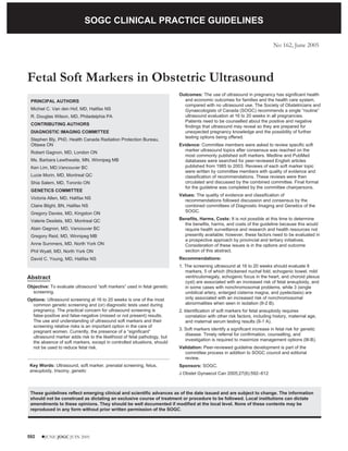
- 5. SOGC CLINICAL PRACTICE GUIDELINES Figure 2. Bilateral renal pyelectasis with anterior/posterior measurement 17. Nyberg DA, Souter VL, El-Bastawissi A, Young S, Luthhardt F, Luthy DA. MILD PYELECTASIS (Figure 2) Isolated sonographic markers for detection of fetal Down syndrome in the second trimester of pregnancy. J Ultrasound Med 2001;20:1053–63. Definition and Imaging Criteria 18. Petrikovsky B, Challenger M, Gross B. Unusual appearances of echogenic foci within the fetal heart: are they benign? Ultrasound Obstet Gynecol Mild pyelectasis is defined as a hypoechoic spherical or 1996;8:229–31. elliptical space within the renal pelvis that measures $ 5 mm and # 10 mm.1–3 The measurement is taken on a transverse 19. Wax JR, Philput C. Fetal intracardiac echogenic foci: does it matter which ventricle? J Ultrasound Med 1998;17:141–4. section through the fetal renal pelvis using the maximum anterior-to-posterior measurement.4 Measurements < 5 20. Bettelheim D, Deutinger J, Bernashek G. The value of echogenic foci (“golf mm are normal, should not be designated as pyelectasis, and balls”) in the fetal heart as a marker of chromosomal abnormalities. Ultra- sound Obstet Gynecol 1999;14:98–100. should not be reported. Pyelectasis may also be referred to as “mild renal pelvic dilatation” or “mild hydronephrosis.” 21. Bromley B, Lieberman E, Shipp TD, Richardson M, Benacceraf BR. Signifi- cance of an echogenic intracardiac focus in fetuses at high and low risk for aneuploidy. J Ultrasound Med 1998;17:127–31. Association With Fetal Aneuploidy 22. Wolman I, Jaffa A, Geva E, Diamant S, Strauss S, Lessing JB, et al. Isolated pyelectasis is seen in 0.7% of fetuses at 16 to 26 Intracardiac echogenic focus: no apparent association with structural cardiac weeks’ gestation.5 It is an isolated finding in fetal Down syn- abnormality. Fetal Diagn Ther 2000;15(4):216–8. drome in approximately 2%.6 Although the likelihood ratio 23. Barsoom MJ, Feldman DM, Borgida AF, Esters D, Diana D, Egan JF. Is an for Down syndrome is approximately 1.9, the 95% CI does isolated cardiac echogenic focus an indication for fetal echocardiography? J cross 1 (0.7–5.1), indicating lack of significance.6 In the Ultrasound Med 2001;20(10):1043–6. absence of other risk factors, the chance of Down syn- 24. Homola J. Are echogenic foci in fetal heart ventricles insignificant findings? drome in the presence of isolated mild pyelectasis remains Ceska Gynekol 1997;62(5):280–2. small and does not justify an invasive diagnostic procedure. 25. Degani S, Leibovitz Z, Shapiro I, Gonen R, Ohel G. Cardiac function in fetuses with intracardiac echogenic foci. Ultrasound Obstet Gynecol Association With Nonchromosomal Abnormalities 2001;18(2):131–4. Fetal pyelectasis is associated with congenital 26. Shipp TD, Bromley B, Lieberman E, Benacerraf BR. The frequency of the hydronephrosis, which is a commonly encountered birth detection of fetal echogenic intracardiac foci with respect to maternal race. defect.7 Renal pelvis measurements > 10 mm should be Ultrasound Obstet Gynecol 2000;15(6):460–2. considered equivalent to congenital hydronephrosis with 27. Van den Hof MC, Demianczuk NN. Contents of a complete ultrasound appropriate follow-up. All fetuses with renal pelvic mea- report. J Soc Obstet Gynaecol Can 2001;23(5):827–8. surements $ 5 mm should have a neonatal ultrasound, and 596 l JUNE JOGC JUIN 2005
- 6. Fetal Soft Markers in Obstetric Ultrasound those having measurements > 10 mm should also have a 7. Aviram R, Pomeran A, Sharony R, Beyth Y, Rathaus V, Tepper R. The increase of renal pelvis dilatation in the fetus and its significance. Ultra- third trimester ultrasound.2 sound Obstet Gynecol 2000; 16:60–2. 8. Van den Hof MC, Demianczuk NN. Content of a complete obstetrical Summary ultrasound report. J Soc Obstet Gynaecol Can 2001;23(5):427–8. Evaluation of fetal kidneys, which includes possible SINGLE UMBILICAL ARTERY (Figure 3) pyelectasis, is considered part of the routine screening ultra- sound at 16 to 20 weeks’ gestation and should be reported.8 Definition and Imaging Criteria The finding of isolated pyelectasis does not appear to signif- Single umbilical artery (SUA) is the absence of one of the icantly increase the risk of fetal aneuploidy in low-risk arteries surrounding the fetal bladder and in the fetal umbil- women and does not justify invasive prenatal testing, but ical cord. Assessment of the umbilical arteries can be made noninvasive maternal serum screening may assist in risk from the cord itself in either transverse or longitudinal sec- assessment. Owing to the increased risk of fetal tions.1–3 The umbilical arteries can also be assessed at the hydronephrosis, a neonatal follow-up scan should be cord insertion site into the fetal abdomen and on either side arranged in all cases of mild isolated pyelectasis. A third tri- of the fetal bladder as the vessels originate from the iliac mester follow-up ultrasound should only be considered if arteries. If needed, the assessment can be enhanced with pyelectasis is $ 10 mm. Referrals should be considered for colour flow Doppler. women aged over 35 years and for women who have addi- tional ultrasound findings, renal pelvis measurements > 10 Association With Fetal Aneuploidy mm, or maternal serum screening results showing increased Isolated SUA has not been found to be significantly associ- chromosomal risks. ated with fetal aneuploidy.1–6 Recommendations Association With Nonchromosomal Abnormalities 1. Evaluation of fetal kidneys is a part of the screening ultra- Isolated SUA has been associated with both underlying fetal sound at 16 to 20 weeks,’ and if pyelectasis is visualized, the renal and cardiac abnormalities,1,7–9 as well as low birth renal pelvis should be measured in the anterior/posterior weight.2,3,5 diameter (III-B). 2. All fetuses with renal pelvic measurements $ 5 mm Summary should have a neonatal ultrasound, and those having mea- Assessment of cord vessels is considered a part of the rou- surements > 10 mm should be considered for a third tri- tine obstetric ultrasound at 16 to 20 weeks.10 The finding of mester scan (II-2 A). a single umbilical artery warrants a detailed review of fetal 3. Isolated mild pyelectasis does not require fetal anatomy, including kidneys and heart (fetal echo). Appro- karyotyping (II-2 E). priate fetal growth should be confirmed through clinical evaluation with follow-up ultrasound for clinical concerns. 4. Referral for pyelectasis should be considered with addi- An isolated SUA does not warrant invasive testing for fetal tional ultrasound findings and (or) in women at increased aneuploidy. risk for fetal aneuploidy owing to maternal age or maternal serum screen results (II-2 A). Recommendations 1. Assessment of cord vessels is considered a part of the References routine obstetric ultrasound at 16 to 20 weeks (III-A). 1. Arger PH, Coleman BH, Mintz MC, Snyder HP, Camardese T, Arensen RL, 2. The finding of a single umbilical artery requires a more et al. Radiology 1985;156:485–9. detailed review of fetal anatomy, including kidneys and 2. Langer B, Simeoni U, Montoya Y, Casanova R, Schlaeder G. Antenatal diagnosis of upper urinary tract dilation by ultrasonography. Fetal Diagn heart (fetal echo) (II-2 B). Ther 1996;11:191–8. 3. An isolated single umbilical artery does not warrant inva- 3. Wilson RD, Lynch S, Lessoway VA. Fetal pyelectasis: comparison of sive testing for fetal aneuploidy (II-2 A). postnatal renal pathology with unilateral and bilateral pyelectasis. Prenat Diagn 1997;17:451–5. 4. Devore, GR. Trisomy 21: 91% detection rate using second-trimester ultra- References sound markers. Ultrasound Obstet Gynecol 2000;16:133–41. 1. Budorick NE, Kelly TE, Dunn JA, Scioscia AL. The single umbilical artery 5. Chudleigh PM, Chitty LS, Pembrey M, Campbell S. The association of in a high-risk patient population. What should be offered? J Ultrasound aneuploidy and mild fetal pyelectasis in an unselected population: the result Med 2001;20:619–27. of a multicenter study. Ultrasound Obstet Gynecol 2001;17:197–202. 2. Farrell T, Leslie J, Owen P. Accuracy and significance of prenatal diagnosis of single umbilical artery. Ultrasound Obstet Gynecol 2000;16:667–8. 6. Smith-Bindman R, Hosmer W, Feldstein VA, Deeks JJ, Goldberg JD. Sec- ond-trimester ultrasound to detect fetuses with Down syndrome. A 3. Geipel A, Germer U, Welp T, Schwinger E, Gembruch U. Prenatal diagno- meta-analysis. JAMA 2001;285:1044–55. sis of single umbilical artery: determination of the absent side, associated JUNE JOGC JUIN 2005 l 597
- 7. SOGC CLINICAL PRACTICE GUIDELINES Figure 3. Single umbilical artery on cross-section of cord anomalies, Doppler findings and perinatal outcome. Ultrasound Obstet echogenicity of fetal bowel and surrounding bone relative Gynecol 2000;15:114–7. to the ultrasound machine gain setting minimizes observer 4. Pierce BT, Dance VD, Wagner RK, Apodaca CC, Nielsen PE, Calhoun BC. variability and should be used. Grade 2 suggests that J Matern Fetal Med 2001;10:59–63. echogenicity is equal to bone whereas grade 3 suggests that 5. Rinehart BK, Terrone DA, Taylor CW, Isler CM, Larmon JE, Roberts WE. Single umbilical artery is associated with an increased incidence of structural it is greater.3 Whenever echogenic bowel is suspected, the and chromosomal anomalies and growth restriction. Am J Perinatol gain setting should be lowered to enable this comparison 2000;17(5):229–32. and to ensure that bowel hyperechogenicity is real.3 This 6. Murphy-Kaulbeck L, Van den Hof M. Single umbilical artery (SUA) and fetal aneuploidy. Ultrasound Obstet Gynecol 2002;20(Suppl1):67. should help to minimize a false-positive diagnosis of 7. Abuhamad AZ, Shaffer W, Mari G, Copel J, Hobbins J, Evans A. Single hyperechogenicity. umbilical artery: does it matter which artery is missing? Am J Obstet Gynecol 1995;173:728–32. Association With Fetal Aneuploidy 8. Persutte W, Hobbins J. Single umbilical artery: a clinical enigma in modern prenatal diagnosis. Ultrasound Obstet Gynecol 1995;6:216–29. The presence of echogenic bowel is associated with an 9. Van den Hof M, Murphy-Kaulbeck L. Single umbilical artery (SUA) and increased risk for fetal aneuploidy, including trisomy 13, 18, risk of congenital heart disease (CHD). Ultrasound Obstet Gynecol 21, and the sex chromosomes. It has been detected in 0.6% 2002;20(Suppl1):83. to 2.4% of all second trimester fetuses2,4–9 and as an isolated 10. Van den Hof MC, Demianczuk NN. Content of a complete obstetrical ultra- finding in 9% of fetuses with aneuploidy (2.8% to 25%).2–19 sound report. J Soc Obstet Gynaecol Can 2001;23(5):427–8. As a result, it has been suggested that the likelihood ratio for ECHOGENIC BOWEL (Figure 4) this marker is 6 (CI 2.7–6.8).6 Definition and Imaging Criteria Association With Nonchromosomal Abnormalities Echogenic bowel is defined as fetal bowel with homoge- The presence of echogenic bowel has been associated with nous areas of echogenicity that are equal to or greater than an increased risk for cystic fibrosis in the fetus, congenital that of surrounding bone.1 The echogenicity has been clas- infection, intra-amniotic bleeding, congenital malforma- sified as either focal or multifocal.2 There have been various tions of the bowel, and other perinatal complications, techniques used to define echogenic bowel, partially including intrauterine growth restriction. The risk of cystic because of concerns raised about intra- and interobserver fibrosis in the fetus with echogenic bowel is approximately variability.3 A grading system based on comparison of the 2% (0 to 13%).3,10–13,18–21 The a priori risk will change if the 598 l JUNE JOGC JUIN 2005
- 8. Fetal Soft Markers in Obstetric Ultrasound Figure 4. Fetal bowel that is as echogenic as surrounding bone parental carrier status is known. The association between investigations may include a fetal karyotype, DNA testing congenital infection and hyperechogenic bowel has been for cystic fibrosis, and testing for congenital infections noted for the most common pathogens known to cause (maternal serum titres, fetal amniotic culture, or polymerase fetal infections (cytomegalovirus [CMV], herpes, parvovi- chain reaction [PCR] for viral DNA). A maternal serum rus, rubella, varicella, and toxoplasmosis).3,4,6,11,12,14,18,19 screen may be considered because elevations in alpha Intra-amniotic bleeding has also been identified as an etiol- fetoprotein and hCG in the presence of echogenic bowel ogy of echogenic bowel. This can result from intra-amniotic may further define a population at increased risk for bleeding owing to placental abruptions or invasive proce- perinatal morbidity and mortality. Obstetric and ultrasound dures.18,19,22–24 Congenital malformations of the fetal bowel follow-up may also be important. can lead to increased echogenicity. Studies have suggested Recommendations that this is more likely with upper gastrointestinal (GI) 1. Evaluation of the fetal bowel should be done routinely lesions. Other ultrasound features, such as ascites and during the 16- to 20-week obstetric ultrasound (III-B). dilated loops of bowel, will often be present in this circum- stance.18,19,25–27 Echogenic bowel has also been reported 2. Echogenic bowel should be identified by comparison with poor fetal growth, which is associated with an increase with the echogenicity of surrounding bone using an appro- in perinatal morbidity and mortality.4–6,10–14,18,19,28 priate transducer and gain setting. Bowel echogenicity equal to or greater than bone is significant (grade 2 or 3) (II-2 A). Summary 3. No further investigations are required for grade 1 Evaluation of the fetal abdomen is an established compo- echogenic bowel (II-2 D). nent of the screening obstetric ultrasound at 16 to 20 4. Grade 2 and 3 echogenic bowel is associated with both weeks.29 This includes an evaluation of bowel echogenicity chromosomal and nonchromosomal abnormalities. Expert using an appropriate transducer (5 MHZ or less) and ultra- review is recommended to initiate the following: a. detailed sound gain setting. Echogenic bowel is associated with a ultrasound evaluation looking for additional structural significantly increased risk for both chromosomal and anomalies or other soft markers of aneuploidy (II-2 A); b. nonchromosomal fetal abnormalities. Timely referral for detailed evaluation of the fetal abdomen looking for signs validation, consultation, and further investigation is of bowel obstruction or perforation (II-2 B); and c. detailed important. evaluation of placental characteristics (echogenicity, thick- Further evaluations may include a detailed review of fetal ness, position, and placental cord insertion site) (II-2 B); d. anatomy, growth, and placental characteristics. Laboratory genetic counselling (II-2 A); e. laboratory investigations that JUNE JOGC JUIN 2005 l 599
- 9. SOGC CLINICAL PRACTICE GUIDELINES should be offered, including fetal karyotype, maternal 20. Sepulveda W, Leung KY, Robertson ME, Kay E, Mayall ES, Fisk NM. Prev- alence of cystic fibrosis mutations in pregnancies with fetal echogenic serum screening, DNA testing for cystic fibrosis (if appro- bowel. Obstet Gynecol 1996;87:103–6. priate), and testing for congenital infection (II-2 A). 21. Berlin BM, Norton ME, Sugarman EA, Tsipis JE, Allitto BA. Cystic fibrosis and chromosome abnormalities associated with echogenic fetal bowel. References Obstet Gynecol 1999;94:135–8. 1. Sepulveda W, Sebire NJ. Fetal echogenic bowel: a complex scenario. Ultra- sound Obstet Gynecol 2000;16:510–4. 22. Sepulveda W, Reid R, Nicolaidis P, Prendiville On, Chapman RS, Fisk N. Second trimester echogenic bowel and intraamniotic bleeding: association 2. Al-Kouatly HB, Chasen ST, Streltzoff J, Chervenak FA. The clinical signifi- between fetal bowel echogenicity and amniotic fluid spectrophotometry at cance of fetal echogenic bowel. Am J Obstet Gynecol 2001;185:1035–8. 410 nm. Am J Obstet Gynecol 1996;174:839–42. 3. Slotnick RN, Abuhamad AZ. Prognostic implications of fetal echogenic bowel. Lancet 1996;347:85–7. 23. Sepulveda W. Harris Birthright Research Center, King’s College Hospital School London. Fetal echogenic bowel. Lancet 1996;(34):1043. 4. Nyberg DA, Dubinsky T, Resta RG, Mahony BS, Hickock D, Luthy DA. Echogenic fetal bowel during the second trimester: clinical importance. 24. Petrikovsky B, Smith-Levitin M, Hosten N. Intra-amniotic bleeding and fetal Radiology 1993;188:527–31. echogenic bowel. Obstet Gynecol 1999;93:684–6. 5. Bromley B, Doubilet P, Frigoletto F, Krauss C, Estroff J, Benacerraf B. Is 25. Phelps S, Fisher R, Partington A, Dykes E. Prenatal ultrasound diagnosis of fetal hyperechoic bowel on second trimester sonogram an indication for gastrointestinal malformations. J Ped Surgery 1997;32:438–40. amniocentesis? Obstet Gyneacol 1994;83:647–51. 6. Hill LM, Fries J, Hecker J, Grzybek P. Second trimester echogenic small 26. Font GE, Solari M. Prenatal diagnosis of bowel obstruction initially mani- bowel: an increased risk of adverse perinatal outcome. Prenat Diagn fested as isolated hyperechoic bowel. J Ultrasound Med 1998;17:721–3. 1994;14:845–50. 27. Shyu MK, Shih JC, Lee CN, Hwa HL, Chow SN, Hsieh FJ. Correlation of 7. Shohl BD, Scioscia AL, Budorick NE, Moore TR. Utility of minor prenatal ultrasound and postnatal outcome in meconium peritonitis. Fetal ultrasonographic markers in the prediction of abnormal fetal karyotype at a Diagn Ther 2003;18:255–61. prenatal diagnostic center. Am J Obstet Gynecol 1999;181:898–903. 8. Nyberg DA, Souter VL, Bastawissi AE, Young S, Luthhardt F, Luthy D. 28. Achiron R, Mazkereth R, Orvieto R, Kuint J, Lipitz S, Rotstein Z. Isolated sonographic markers for detection of fetal down syndrome in the Echogenic bowel in intrauterine growth restriction fetuses: does this jeopar- second trimester of pregnancy. J Ultrasound Med 2001;20:1053–63. dize the gut. Am Obstet Gynecol 2002;100:120–5. 9. Bromley B, Lieberman E, Shipp TD, Benacerraf BR. The genetic 29. Van den Hof MC, Demianczuk NN. Contents of a complete ultrasound sonogram. A method of risk assessment for down syndrome in the second report. J Soc Obstet Gynaecol Can 2001;23(5):827–8. trimester. J Ultrasound Med 2002;21:1087–96. 10. Dicke JM, Crane JP. Sonographically detected hyperechoic fetal bowel: sig- THICKENED NUCHAL FOLD (Figure 5) nificance and implications for pregnancy management. Obstet Gynecol 1992;80:778–82. Definition and Imaging Criteria 11. Muller F, Dommergues M, Aubry MC, Simon-Bouy B, Gautier E, Oury JF, et al. Hyperechogenic fetal bowel: an ultrasonographic marker for adverse The nuchal fold is the skin thickness in the posterior aspect fetal and neonatal outcome. Am J Obstet Gynecol 1995;173:508–13. of the fetal neck. A nuchal fold measurement is obtained in 12. Yaron Y, Hassan S, Geva E, Kupferminc MJ, Yavetz H, Evans MI. Evalua- a transverse section of the fetal head at the level of the tion of fetal echogenic bowel in the second trimester. Fetal Diagn Ther cavum septum pellucidum and thalami, angled posteriorly 1999;14:176–80. to include the cerebellum. The measurement is taken from 13. Ghose I, Mason GC, Martinez D, Harrison KL, Evans JA, Ferriman EL, et the outer edge of the occiput bone to the outer skin limit al. Hyperechogenic fetal bowel: a prospective analysis of sixty consecutive directly in the midline.1 The definition of a thickened nuchal cases. Br J Obstet Gynaecol 2000;107:426–9. fold has varied,1,2 although many researchers and centres 14. Stocker AM, Snijders RJ, Carlson DE, Greene N, Gregory KD, Walla CA, et now use gestational-age specific criteria.3,4 Consensus for al. Fetal echogenic bowel: parameters to be considered in differential diag- nosis. Ultrasound Obstet Gynecol 2000;16:519–23. this document is that a measurement $6 mm be considered significant between 18 and 24 weeks and a measurement of 15. Rotmensch S, Liberati M, Bronshtein M, Schoenfeld-Dimaio M, Shalev J, Ben-Rafael Z, et al. Pernatal sonographic findings in 187 fetuses with down $ 5 mm be considered significant at 16 to 18 weeks.1–5 A syndrome. Prenat Diagn 1997;17:1001–9. thickened nuchal fold should be distinguished from cystic 16. Smith-Bindman R, Hosmer W, Feldstein VA, Deeks JJ, Goldberg JD. Sec- hygroma, in which the skin in this area has fluid-filled ond-trimester ultrasound to detect fetuses with down syndrome: a loculations. A thickened nuchal fold should not be con- meta-analysis. JAMA 2001;285:1044–55. fused with nuchal translucency, which is a specific measure- 17. Shipp TD, Benacerraf BR. Second-trimester ultrasound screening for ment of fluid in the posterior aspect of the neck at 11 to 14 aneuploidy. Prenat Diagn 2002;22:296–307. weeks’ gestation. 18. Kesrouani AK, Guibourdenche J, Muller F, Denamur E, Vuillard E, Garel C, et al. Etiology and outcome of fetal echogenic bowel. Fetal Diagn Ther Association With Fetal Aneuploidy 2003;18:240–6. A meta-analysis reviewed the performance of a thick nuchal 19. Simon-Bouy B, Satre V, Ferec C, Malinge MC, Girodon E, Denamur E, et al. Management of prenatally diagnosed hyperechogenic bowel. Am J Med fold at 6 mm or greater and showed that the risk for Down Genet 121A:209,2003. syndrome increased by approximately 17-fold (CI 8–35).6 600 l JUNE JOGC JUIN 2005
- 10. Fetal Soft Markers in Obstetric Ultrasound Figure 5. Increased nuchal fold Association With Nonchromosomal Abnormalities increased nuchal fold justifies a directed, detailed anatomic A thickened nuchal fold can be associated with single gene survey of the fetus and a careful newborn examination.12 abnormalities, such as Noonan syndrome, multiple Recommendations pterygium syndrome, and skeletal dysplasias.7,8 Thickened nuchal fold has also been associated with congenital cardiac 1. Nuchal fold measurement should be a part of the screen- defects.7,9,10 ing obstetric ultrasound at 16 to 20 weeks (III-B). 2. A thickened nuchal fold significantly increases the risk of Summary fetal aneuploidy. Expert review is recommended, and karyotyping should be offered (II-1 A). Evaluation of the nuchal fold should be considered during the screening ultrasound at 16 to 22 weeks’ gestation. A 3. A thickened nuchal fold is associated with congenital nuchal fold of 6 mm or greater at 18 to 24 weeks or of 5 mm heart disease and rarely with other genetic syndromes. or greater at 16 to 18 weeks should be considered significant Expert review is recommended (II-2 B). and should prompt referral for validation and consultation. References The finding of an isolated thickened nuchal fold signifi- cantly increases the risk for fetal aneuploidy, and fetal 1. Benacerraf BR, Frigoletto FD. Soft tissue nuchal fold in the second trimes- ter fetus: standards for normal measurements compared with those with karyotyping should be offered. Centres may use alternate Down syndrome. Am J Obstet Gynecol 1987;157(5):1146–9. definitions, taking into account gestational age and other 2. Nyberg DA, Souter VL, El-Bastawissi A, Young S, Luthhardt F, Luthy DA. risk factors. Nuchal index has been described as an effective Isolated sonographic markers for detection of fetal Down syndrome in the second trimester of pregnancy. J Ultrasound Med 2001;20:1053–63. method to deal with the normal increase in nuchal fold mea- surement that accompanies advancing gestational age. 3. Locatelli A, Piccoli MG, Vergani P, Mariani E, Ghidini A, Mariana S, et al. Critical appraisal of the use of nuchal fold thickness measurements for the Nuchal index is the mean nuchal fold/mean biparietal prediction of Down syndrome. Am J Obstet Gynecol 2000;82(1)192–8. diameter (BPD) × 100. A value of 11 or greater has a sensi- 4. Bahado-Singh RO, Oz UA, Kovanci E, Deren O, Feather M, Hsu CD, et tivity of 50% and a specificity of 96%.11 al. Gestational age standardized nuchal thickness values for estimating mid-trimester Down syndrome risk. J Matern Fetal Med 1999;8(2):37–43. The suggested association of nuchal fold thickening and 5. Gray DL, Crane JP. Optimal nuchal skin-fold thresholds based on gesta- congenital heart defect is based on small studies. Careful tional age for prenatal detection of Down syndrome. Am J Obstet Gynecol 1994;171:1282–6. detailed ultrasound examination, including the 4-chamber 6. Smith-Blindman R, Hosmer W, Feldstein VA, Deeks JJ, Goldberg JD. Sec- view and outflow tracts, should be performed. The rare ond trimester ultrasound to detect fetuses with Down syndrome: a occurrence of an underlying syndromic etiology for the meta-analysis. JAMA 2001;285(8):1044–55. JUNE JOGC JUIN 2005 l 601
- 11. SOGC CLINICAL PRACTICE GUIDELINES Figure 6. Slightly enlarged posterior horn of the lateral ventricle 7. Souter VL, Nyberg DA, El-Bastawissi A, Zebelman A, Luthhardt F, Luthy Association With Fetal Aneuploidy DA. Correlation of ultrasound findings and biochemical markers in the sec- ond trimester of pregnancy in fetuses with trisomy 21. Prenat Diagn 2002;22(3):175–82. When MVM is isolated, the incidence of abnormal fetal 8. Shipp TD, Benacerraf BR. Second trimester ultrasound screening for aneuploidy. Prenat Diagn 2002;22:296–307. karyotype is estimated at 3.8% (0 to 28.6%).2 Idiopathic lat- 9. DeVore GR, Alfi O. The association between an abnormal nuchal skin fold, eral ventriculomegaly is found in approximately 0.15% of trisomy 21, and ultrasound abnormalities identified during the second tri- chromosomally-normal fetuses,3 whereas 1.4% of trisomy mester of pregnancy. Ultrasound Obstet Gynecol 1993;3:387–94. 21 fetuses in the second trimester have idiopathic 10. Dahlgren LS, Sandor GS, Lim KI. Is the nuchal index increased in fetuses ventriculomegaly.4 This suggests a likelihood ratio of 9 for with congenital structural heart defects? Am J Obs Gynecol 2002;(Suppl the risk of karyotype abnormality. 187);(6):5191. 11. Lim KI, Pugash D, Dansereau J, Wilson RD. Nuchal index: a gestational age independent ultrasound marker for the detection of Down syndrome. Prenat Diagn 2002;22(13):1233–7. Association With Nonchromosomal Abnormalities 12. Baumann C, Delagarde R, Vuillard E, Oury JF. Pregnancy outcome and infant follow-up after diagnosis of nuchal anomalies at the 1st or 2nd tri- mester ultrasound examination. J Gynecol Obstet Biol Reprod 2001;(30 Fetal ventriculomegaly is the most commonly detected Suppl 1):68–74. ultrasonographic abnormality of the central nervous sys- tem.5 Ventriculomegaly can arise from agenesis of the cor- MILD VENTRICULOMEGALY (Figure 6) pus callosum, cerebral maldevelopment or destruction, vas- cular anomalies, or an obstruction within the ventricular Definition and Imaging Criteria system.6 Children with a prenatal diagnosis of MVM have Cerebral ventriculomegaly is defined by atrial measure- abnormal neurodevelopment in 10% to 36% of cases ments $ 10 mm. Mean atrial measurements are 7.6 mm, dependent on associated anomalies, etiology,7,8 and ventric- standard deviation (SD) 0.6 mm. Mild ventriculomegaly ular measurement. In combined case series, mortality is (MVM) is defined as measurements $ 10 to # 15 mm.1 Mea- reported at 3.7%.2 When MVM resolves, abnormal out- surements are obtained from an axial plane at the level of come has been reported but is infrequent (< 10%).9,10 Uni- the thalamic nuclei just below the standard image to mea- lateral MVM also carries a favourable prognosis when iso- sure the BPD. Ventricular measurements are usually lated.11,12 After the prenatal diagnosis of MVM, maternal obtained in the far image field because of “typical” evaluation for congenital infection is recommended. near-field artifacts. Cursors are positioned perpendicular to Amniocentesis should be offered for karyotype and con- the long axis of the ventricle at the edges of the ventricular genital infection assessment. Other imaging modalities such lumen, near the posterior portion of the choroid plexus. as magnetic resonance imaging (MRI) might be considered.13,14 602 l JUNE JOGC JUIN 2005
- 12. Fetal Soft Markers in Obstetric Ultrasound Summary 9. Signorelli M, Tiberti A, Valseriati D, Molin E, Cerri V, Grali C, et al. Width of the fetal lateral ventricular atrium between 10 and 12 mm: a simple varia- tion of the norm? Ultrasound Obstet Gynecol 2004;23:14–8. Lateral ventriculomegaly can be detected on standard cra- nial biometry planes and should be evaluated on both 10. Patel HD, Filly AL, Hersh DR, Goldstein RB. Isolated mild fetal cerebral ventriculomegaly: clinical course and outcome. Radiology 1994;192:759–64. screening ultrasounds as well as detailed ultrasound for 11. Lipitz S, Yagel S, Malinger G, Meizner I, Zalel Y, Achiron R. Outcome of higher risk women.15 The ventricles should be measured if fetuses with isolated borderline unilateral ventriculomegaly diagnosed at they appear to be larger than the choroid plexus. The find- mid-gestation. Ultrasound Obstet Gynecol 1998;12(1):23–6. ing of ventriculomegaly should prompt a timely referral for 12. Senat MV, Bernard JP, Schwarzler P, Britten J, Ville Y. Prenatal diagnosis consultation and validation. Evaluation of lateral and follow-up of 14 cases of unilateral ventriculomegaly. Ultrasound Obstet ventriculomegaly should include a detailed examination of Gynecol 1999;14(5):327–32. fetal anatomy, including the heart. Neonatal assessment and 13. Levine D, Barnes PD, Madsen JR, Abbott J, Mehta T, Edelman RR. Central follow-up are important to rule out associated abnormali- nervous system abnormalities assessed with prenatal magnetic resonance imaging. Obstet Gynecol 1999;94(6):1011–9. ties because of the potential for abnormal 14. Launay S, Robet Y, Valat AS, Thomas D, Devisme L, Rocourt N, et al. neurodevelopment. Cerebral fetal MRI and ventriculomegaly. J Radiol 2002;83(6 pt 1):723–30. Recommendations 15. Van den Hof, MC, Deminaczuk NN. Content of a complete obstetrical ultrasound report. J Soc Obstet Gynaecol Can 2001;23(5):427–8. 1. Fetal cerebral ventricles should be measured if they sub- jectively appear larger than the choroid plexus (III-B). CHOROID PLEXUS CYSTS (Figure 7) 2. Cerebral ventricles greater than or equal to 10 mm are associated with chromosomal and central nervous system Definition and Imaging Criteria pathology. Expert review should be initiated to obtain the Choroid plexus cysts (CPCs) are sonographically discrete, following: a. a detailed anatomic evaluation looking for small cysts ($ 3 mm) found in the choroid plexus within the additional malformations or soft markers (III-B); b. labora- lateral cerebral ventricles of the developing fetus at 14 to 24 tory investigation for the presence of congenital infection weeks’ gestation.1 Imaging of the choroid plexus is per- or fetal aneuploidy (III-B); and c. MRI as a potential addi- formed in the transverse plane of the fetal head at the same tional imaging technique (II-2 C). level that the lateral cerebral ventricle is evaluated. The choroid plexus should be inspected bilaterally for the pres- 3. Neonatal assessment and follow-up are important to rule ence of cysts. The size of CPCs is not of clinical relevance.2 out associated abnormalities and are important because of Evaluation of the choroid plexus in the near field ventricle the potential for subsequent abnormal neurodevelopment will be more difficult owing to imaging artifact. (II-2 B). Association With Fetal Aneuploidy References CPCs have been identified in 1% of fetuses during the sec- 1. Cardoza JD, Goldstein RB, Filly RA. Exclusion of fetal ventriculomegaly ond trimester screening ultrasound.3–10 The incidence of with a single measurement: the width of the lateral ventricular atrium. Radi- ology 1988;169:711–4. CPCs is 50% in fetuses with trisomy 185,11,12; however, only 2. Pilu G, Falco P, Gabrielli S, Perolo A, Sandri F, Bovicelli L. The clinical sig- 10% of fetuses with trisomy 18 will have CPCs as the only nificance of fetal isolated cerebral borderline ventriculomegaly: report of 31 identifiable sonographic marker on ultrasound screen- cases and review of the literature. Ultrasound Obstet Gynecol ing.3,4,6–9,12–16 The likelihood ratio for trisomy 18 when an 1999;14:320–6. isolated CPC is identified is 7 (95% CI 4–12).9 The number 3. Achiron R, Schimmel M, Achiron A, Mashiach S. Fetal mild idiopathic lat- eral ventriculomegaly: is there a correlation with fetal trisomy? Ultrasound of cysts and the cysts’ distribution or size does not change Obstet Gynecol 1993;3:89–92. the risk.2 Although it has been suggested that an isolated 4. Nyberg DA, Resta RG, Luthy DA, Hickox DE, Mahony BS, Hirsch JH. CPC may increase the risk for trisomy 21 with a likelihood Prenatal sonographic findings in Down syndrome. Review of 94 cases. ratio of 1.9, the 95% CI crosses 1 (0.78–4.46) and lacks sta- Obstet Gynecol 1990;76:370–7. tistical significance.17,18 5. Filly RA, Cardoza JD, Goldstein RB, Barkovich AJ. Detection of fetal cen- tral nervous system anomalies: a practical level of effort for a routine sonogram. Radiology 1989;172:403–8. Association With Nonchromosomal Abnormalities The presence of CPCs in chromosomally normal fetuses is 6. Tsao PN, Teng RJ, Wu TJ, Yau KIT, Wang PJ. Nonprogressive congenital unilateral ventriculomegaly. Pediatr Neur 1996;14:66–8. not associated with other fetal abnormalities or abnormal 7. Nicolaides KH, Berry S, Snijders RJ, Thorpe-Beeston JG, Gosden C. Fetal postnatal development.15 lateral cerebral ventriculomegaly: associated malformations and chromo- somal defects. Fetal Diagn Ther 1990;5(1):5–14. Summary 8. Tomlinson MW, Treadwell MC, Bottoms SF. Isolated mild ventriculomegaly: associated karyotypic abnormalities and in utero observa- Evaluation of the fetal cranium, including the ventricles and tions. J Matern Fetal Med 1997;6:241–4. choroid plexus, is considered part of the routine screening JUNE JOGC JUIN 2005 l 603
- 13. SOGC CLINICAL PRACTICE GUIDELINES Figure 7. Choroid plexus cyst ultrasound at 16 to 20 weeks’ gestation.19 Identification and 2. Gratton RJ, Hogge WA, Aston CE. Choroid plexus cysts and trisomy 18: risk of modification based on maternal age and multiple-marker screening. reporting of CPCs should be a part of this screening exami- Am J Obstet Gynecol 1996;175(6):1493–7. nation. With the presence of CPCs, caregivers should next 3. Walkinshaw S, Pilling D, Spriggs A. Isolated choroid plexus cysts – the evaluate maternal age risk and, if available, the maternal need for routine offer of karyotyping. Prenat Diagn 1994;14(8):663–7. serum screen.2 CPCs increase the risk for trisomy 18. 4. Kupferminc MJ, Tamura RK, Sabbagha RE, Parilla BV, Cohen LS, Pergament E. Isolated choroid plexus cyst(s): an indication for amniocente- Follow-up ultrasound is not necessary for isolated CPCs. sis. Am J Obstet Gynecol 1994;171(4):1068–71. Referral for counselling and possible invasive testing is only 5. Gray DL, Winborn RC, Suessen TL, Crane JP. Is genetic amniocentesis necessary if maternal age is 35 years or older or the maternal warranted when isolated choroid plexus cysts are found? Prenat Diagn serum screen is positive for either trisomy 18 or 21.2,20 1996;16(11):983–90. 6. Reinsch RC. Choroid plexus cysts – association with trisomy: prospective Recommendations review of 16,059 patients. Am J Obstet Gynecol 1997;176(6):1381–3. 1. Choroid plexus should be evaluated for the presence of 7. Geary M, Patel S, Lamont R. Isolated choroid plexus cysts and association with fetal aneuploidy in an unselected population. Ultrasound Obstet discrete cysts during the 16- to 20-week ultrasound (III-B). Gynecol 1997;10(3):171–3. 2. Isolated CPCs require no further investigation when 8. Sohn C, Gast AS, Krapfl E. Isolated fetal choroid plexus cysts: not an indi- cation for genetics diagnosis? Fetal Diagn Ther 1997;12(5):255–9. maternal age or the serum screen equivalent is less than the risk of a 35-year-old (II-2 E). 9. Ghidini A, Strobelt N, Locatelli A, Mariani E, Piccoli MG, Vergani P. Iso- lated fetal choroid plexus cysts: role of ultrasonography in establishment of 3. Fetal karyotyping should only be offered if isolated CPCs the risk of trisomy 18. Am J Obstet Gynecol 2000;182(4):972–7. are found in women 35 years or older or if the maternal 10. Snijders RJ, Shawa L, Nicholaides KH. Fetal choroid plexus cysts and trisomy 18: assessment of risk based on ultrasound findings and maternal serum screen is positive for either trisomy 18 or 21 (II-2 A). age. Prenat Diagn 1994;14(12):1119–27. 4. All women with fetal CPCs and additional malformation 11. Denis E, Dufour P, Valat AS, Vaast P, Subtil D, Bourgeot P, et al. Choroid should be offered referral and karyotyping (II-2 A). plexus cysts and risk of chromosome anomalies. Review of the literature and proposed management. J Gynecol Obstet Biol Reprod 1998;27(2):144–9. 5. All women with CPCs and additional soft markers should 12. Gonen R, Kar H, Degani S. The karyotype of fetuses with anomalies be offered additional counselling and further ultrasound detected by second trimester ultrasonography. Europ J Obstet Gynecol review (III-B). Reprod Biol 1995;58(2):153–5. 13. Maieron A, Rustico M, Pecile V, Natale R, D’Ottavio G, Fischer Tamaro L, References et al. The indications of the management of fetuses with choroid plexus cysts. Minerva Ginecol 1996;48(4):125–33. 1. Chitty LS, Chudleigh RP, Wright E, Campbell S, Pembrey M. The signifi- cant of choroid plexus cysts in an unselected population: results of a 14. Digiovanni LM, Quinlan MP, Verp MS. Choroid plexus cysts: infant and multicenter study. Ultrasound Obstet Gynecol 1998;12(6):391–7. early childhood development outcome. Obstet Gynecol 1997;90(2):191–4. 604 l JUNE JOGC JUIN 2005
- 14. Fetal Soft Markers in Obstetric Ultrasound Figure 8. Enlarged cisterna magna 15. Morcos CL, Platt LD, Carlson DE, Gregory KD, Greene NH, Korst LM. The isolated choroid plexus cyst. Obstet Gynecol 1998;92(2):232–6. association with aneuploidy appears to be strongest in the absence of ventricular dilatation but in the presence of 16. Sullivan A, Giudice T, Vavelidis F, Thiagaraja S. Choroid plexus cysts: is bio- chemical testing a valuable adjunct to targeted ultrasonography? Am J other anomalies.4–6 Isolated enlarged cisterna magna does Obstet Gynecol 1999;181(2):260–5. not appear to be strongly associated with aneuploidy.2 17. Yoder PR, Sabbagha RE, Gross SJ, Zelop CM. The second-trimester fetus There are no large prospective studies to evaluate this with isolated choroid plexus cysts: a meta-analysis of risk of trisomies 18 marker. and 21. Obstet Gynecol 1999;93:869–72. 18. Bromley B, Lieberman R, Benacerraf BR. Choroid plexus cysts: not associ- Association With Nonchromosomal Abnormalities ated with Down syndrome. Ultrasound Obstet Gynecol 1996;8(4):232–5. An enlarged cisterna magna is commonly seen in associa- 19. Van den Hof MC, Demianczuk NN. Content of a complete obstetrical ultra- tion with other anatomic (arachnoid cyst, Dandy Walker sound report. J Soc Obstet Gynaecol Can 2001;23(5):427–8. malformation, and Dandy Walker variant)8–10 and syn- 20. Demasio K, Canterino J, Ananth C, Fernandez C, Smulian J, Vintzileos A. dromic (oro-facial–digital syndrome, Meckel-Gruber syn- Isolated choroid plexus cyst in low-risk women less than 35 years old. Am J Obstet Gynecol 2002;187:1246–9. drome, and DiGeorge syndrome)4 abnormalities. ENLARGED CISTERNA MAGNA (Figure 8) Summary Review of the fetal cerebellum and cisterna magna is a rou- Definition and Imaging Criteria tine part of the screening ultrasound at 16 to 20 weeks’ ges- The cisterna magna is measured on a transaxial view of the tation.11,12 If the cisterna magna is subjectively increased, a fetal head angled 15 degrees caudal to the canthomeatal line. measurement should be undertaken. The mean diameter of The anterior/posterior diameter is taken between the a normal cisterna magna is 5 mm, SD 3 mm.3 A measure- inferior/posterior surface of the vemis of the cerebellum to ment $ 10 mm is considered an abnormality and appropri- the inner surface of the cranium. An enlarged cisternal ate referral for consultation and validation should be magna is defined by an anterior/posterior diameter $ 10 initiated. A detailed fetal examination should be performed mm.1,2 The measurement will be falsely exaggerated by a looking for other anomalies, growth restriction, or abnor- steep scan angle through the posterior fossa or mal amniotic fluid volume. An isolated enlarged cisterna dolichocephaly.3,4 magna is not an indication for fetal karyotyping. Association With Fetal Aneuploidy Recommendations An enlarged cisterna magna has been described in associa- 1. Review of the fetal cerebellum and cisterna magna is a tion with fetal aneuploidy, particularly trisomy 18.5–7 The routine part of the screening ultrasound at 16 to 20 weeks. JUNE JOGC JUIN 2005 l 605
- 15. SOGC CLINICAL PRACTICE GUIDELINES If the cisterna magna is subjectively increased, a measure- 4. Nyberg D, Mahony B, Hegge F, Hickok D, Luthy D, Kapur R. Enlarged cisterna magna and the Dandy-Walker malformation: factors associated ment should be taken (III-B). with aneuploidy. Obstet Gynecol 1991;77:436. 2. An isolated enlarged cisterna magna is not an indication 5. Laing FC, Frates MC, Brown DL,Bensen CB, Di Salvo D, Doubilet P. Sonography of the fetal cisterna magna: false appearance of mega-cisterna for fetal karyotyping (III-D). magna and Dandy-Walker variant. Radiology 1994;192:274. 3. With an enlarged cisterna magna, expert review is recom- 6. Chen CP, Hung TH, Jan SW, Jeng CJ. Enlarged cisterna magna in the third trimester as a clue to fetal trisomy 18. Fetal Diagn Ther 1998;13:29–34. mended for follow-up ultrasounds and possible other imag- 7. Nyberg DA, Kramer D, Resta RG, Kapur R, Mahony BS, Luthy DA, et al. ing modalities (for example, MRI) and investigations Prenatal sonographic findings of trisomy 18: review of 47 cases. J Ultra- (III-B). sound Med 1993;2:103–13. 8. Ecker JL, Shipp TD, Bromley B, Benacerraf B. The sonographic diagnosis 4. If the enlarged cisterna magna is seen in association with of Dandy-Walker and Dandy-Walker variant: associated findings and out- other abnormal findings, fetal karyotyping should be comes. Prenat Diagn 2000;20:328–32. offered (III-B). 9. Aletebi FA, Fung Kee Fung K. Neurodevelopmental outcome after antena- tal diagnosis of posterior fossa abnormalities. J Ultrasound Med 1999;18:683–9. REFERENCES 10. Ulm MR, Ulm J, Bernaschek G. Dandy-Walker malformation diagnosed 1. Comstock C, Boal D. Enlarged fetal cisterna magna: appearance and signifi- before 21 weeks of gestation: associated malformations and aneuploidy. cance. Obstet Gynecol 1985;66:25S. Ultrasound Obstet Gynecol 1997;10:167–70. 2. Haimovici J, Doubilet P, Benson C, Frates MC. Clinical significance of iso- lated enlargement of the cisterna magna (>10mm) on prenatal sonography. 11. Van den Hof MC, Demianczuk NN. Contents of a complete ultrasound J Ultrasound Med 1997;16:731–4. report. J Soc Obstet Gynaecol Can 2001;23(5):827–8. 3. Mahoney B, Callen P, Filly R, Hoddick W. The fetal cisterna magna. Radiol- 12. Society of Obstetricians and Gynaecologists of Canada. Guidelines for Per- ogy 1984;153:773. formance of ultrasound. J Soc Obstet Gynaecol Can 1995;17:263–6. FETAL SOFT MARKERS USEFUL FOR COMPREHENSIVE ULTRASOUND SHORT FEMUR LENGTH If a femur appears abnormal or its length is found to be below the 2.5th percentile for gestational age, it may be Definition and Imaging Criteria indicative of fetal growth restriction or a more general skel- A short femur length is defined as either a measurement etal malformation. In this circumstance, other long bones below the 2.5th percentile for gestational age or a measure- should be assessed and referral with follow-up ultrasound ment that is less than 0.9 of that predicted by the measured considered. biparietal diameter.1 The femur should be measured with the bone perpendicular to the ultrasound beam and with Recommendations epiphyseal cartilages visible but not included in the mea- 1. Although femur length is standard biometry on the 16- to surement. The relation between bone length and head size 20-week ultrasound, the assessment for relative shortness is may differ across racial groups.2 not part of the screening evaluation (III-C). Association With Fetal Aneuploidy 2. Relative femur shortening is an ultrasound marker for Short femur length has been found to have a sensitivity of trisomy 21 and should be considered during tertiary level 16% in the prediction of Down syndrome with a false- evaluation (II-1 A). positive rate of 4%. A meta-analysis showed a likelihood 3. If a femur appears abnormal or measures short on ratio of 2.7 (95% CI 2.1–6.0).3 screening ultrasound, other long bones should be assessed and referral with follow-up ultrasound considered (III-B). Association With Nonchromosomal Abnormalities Short femur length can also be associated with skeletal SHORT HUMERUS LENGTH dysplasias or fetal growth restriction.4 Definition and Imaging Criteria Summary A short humerus length is defined as a length below the Short femur length is an ultrasound marker for fetal 2.5th percentile for gestational age or as a measurement less aneuploidy, particularly trisomy 21. The mathematical than 0.9 of that predicted by the measured biparietal diame- model used to determine a positive result is not amenable to ter.1 The humerus should be measured with the bone screening ultrasound; however, it should be included in the perpendicular to the ultrasound beam and with epiphyseal panel of markers used by tertiary centres. cartilages visible but not included in the measurement. 606 l JUNE JOGC JUIN 2005
- 16. Fetal Soft Markers in Obstetric Ultrasound Association With Fetal Aneuploidy echogenic line within the bridge of the fetal nose. The fetus Short humeral length has been found to have a sensitivity of is imaged facing the transducer with the fetal face strictly in 9% with a false-positive rate of 3%. A meta-analysis showed the midline. The angle of insonation is 90 degrees, with the a likelihood ratio of 7.5 (95% CI 4.5–12).3 longitudinal axis of the nasal bone as the reference line. Calibres are placed at each end of the nasal bone. Absence Association With Nonchromosomal Abnormalities of the nasal bone or measurements below 2.5th percentile Short humeral length can also be associated with skeletal are considered significant.2–4 dysplasias or fetal growth restriction.4 Humeral length has also been recorded as multiples of the median for gesta- Association With Fetal Aneuploidy tional age. This allows for a graded response including a Preliminary second trimester studies appear to confirm that negative predictor for the relatively longer humerus.5 hypoplastic or absent nasal bone is an ultrasound marker for fetal Down syndrome, while, conversely, a normal nasal Summary bone would reduce significantly the risk.5–7 The likelihood Short humeral length is an ultrasound marker for fetal ratio for this finding varies depending on ethnic back- aneuploidy, particularly trisomy 21. Humeral length is not ground. Although a hypoplastic nasal bone was associated currently part of the screening obstetric ultrasound; how- with an overall likelihood ratio for Down syndrome at 51, it ever, it should be included in the panel of markers used by was found to be 132 for Caucasians and 8.5 for African tertiary centres. During screening ultrasound, if the Caribbeans. The negative likelihood ratio was 0.39 for Cau- humerus appears abnormal or its length is short, other long casians and 0.27 for African Caribbeans.7 Nasal hypoplasia bones should be assessed and referral with follow-up ultra- has not been associated with other aneuploidy. sound considered. Association With Nonchromosomal Abnormalities Recommendations An absent or hypoplastic nasal bone has not been found to 1. Humeral length is not part of the current screening ultra- be associated with chromosomal abnormalities. sound at 16 to 20 weeks but should be considered for future inclusion (III-B). Summary 2. Relative humeral shortening is an ultrasound marker for Hypoplastic or absent nasal bone is an ultrasound marker trisomy 21 and should be considered during tertiary level for fetal Down syndrome, and a normal nasal bone length evaluation (II-1 A). significantly reduces the risk. Although views of the fetal 3. If the humerus is evaluated and appears abnormal or nasal bone are readily obtained by imaging the facial profile, short, other long bones should be assessed and referral with this is not considered a part of the routine screening ultra- follow-up ultrasound considered (III-B). sound.8 In circumstances where the facial profile is seen and the nasal bone is felt to be absent or hypoplastic, referral is References recommended. Assessment of the nasal bone should be 1. Nyberg DA, Resta RG, Luthy MA, Hickok DE, Williams MA. Humerus considered for research or tertiary level evaluation. and femur length shortening in the detection of Down syndrome. Am J Obstet Gynecol 1993;168:534–8. Recommendations 2. Shipp TD, Bromley B, Mascola M, Benacerraf B. Variation in fetal femur 1. Assessment of the fetal nasal bone is not considered a length with respect to maternal race. J Ultrasound Med 2001;20:141–4. part of the screening ultrasound at 16 to 20 weeks (III-B). 3. Smith-Bindman R, Hosmer W, Feldstein VA, Deeks JJ, Goldberg JD. Sec- ond-trimester ultrasound to detect fetuses with Down syndrome. JAMA 2. Hypoplastic or absence nasal bone is an ultrasound 2001;285:1044–55. marker for fetal Down syndrome, and if suspected, expert 4. Pilu G, Nicolaides KH. Diagnosis of fetal abnormalities: the 18-23-week review is recommended (II-2 B). scan. London: The Parthenon Publishing Group Inc; 1999. 5. Bahado-Singh RO, Oz AU, Kovanci E, Deren O, Copel J, Baumgarten A, References et al. New Down syndrome screening algorithm: ultrasonographic biometry and multiple serum markers combined with maternal age. Am J Obstet 1. Down LJ. Observations on an ethnic classification of idiots. Clinical Lec- Gynecol 1998;179:1627–31. tures and Reports, London Hospital 1866;3:259–62. 2. Cicero S, Curcio P, Papageorghiou A, Sonek J, Nicolaides K. Absence of NASAL BONE nasal bone in fetuses with trisomy 21 at 11-14 weeks of gestation: an obser- vational study. Lancet 2001;358:1665–7. Definition and Imaging Criteria 3. Sonek JD. Nasal bone evaluation with ultrasonography: a marker for fetal aneuploidy. Ultrasound Obstet Gynecol 2003;22:11–5. Nasal hypoplasia has been recognized as a feature of 4. Minderer S, Gloning KP, Henrich W, Stoger H. The nasal bone in fetuses postnatal trisomy 21.1 This has led to prenatal evaluation of with trisomy 21: sonographic versus pathomorphological findings. Ultra- the nasal bone, which has been shown to be a thin sound Obstet Gynecol 2003;22:16–21. JUNE JOGC JUIN 2005 l 607