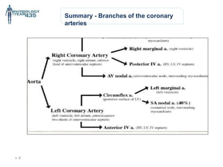
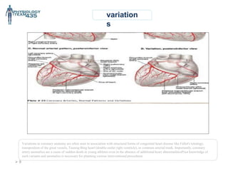
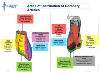
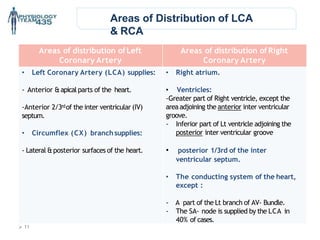
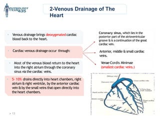
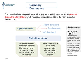
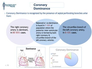
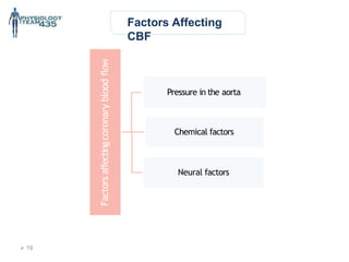
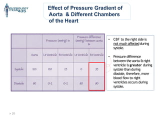
Very important
Extra
information
* Guyton corners, anything that is colored with grey is EXTRA
explanation
 2
Objectives :
• Define autoregulation and its mechanism (metabolic & myogenic).
• Identify the vasodilator metabolites.
• List vasodilator and vasoconstrictor agents and those secreted
by the endothelium.
• Identify vessels supplying the heart (coronaries).
• Understand and list factors affecting coronary blood flow (cardiac
cycle, autoregulation, chemical factors & neural factors.
* We recommend studying anatomy lecture (Blood supply of the
heart) first.
Coronary Circulation
Contact us :Physiology435@gmail.com
Coronary
Circulation
It is important!Why?
One thirdofalldeathsintheworldresultfromcoronaryarterydisease.
Alsoit’smostlikelytobeseeninelderly patients.
Consists of :
1. Arterial supply. (supplyofO2 tothecardiacmuscle.)
2. Venous drainage. (removalofwastesand CO2)
3. Lymphatic drainage. (wastes removal)
- Anterior descending > left side of the
heart
- Posterior descending > right side of the
heart
 3
1-Arterial
Supply
Cardiac muscle is supplied by two coronary arteries:
A. Right coronary artery (RCA.)
B. Left coronary artery (LCA.)
*both arisefromthecoronarysinusesjustsuperiortothe
aorticvalvecusps attheaorticroot.
- Coronary arteries deliver oxygenated blood to the cardiac muscle.
The aortic valvehas three cusps:
A. leftcoronary (LC) cusp.
B. Right coronary (RC) cusp.
C. Posterior non-coronary (NC) cusp.
*There maybe variations in the number, shape & location
of coronary ostia or origins of the coronary arteries,
most of which are of no clinical significance. Right coronary cusp gives rise to right coronary
artery.
Left coronary cusp gives rise to left coronary
artery.
 4
Guyton corner : Figure 21-3 shows the heart and its
coronary blood supply. Note that the main coronary arteries
lie on the surface of the heart and smaller arteries then
penetrate from the surface into the cardiac muscle mass. It
is almost entirely through these arteries that the heart
receives its nutritive blood supply. Only the inner 1/10
millimeter of the endocardial surface can obtain significant
nutrition directly from the blood inside the cardiac chambers,
so this source of muscle nutrition is minuscule. The left
coronary artery supplies mainly the anterior and left lateral
portions of the left ventricle, whereas the right coronary
artery supplies most of the right ventricle, as well as the
posterior part of the left ventricle in 80 to 90 percent of
people.
Most of the coronary venous blood flow from the left
ventricular muscle returns to the right atrium of the heart by
way of the coronary sinus, which is about 75 percent of the
total coronary blood flow. On the other hand, most of the
coronary venous blood from the right ventricular muscle
returns through small anterior cardiac veins that flow directly
into the right atrium, not by way of the coronary sinus. A very
small amount of coronary venous blood also flows back into
the heart through very minute thebesian veins, which empty
directly into all chambers of the heart. Page 262
1-Arterial Supply-
EXTRA
 5
 6
Coronary
arteries
Left coronary artery (LCA) Right coronary artery (RCA)
Size larger smaller
origin* Left posterior aortic sinus Anterior aortic sinus
Termination
By anastomosing with the right
coronary artery
By anastomosing with the left coronary
artery
Branches
• Left Anterior Descending
(LAD) ( = Anterior
interventricular)
• Marginal artery
• Circumflex artery (CX)
• Posterior Descending Branch
( = posterior interventricular)
• Marginal artery
*Regarding the origin of coronary arteries, we are not quitesure about the points written in this physiology lecture
“not mentioned in Guyton” So, we prefer you cover this from anatomy(read more from the box below).
• SNELL Corner :
The aortic valve guards the aortic orifice and is precisely
similar in structure to the pulmonary valve. One cusp is
situated on the anterior wall (right cusp), and two cusps are
located on the posterior wall (left and posterior cusps).
Behind each cusp, the aortic wall bulges to form an aortic
sinus. The anterior aortic sinus gives origin to the right
coronary artery, and the left posteriorsinus gives origin to the
left coronaryartery.
“Page 143 from Clinical Anatomy By Systems by Richard S.
LCA
RCA
Left coronary artery branches :
1) Left anterior ascending
“LAD”
2) Left marginal artery “LMA”
3)Circumflex artery “CX”
Right coronary artery
branches :
1) Right marginal artery “RMA”
2) Posterior descending artery
(posterior interventricular artery)
“PIv”
Coronary
arteries
 7
Summary - Branches of the coronary
arteries
 8
variation
s
Variations in coronary anatomy are often seen in association with structural forms of congenital heart disease like Fallot's tetralogy,
transposition of the great vessels, Taussig-Bing heart (double-outlet right ventricle), or common arterial trunk. Importantly, coronary
artery anomalies are a cause of sudden death in young athletes even in the absence of additional heart abnormalitiesPrior knowledge of
such variants and anomalies is necessary for planning various interventional procedures
 9
Areas of Distribution of Coronary
Arteries
Left Circumflex
Artery Supplies
the left atrium and
left ventricles
Right
Coronary
Artery
Supplies the
right atrium
and right
ventricles
Right
Coronary
Artery
Supplies the
right atrium
and right
ventricles
Left Anterior Descending
Artery
Supplies the right
ventricles ,left
ventricles and
interventricular
septum
Left Marginal
Artery Supplies
left ventricles
Posterior Interventricular
Artery
Supplies the right and left
ventricles and
interventricular septum
Right Marginal
Artery Supplies the
right ventricles and
the apex
Right Marginal
Artery Supplies the
right ventricles and
the apex
Left
Circumflex
Artery
Supplies the
left atrium
and left
ventricles
Left Marginal
Artery Supplies
left ventricles
 10
Areas of distribution ofLeft
Coronary Artery
Areas of distribution ofRight
Coronary Artery
• Left Coronary Artery (LCA) supplies:
- Anterior &apicalparts of the heart.
-Anterior 2/3rdof the inter ventricular (IV)
septum.
• Circumflex (CX) branchsupplies:
- Lateral &posterior surfaces of the heart.
• Right atrium.
• Ventricles:
-Greater part of Right ventricle, except the
area adjoining the anterior inter ventricular
groove.
- Inferior part of Lt ventricle adjoining the
posterior inter ventricular groove
• posterior 1/3rd of the inter
ventricular septum.
• The conducting system of the heart,
except :
- A part of the Lt branch of AV- Bundle.
- The SA- node is supplied by the LCA in
40% of cases.
Areas of Distribution of LCA
& RCA
 11
Collateral
Circulation
CardiacAnastomosis:
The two coronary arteries anastomose in the myocardium
Extra cardiac anastomosis:
the two coronary arteries anastomose with:
1. Vasa vasorum of the aorta.
2. Vasa vasorum of pulmonary arteries.
3. Internal thoracic arteries.
4. The bronchial arteries.
5. Phrenic arteries
Extra cardiac channels open up in emergencies when the coronary arteries
are blocked.
• Collateral circulation = anastomosis
• What’s the benefit of the extra anastomosis?
It just open in emergency, when the coronary arteries have poor supply of the O2 and we need more O2 it’ll
open
• It’s found originally in the heart, but it won’t be activated unless there’semergency
 12
2-Venous Drainage of The
Heart
Coronary sinus, which lies in the
posterior part of the atrioventricular
groove &is acontinuation of the great
cardiac vein.
Anterior, middle &small cardiac
veins.
Venae Cordis Minimae
(smallest cardiac veins.)
• Venous drainage brings deoxygenated cardiac
blood back to the heart.
• 5- 10% drains directly into heart chambers, right
atrium &right ventricle, by the anterior cardiac
vein &by the small veins that open directly into
the heart chambers.
• Most of the venous blood return to the heart
into the right atrium through the coronary
sinus via the cardiac veins.
 13
• Cardiac venous drainage occur through:
3-Lymphatic Drainage of The
Heart
Lymphatics of the heart
accompany the two coronary
arteries &form two trunks
The right trunk, ends inthe
brachiocephalic node.
The left trunk, ends into the
tracheo-bronchial lymph nodes at
the bifurcation of the trachea.
 14
Coronary
Dominance
Coronary dominance depends on which artery (or arteries) gives rise to the posterior
descending artery (PDA), which runs along the posterior side of the heart &supplies
theAV- node.
A person can be:
Right dominant
Co-dominant
Left dominant
Clinical importance
In leftdominance, a
block in left
coronary artery
affectthe entire Lt
ventricle &IV
septum.
Guyton corner
:
P 246, 12TH
edition
The left coronary
artery supplies mainly
the anterior and left
lateral portions of the
left
ventricle, whereas the
right coronary artery
supplies most of the
right ventricle, as well
as the posterior part
of the left ventricle in
80 to 90
percent of people.
In right or balances
dominance, ablock in
right coronary artery
atleast spares part
(2/3)of the septum &
left ventricle.
 15
Coronary
Dominance
Coronary Dominance
Right
dominan
t
left
dominan
t
Co-dominant
Coronary Dominance is recognized by the presence of septal perforating branches arise
from:
The right coronary
artery is dominant,
in 80–85% cases.
The circumflex branch of
the leftcoronary artery,
in 8-10% cases.
Balanced or co-dominance
is found in 7-10% of
population where the
posterior inter ventricular
artery is formed by both
right coronary &
circumflex branch of the
leftcoronary arteries.
 16
 1
7
Coronary Blood Flow
(CBF)
• Guyton corner :
During strenuous exercise, the heart in the young adult increases its cardiac output fourfold to
sevenfold, and it pumps this blood against a higher than normal arterial pressure. Consequently, the
work output of the heart under severe conditions may increase sixfold to ninefold. At the same time, the
coronary blood flow increases threefold to fourfold to supply the extra nutrients needed by theheart.
This increase is not as much as the increase in workload, which means that the ratio of energy
expenditure by the heart to coronary blood flow increases. Thus, the "efficiency" of cardiac utilization of
energy increases to make up for the relative deficiency of coronaryblood supply.
- CBF is about 225-250
mL/min (5% of cardiac
output.)
- Heart extracts 60-70% of
O2, due to presence of
more mitochondria which
generate energy for
contraction by aerobic
metabolism (other tissue
extract only 25%.)
At rest
CBF increases during
exercise or work output.
Phasic changes in CBF during systole &
diastole
Guyton corner: Figure shows the changes in blood flow through the nutrient capillaries of the left ventricular coronary system
in ml/min in the human heart during systole and diastole, as extrapolated from studies in experimental animals. Note from this diagram
that the coronary capillary blood flow in the left ventricle muscle falls to a low value during systole, which is opposite to flow in vascular
beds elsewhere in the body. The reason for this is strong compression of the left ventricular muscle around the intramuscular vessels
during systolic contraction.
During diastole, the cardiac muscle relaxes and no longer obstructs blood flow through the left ventricular muscle capillaries, so blood
flows rapidly during all of diastole.
During Systole
• Coronary arteries
are compressed.
• Blood flow to the
leftventricle is
reduced.
Blood flows to the
subendocardial portion of
the leftventricle occurs
only during diastole, and
is not there during
systole. Therefore, this
region (subendocardial) is
prone to ischemic
damage &it is the most
common site of
myocardial infarction.
Notes:
- Less flow of blood in the systole, and more in the diastole.
-The blood reaches the subendocardial portion only during diastole.
During Diastole
 18
Factors Affecting
CBF
Factors
affecting
coronary
blood
flow
Pressure in the aorta
Chemical factors
Neural factors
 19
Effect of Pressure Gradient of
Aorta & Different Chambers
of the Heart
• CBF to the right side is
not much affectedduring
systole.
• Pressure difference
between the aorta &right
ventricle is greater during
systole than during
diastole, therefore, more
blood flow to right
ventricles occurs during
systole.
 20
Factors Affecting Coronary
Blood Flow
Chemical factors causing Coronary vasodilatation (Increased coronary blood flow):
• Lack of oxygen.
• Increased local concentration of Co2.
• Increased local concentration of H+ ion.
• Increased local concentration of k + ion.
• Increased local concentration of Lactate,Prostaglandin,Adenosine, Adeninenucleotides.
Neural Factors Affecting CoronaryBlood Flow:
• Sympathetic stimulation.
• Parasympathetic stimulation.
 21
Effect of Sympathetic
Stimulation on
Coronary Blood Flow
Coronary arteries have:
• Alpha Adrenergic receptors, which mediatevasoconstriction (Epicardial.)
• BetaAdrenergic receptors, which mediate vasodilatation(Intramuscular.)
Sympatheticstimulationcan eitherbeIndirectorDirect.
Indirect effect of sympathetic stimulation:
Sympathetic stimulation in intact body will leadto release of adrenaline&nor
adrenaline,which in turn increase heart rate &force of contraction.
Vasodilator metabolites will be released leading to coronary vasodilatation.
Direct effect of sympathetic stimulation:
Experimentally, injectionof noradrenalin after blocking of the Beta adrenergic
receptors in un anesthetized animals elicits coronary vasoconstriction.
 22
As aReflex nor adrenergic discharge will increase.
Coronary Blood Flow will increase secondary to metabolic changes in the myocardium.
Benefits of the indirect effect of Nor adrenergic
discharge
What are the benefits of the indirect effect of Nor adrenergic
discharge?
• The answer is :to preserve circulation of the heart while the flow to other
organs is compromised.
When systemic blood pressure gets low.
 23
Effect of Parasympathetic
Stimulation on Coronary
Blood Flow
• Vagalstimulation (Parasympathetic) causes coronary
vasodilatation.
• However, parasympathetic distribution is not great.
• There is more sympathetic innervation of coronary
vessels.
 24
Coronary Blood
Flow
T
achycardia
The diastolic
period will be
shortened
Coronary
Blood Flow is
reduced
Control of Coronary Blood Flow
• Coronary Blood Flow shows considerable auto regulation.
• Local muscle metabolism is the primary controller:
1. Oxygen demand is a major factor in local coronary blood flow regulation.
• Nervous control of Coronary Blood Flow:
1. Direct effectsof nervous stimuli on the coronary vasculature.
2. Sympathetic greater effectsthan parasympathetic.
Effectof Tachycardiaon Coronary BloodFlow
 25
Extra “just read it, you will get the
idea”
• Guyton corner :
Nervous Control of Coronary Blood Flow
Stimulation of the autonomic nerves to the heart can affect coronary blood flow both directly and indirectly. The direct effects
result from action of the nervous transmitter substances acetylcholine from the vagus nerves and norepinephrine from the
sympathetic nerves on the coronary vessels. The indirect effects result from secondary changes in coronary blood flow caused
by increased or decreased activity of the heart.
The indirect effects, which are mostly opposite to the direct effects, play a far more important role in normal control of coronary blood
flow. Thus, sympathetic stimulation, which releases norepinephrine from the sympathetic nerves and epinephrine as well as
norepinephrine from the adrenal medullae, increases both heart rate and heart contractility and increases the rate of metabolism of
the heart. In turn, the increased metabolism of the heart sets off local blood flow regulatory mechanisms for dilating the coronary
vessels, and the blood flow increases approximately in proportion to the metabolic needs of the heart muscle. In contrast, vagal
stimulation, with its release of acetylcholine, slows the heart and has a slight depressive effect on heart contractility. These effects
decrease cardiac oxygen consumption and, therefore, indirectly constrict the coronary arteries
Direct Effects of Nervous Stimuli on the Coronary Vasculature.
The distribution of parasympathetic (vagal) nerve fibers to the ventricular coronary system is not very great. However, the
acetylcholine released by parasympathetic stimulation has a direct effect to dilate the coronaryarteries.
Much more extensive sympathetic innervation of the coronary vessels occurs. In Chapter 61, we see that the sympathetic
transmitter substances norepinephrine and epinephrine can have either vascular constrictor or vascular dilator effects, depending
on the presence or absence of constrictor or dilator receptors in the blood vessel walls. The constrictor receptors are called alpha
receptors and the dilator receptors are called beta receptors. Both alpha and beta receptors exist in the coronary vessels. In
general, the epicardial coronary vessels have a preponderance of alpha receptors, whereas the intramuscular arteries may have a
preponderance of beta receptors. Therefore, sympathetic stimulation can, at least theoretically, cause slight overall coronary
constriction or dilation, but usually constriction. In some people, the alpha vasoconstrictor effects seem to be disproportionately
severe, and these people can have vasospastic myocardial ischemia during periods of excess sympathetic drive, often with
resultant anginalpain.
Metabolic factors, especially myocardial oxygen consumption, are the major controllers of myocardial blood flow. Whenever the
direct effects of nervous stimulation reduce coronary blood flow, the metabolic control of coronary flow usually overrides the direct
coronary nervous effects within seconds.
 26
Physiology
Leaders :
Khawla
Alammari
Nojood
Alhaidri Rawaf
Alrawaf
Girls team :
• Atheer Alnashwan
• Asrar Batarfi
• Afnan Almalki
• Alhanouf Aljlaoud
• Deema AlFaris
• Elham Alzahrani
• Johara Almalki
• Lojain alsiwat
• Malak Alsharif
• Monirah Alsalouli
• Nora AlRomaih
• Nurah Alqahtani
• Nouf Alabdulkarim
• Nora Albusayes
• Nora Alsomali
• NorahAlakeel
• Reem Alageel
• Rawan Aldhuwayhi
• Reham Al-Obaidan
• Samar AlOtaibi
• Shamma Alsaad
Boys team :
• Abdullah Aljaafar
• Omar Alotaibi
• Abdulrahman Albarakah
• Adel Alshehri
• Abdulaziz Alghanaym
• AbdulmajeedAlotaibi
• Khalil Alduraibi
• Hassan Albeladi
• Omar Alshehri
• Saleh Alshawi
• Abdulaziz Alhammad
• Faisal Alabdulatif
• Abdulnasser Alwabel
• Saad Almutairy

14- Coronary circulation .pptx,Coronary circulation .pptx

  • 1.
    Very important Extra information * Guytoncorners, anything that is colored with grey is EXTRA explanation
  • 2.
     2 Objectives : •Define autoregulation and its mechanism (metabolic & myogenic). • Identify the vasodilator metabolites. • List vasodilator and vasoconstrictor agents and those secreted by the endothelium. • Identify vessels supplying the heart (coronaries). • Understand and list factors affecting coronary blood flow (cardiac cycle, autoregulation, chemical factors & neural factors. * We recommend studying anatomy lecture (Blood supply of the heart) first. Coronary Circulation Contact us :Physiology435@gmail.com
  • 3.
    Coronary Circulation It is important!Why? Onethirdofalldeathsintheworldresultfromcoronaryarterydisease. Alsoit’smostlikelytobeseeninelderly patients. Consists of : 1. Arterial supply. (supplyofO2 tothecardiacmuscle.) 2. Venous drainage. (removalofwastesand CO2) 3. Lymphatic drainage. (wastes removal) - Anterior descending > left side of the heart - Posterior descending > right side of the heart  3
  • 4.
    1-Arterial Supply Cardiac muscle issupplied by two coronary arteries: A. Right coronary artery (RCA.) B. Left coronary artery (LCA.) *both arisefromthecoronarysinusesjustsuperiortothe aorticvalvecusps attheaorticroot. - Coronary arteries deliver oxygenated blood to the cardiac muscle. The aortic valvehas three cusps: A. leftcoronary (LC) cusp. B. Right coronary (RC) cusp. C. Posterior non-coronary (NC) cusp. *There maybe variations in the number, shape & location of coronary ostia or origins of the coronary arteries, most of which are of no clinical significance. Right coronary cusp gives rise to right coronary artery. Left coronary cusp gives rise to left coronary artery.  4
  • 5.
    Guyton corner :Figure 21-3 shows the heart and its coronary blood supply. Note that the main coronary arteries lie on the surface of the heart and smaller arteries then penetrate from the surface into the cardiac muscle mass. It is almost entirely through these arteries that the heart receives its nutritive blood supply. Only the inner 1/10 millimeter of the endocardial surface can obtain significant nutrition directly from the blood inside the cardiac chambers, so this source of muscle nutrition is minuscule. The left coronary artery supplies mainly the anterior and left lateral portions of the left ventricle, whereas the right coronary artery supplies most of the right ventricle, as well as the posterior part of the left ventricle in 80 to 90 percent of people. Most of the coronary venous blood flow from the left ventricular muscle returns to the right atrium of the heart by way of the coronary sinus, which is about 75 percent of the total coronary blood flow. On the other hand, most of the coronary venous blood from the right ventricular muscle returns through small anterior cardiac veins that flow directly into the right atrium, not by way of the coronary sinus. A very small amount of coronary venous blood also flows back into the heart through very minute thebesian veins, which empty directly into all chambers of the heart. Page 262 1-Arterial Supply- EXTRA  5
  • 6.
     6 Coronary arteries Left coronaryartery (LCA) Right coronary artery (RCA) Size larger smaller origin* Left posterior aortic sinus Anterior aortic sinus Termination By anastomosing with the right coronary artery By anastomosing with the left coronary artery Branches • Left Anterior Descending (LAD) ( = Anterior interventricular) • Marginal artery • Circumflex artery (CX) • Posterior Descending Branch ( = posterior interventricular) • Marginal artery *Regarding the origin of coronary arteries, we are not quitesure about the points written in this physiology lecture “not mentioned in Guyton” So, we prefer you cover this from anatomy(read more from the box below). • SNELL Corner : The aortic valve guards the aortic orifice and is precisely similar in structure to the pulmonary valve. One cusp is situated on the anterior wall (right cusp), and two cusps are located on the posterior wall (left and posterior cusps). Behind each cusp, the aortic wall bulges to form an aortic sinus. The anterior aortic sinus gives origin to the right coronary artery, and the left posteriorsinus gives origin to the left coronaryartery. “Page 143 from Clinical Anatomy By Systems by Richard S.
  • 7.
    LCA RCA Left coronary arterybranches : 1) Left anterior ascending “LAD” 2) Left marginal artery “LMA” 3)Circumflex artery “CX” Right coronary artery branches : 1) Right marginal artery “RMA” 2) Posterior descending artery (posterior interventricular artery) “PIv” Coronary arteries  7
  • 8.
    Summary - Branchesof the coronary arteries  8
  • 9.
    variation s Variations in coronaryanatomy are often seen in association with structural forms of congenital heart disease like Fallot's tetralogy, transposition of the great vessels, Taussig-Bing heart (double-outlet right ventricle), or common arterial trunk. Importantly, coronary artery anomalies are a cause of sudden death in young athletes even in the absence of additional heart abnormalitiesPrior knowledge of such variants and anomalies is necessary for planning various interventional procedures  9
  • 10.
    Areas of Distributionof Coronary Arteries Left Circumflex Artery Supplies the left atrium and left ventricles Right Coronary Artery Supplies the right atrium and right ventricles Right Coronary Artery Supplies the right atrium and right ventricles Left Anterior Descending Artery Supplies the right ventricles ,left ventricles and interventricular septum Left Marginal Artery Supplies left ventricles Posterior Interventricular Artery Supplies the right and left ventricles and interventricular septum Right Marginal Artery Supplies the right ventricles and the apex Right Marginal Artery Supplies the right ventricles and the apex Left Circumflex Artery Supplies the left atrium and left ventricles Left Marginal Artery Supplies left ventricles  10
  • 11.
    Areas of distributionofLeft Coronary Artery Areas of distribution ofRight Coronary Artery • Left Coronary Artery (LCA) supplies: - Anterior &apicalparts of the heart. -Anterior 2/3rdof the inter ventricular (IV) septum. • Circumflex (CX) branchsupplies: - Lateral &posterior surfaces of the heart. • Right atrium. • Ventricles: -Greater part of Right ventricle, except the area adjoining the anterior inter ventricular groove. - Inferior part of Lt ventricle adjoining the posterior inter ventricular groove • posterior 1/3rd of the inter ventricular septum. • The conducting system of the heart, except : - A part of the Lt branch of AV- Bundle. - The SA- node is supplied by the LCA in 40% of cases. Areas of Distribution of LCA & RCA  11
  • 12.
    Collateral Circulation CardiacAnastomosis: The two coronaryarteries anastomose in the myocardium Extra cardiac anastomosis: the two coronary arteries anastomose with: 1. Vasa vasorum of the aorta. 2. Vasa vasorum of pulmonary arteries. 3. Internal thoracic arteries. 4. The bronchial arteries. 5. Phrenic arteries Extra cardiac channels open up in emergencies when the coronary arteries are blocked. • Collateral circulation = anastomosis • What’s the benefit of the extra anastomosis? It just open in emergency, when the coronary arteries have poor supply of the O2 and we need more O2 it’ll open • It’s found originally in the heart, but it won’t be activated unless there’semergency  12
  • 13.
    2-Venous Drainage ofThe Heart Coronary sinus, which lies in the posterior part of the atrioventricular groove &is acontinuation of the great cardiac vein. Anterior, middle &small cardiac veins. Venae Cordis Minimae (smallest cardiac veins.) • Venous drainage brings deoxygenated cardiac blood back to the heart. • 5- 10% drains directly into heart chambers, right atrium &right ventricle, by the anterior cardiac vein &by the small veins that open directly into the heart chambers. • Most of the venous blood return to the heart into the right atrium through the coronary sinus via the cardiac veins.  13 • Cardiac venous drainage occur through:
  • 14.
    3-Lymphatic Drainage ofThe Heart Lymphatics of the heart accompany the two coronary arteries &form two trunks The right trunk, ends inthe brachiocephalic node. The left trunk, ends into the tracheo-bronchial lymph nodes at the bifurcation of the trachea.  14
  • 15.
    Coronary Dominance Coronary dominance dependson which artery (or arteries) gives rise to the posterior descending artery (PDA), which runs along the posterior side of the heart &supplies theAV- node. A person can be: Right dominant Co-dominant Left dominant Clinical importance In leftdominance, a block in left coronary artery affectthe entire Lt ventricle &IV septum. Guyton corner : P 246, 12TH edition The left coronary artery supplies mainly the anterior and left lateral portions of the left ventricle, whereas the right coronary artery supplies most of the right ventricle, as well as the posterior part of the left ventricle in 80 to 90 percent of people. In right or balances dominance, ablock in right coronary artery atleast spares part (2/3)of the septum & left ventricle.  15
  • 16.
    Coronary Dominance Coronary Dominance Right dominan t left dominan t Co-dominant Coronary Dominanceis recognized by the presence of septal perforating branches arise from: The right coronary artery is dominant, in 80–85% cases. The circumflex branch of the leftcoronary artery, in 8-10% cases. Balanced or co-dominance is found in 7-10% of population where the posterior inter ventricular artery is formed by both right coronary & circumflex branch of the leftcoronary arteries.  16
  • 17.
     1 7 Coronary BloodFlow (CBF) • Guyton corner : During strenuous exercise, the heart in the young adult increases its cardiac output fourfold to sevenfold, and it pumps this blood against a higher than normal arterial pressure. Consequently, the work output of the heart under severe conditions may increase sixfold to ninefold. At the same time, the coronary blood flow increases threefold to fourfold to supply the extra nutrients needed by theheart. This increase is not as much as the increase in workload, which means that the ratio of energy expenditure by the heart to coronary blood flow increases. Thus, the "efficiency" of cardiac utilization of energy increases to make up for the relative deficiency of coronaryblood supply. - CBF is about 225-250 mL/min (5% of cardiac output.) - Heart extracts 60-70% of O2, due to presence of more mitochondria which generate energy for contraction by aerobic metabolism (other tissue extract only 25%.) At rest CBF increases during exercise or work output.
  • 18.
    Phasic changes inCBF during systole & diastole Guyton corner: Figure shows the changes in blood flow through the nutrient capillaries of the left ventricular coronary system in ml/min in the human heart during systole and diastole, as extrapolated from studies in experimental animals. Note from this diagram that the coronary capillary blood flow in the left ventricle muscle falls to a low value during systole, which is opposite to flow in vascular beds elsewhere in the body. The reason for this is strong compression of the left ventricular muscle around the intramuscular vessels during systolic contraction. During diastole, the cardiac muscle relaxes and no longer obstructs blood flow through the left ventricular muscle capillaries, so blood flows rapidly during all of diastole. During Systole • Coronary arteries are compressed. • Blood flow to the leftventricle is reduced. Blood flows to the subendocardial portion of the leftventricle occurs only during diastole, and is not there during systole. Therefore, this region (subendocardial) is prone to ischemic damage &it is the most common site of myocardial infarction. Notes: - Less flow of blood in the systole, and more in the diastole. -The blood reaches the subendocardial portion only during diastole. During Diastole  18
  • 19.
    Factors Affecting CBF Factors affecting coronary blood flow Pressure inthe aorta Chemical factors Neural factors  19
  • 20.
    Effect of PressureGradient of Aorta & Different Chambers of the Heart • CBF to the right side is not much affectedduring systole. • Pressure difference between the aorta &right ventricle is greater during systole than during diastole, therefore, more blood flow to right ventricles occurs during systole.  20
  • 21.
    Factors Affecting Coronary BloodFlow Chemical factors causing Coronary vasodilatation (Increased coronary blood flow): • Lack of oxygen. • Increased local concentration of Co2. • Increased local concentration of H+ ion. • Increased local concentration of k + ion. • Increased local concentration of Lactate,Prostaglandin,Adenosine, Adeninenucleotides. Neural Factors Affecting CoronaryBlood Flow: • Sympathetic stimulation. • Parasympathetic stimulation.  21
  • 22.
    Effect of Sympathetic Stimulationon Coronary Blood Flow Coronary arteries have: • Alpha Adrenergic receptors, which mediatevasoconstriction (Epicardial.) • BetaAdrenergic receptors, which mediate vasodilatation(Intramuscular.) Sympatheticstimulationcan eitherbeIndirectorDirect. Indirect effect of sympathetic stimulation: Sympathetic stimulation in intact body will leadto release of adrenaline&nor adrenaline,which in turn increase heart rate &force of contraction. Vasodilator metabolites will be released leading to coronary vasodilatation. Direct effect of sympathetic stimulation: Experimentally, injectionof noradrenalin after blocking of the Beta adrenergic receptors in un anesthetized animals elicits coronary vasoconstriction.  22
  • 23.
    As aReflex noradrenergic discharge will increase. Coronary Blood Flow will increase secondary to metabolic changes in the myocardium. Benefits of the indirect effect of Nor adrenergic discharge What are the benefits of the indirect effect of Nor adrenergic discharge? • The answer is :to preserve circulation of the heart while the flow to other organs is compromised. When systemic blood pressure gets low.  23
  • 24.
    Effect of Parasympathetic Stimulationon Coronary Blood Flow • Vagalstimulation (Parasympathetic) causes coronary vasodilatation. • However, parasympathetic distribution is not great. • There is more sympathetic innervation of coronary vessels.  24
  • 25.
    Coronary Blood Flow T achycardia The diastolic periodwill be shortened Coronary Blood Flow is reduced Control of Coronary Blood Flow • Coronary Blood Flow shows considerable auto regulation. • Local muscle metabolism is the primary controller: 1. Oxygen demand is a major factor in local coronary blood flow regulation. • Nervous control of Coronary Blood Flow: 1. Direct effectsof nervous stimuli on the coronary vasculature. 2. Sympathetic greater effectsthan parasympathetic. Effectof Tachycardiaon Coronary BloodFlow  25
  • 26.
    Extra “just readit, you will get the idea” • Guyton corner : Nervous Control of Coronary Blood Flow Stimulation of the autonomic nerves to the heart can affect coronary blood flow both directly and indirectly. The direct effects result from action of the nervous transmitter substances acetylcholine from the vagus nerves and norepinephrine from the sympathetic nerves on the coronary vessels. The indirect effects result from secondary changes in coronary blood flow caused by increased or decreased activity of the heart. The indirect effects, which are mostly opposite to the direct effects, play a far more important role in normal control of coronary blood flow. Thus, sympathetic stimulation, which releases norepinephrine from the sympathetic nerves and epinephrine as well as norepinephrine from the adrenal medullae, increases both heart rate and heart contractility and increases the rate of metabolism of the heart. In turn, the increased metabolism of the heart sets off local blood flow regulatory mechanisms for dilating the coronary vessels, and the blood flow increases approximately in proportion to the metabolic needs of the heart muscle. In contrast, vagal stimulation, with its release of acetylcholine, slows the heart and has a slight depressive effect on heart contractility. These effects decrease cardiac oxygen consumption and, therefore, indirectly constrict the coronary arteries Direct Effects of Nervous Stimuli on the Coronary Vasculature. The distribution of parasympathetic (vagal) nerve fibers to the ventricular coronary system is not very great. However, the acetylcholine released by parasympathetic stimulation has a direct effect to dilate the coronaryarteries. Much more extensive sympathetic innervation of the coronary vessels occurs. In Chapter 61, we see that the sympathetic transmitter substances norepinephrine and epinephrine can have either vascular constrictor or vascular dilator effects, depending on the presence or absence of constrictor or dilator receptors in the blood vessel walls. The constrictor receptors are called alpha receptors and the dilator receptors are called beta receptors. Both alpha and beta receptors exist in the coronary vessels. In general, the epicardial coronary vessels have a preponderance of alpha receptors, whereas the intramuscular arteries may have a preponderance of beta receptors. Therefore, sympathetic stimulation can, at least theoretically, cause slight overall coronary constriction or dilation, but usually constriction. In some people, the alpha vasoconstrictor effects seem to be disproportionately severe, and these people can have vasospastic myocardial ischemia during periods of excess sympathetic drive, often with resultant anginalpain. Metabolic factors, especially myocardial oxygen consumption, are the major controllers of myocardial blood flow. Whenever the direct effects of nervous stimulation reduce coronary blood flow, the metabolic control of coronary flow usually overrides the direct coronary nervous effects within seconds.  26
  • 27.
    Physiology Leaders : Khawla Alammari Nojood Alhaidri Rawaf Alrawaf Girlsteam : • Atheer Alnashwan • Asrar Batarfi • Afnan Almalki • Alhanouf Aljlaoud • Deema AlFaris • Elham Alzahrani • Johara Almalki • Lojain alsiwat • Malak Alsharif • Monirah Alsalouli • Nora AlRomaih • Nurah Alqahtani • Nouf Alabdulkarim • Nora Albusayes • Nora Alsomali • NorahAlakeel • Reem Alageel • Rawan Aldhuwayhi • Reham Al-Obaidan • Samar AlOtaibi • Shamma Alsaad Boys team : • Abdullah Aljaafar • Omar Alotaibi • Abdulrahman Albarakah • Adel Alshehri • Abdulaziz Alghanaym • AbdulmajeedAlotaibi • Khalil Alduraibi • Hassan Albeladi • Omar Alshehri • Saleh Alshawi • Abdulaziz Alhammad • Faisal Alabdulatif • Abdulnasser Alwabel • Saad Almutairy