Pneumocystis carinii
Pneumonia
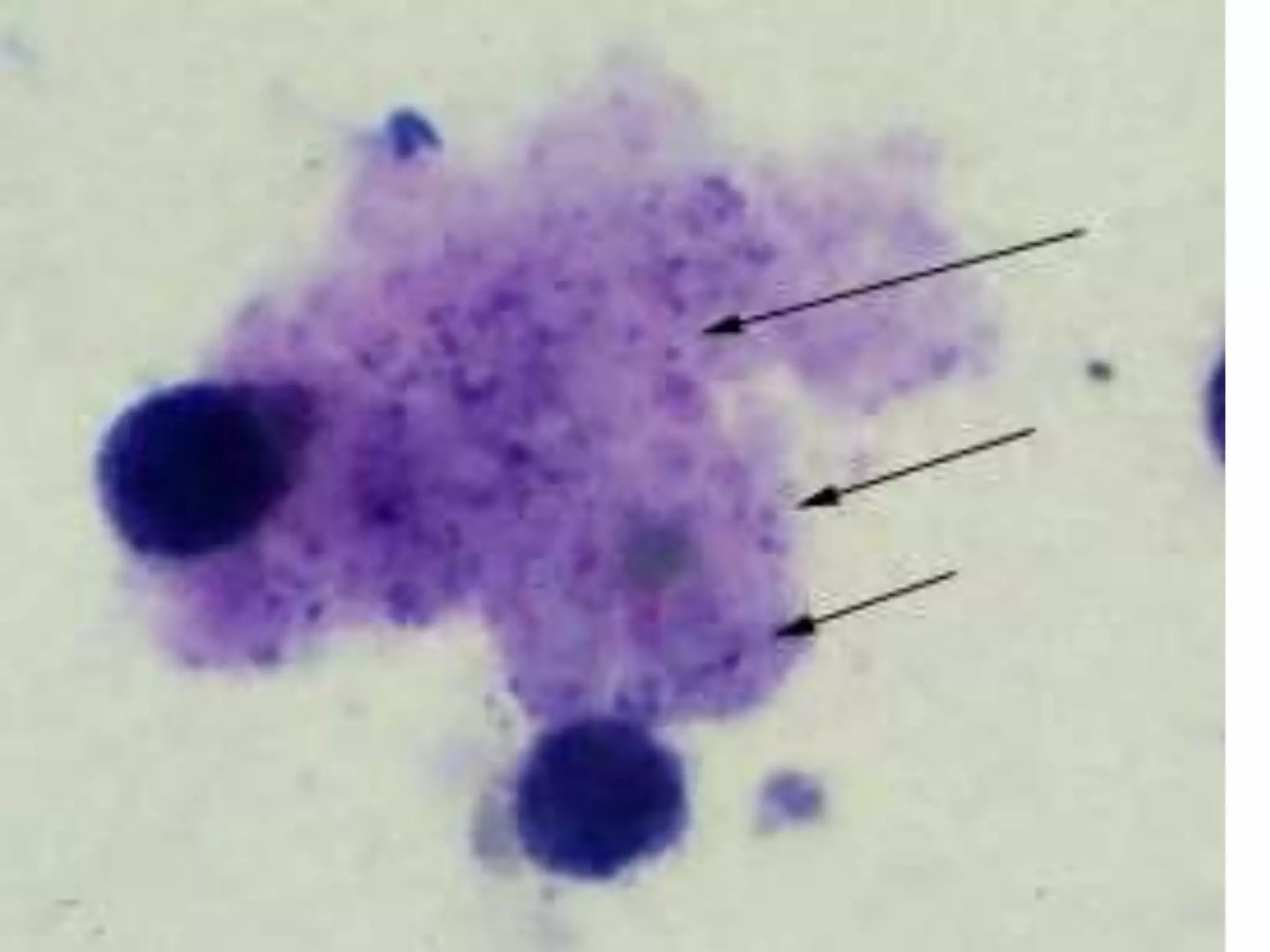
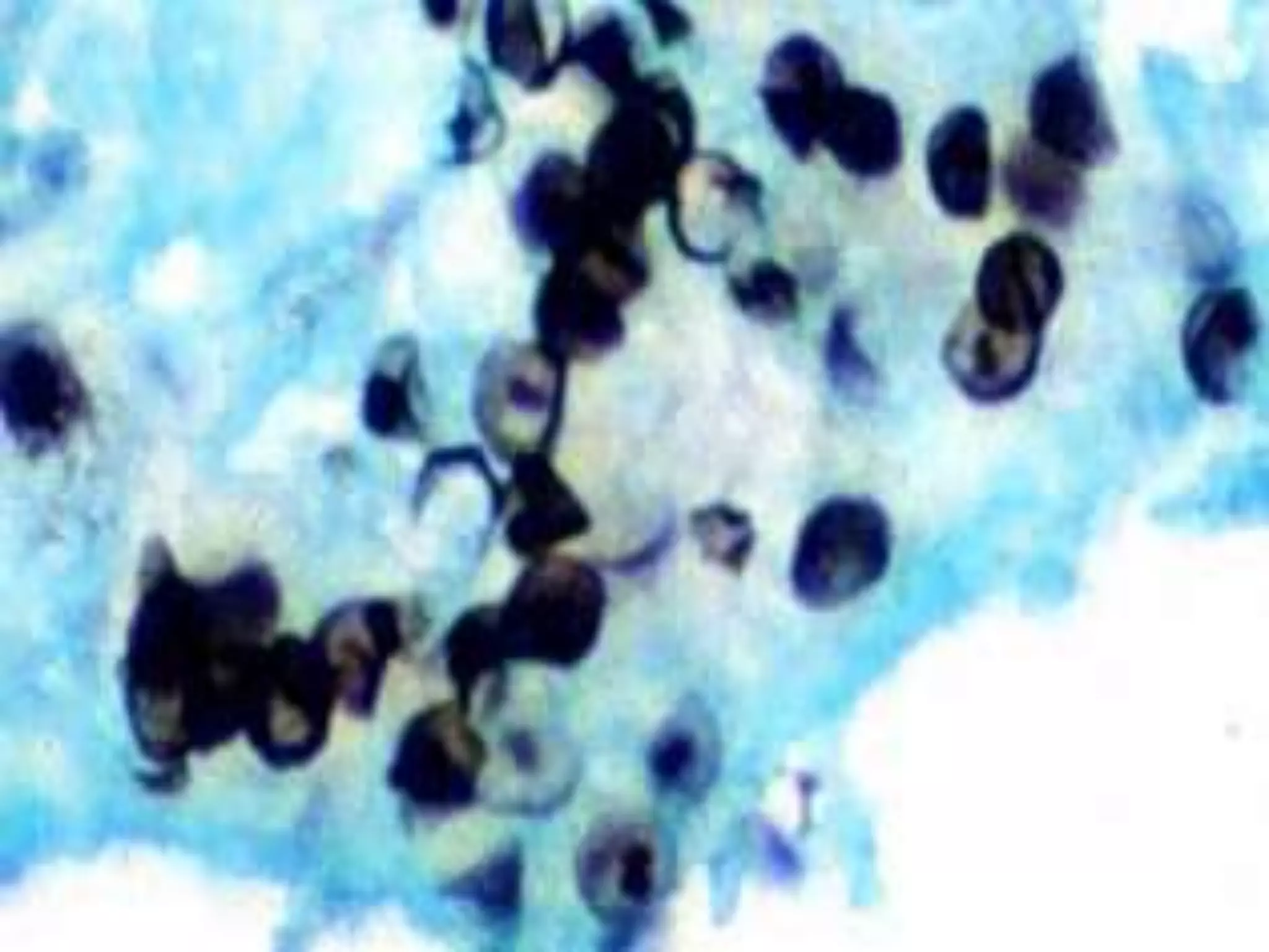
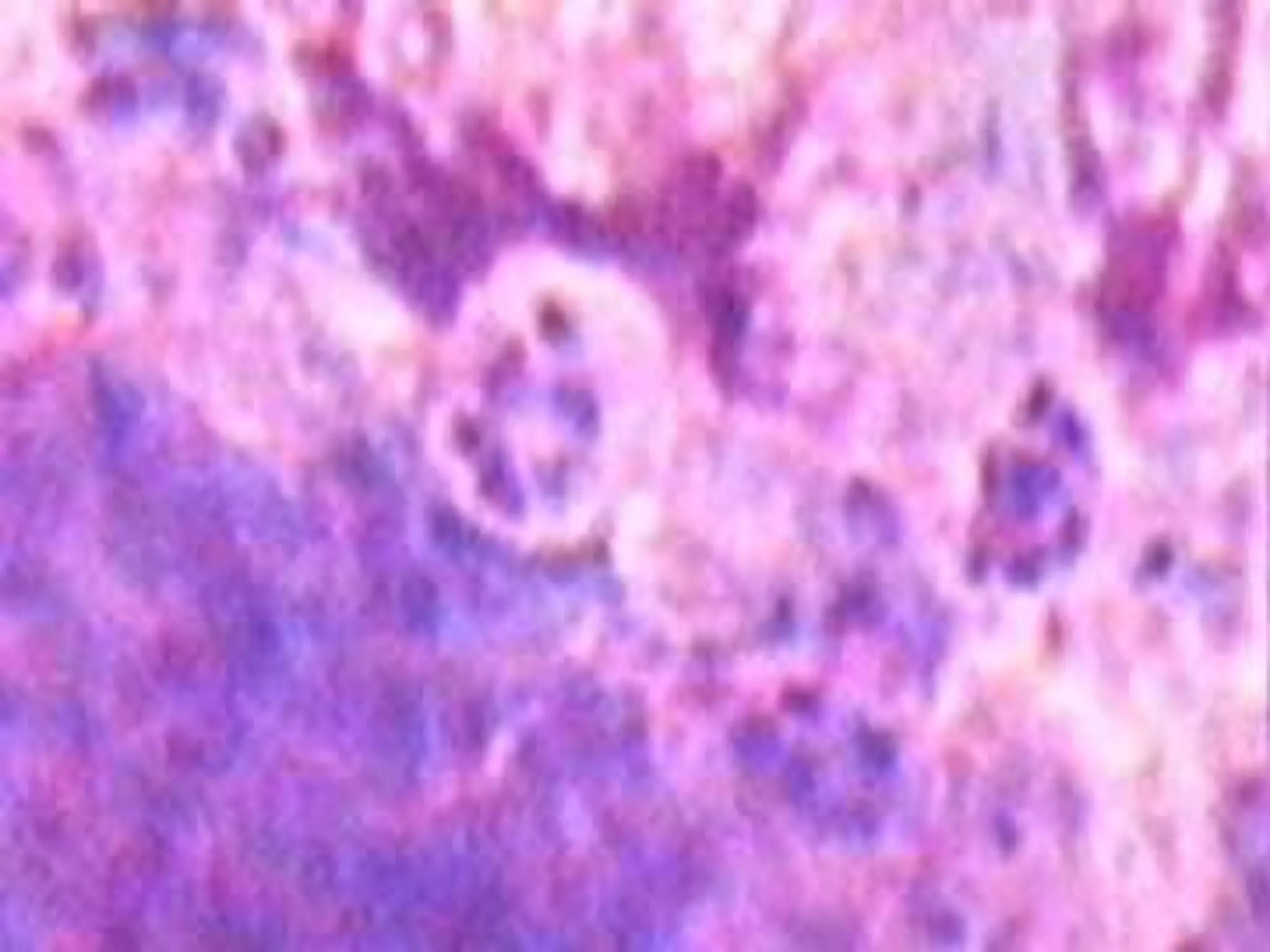
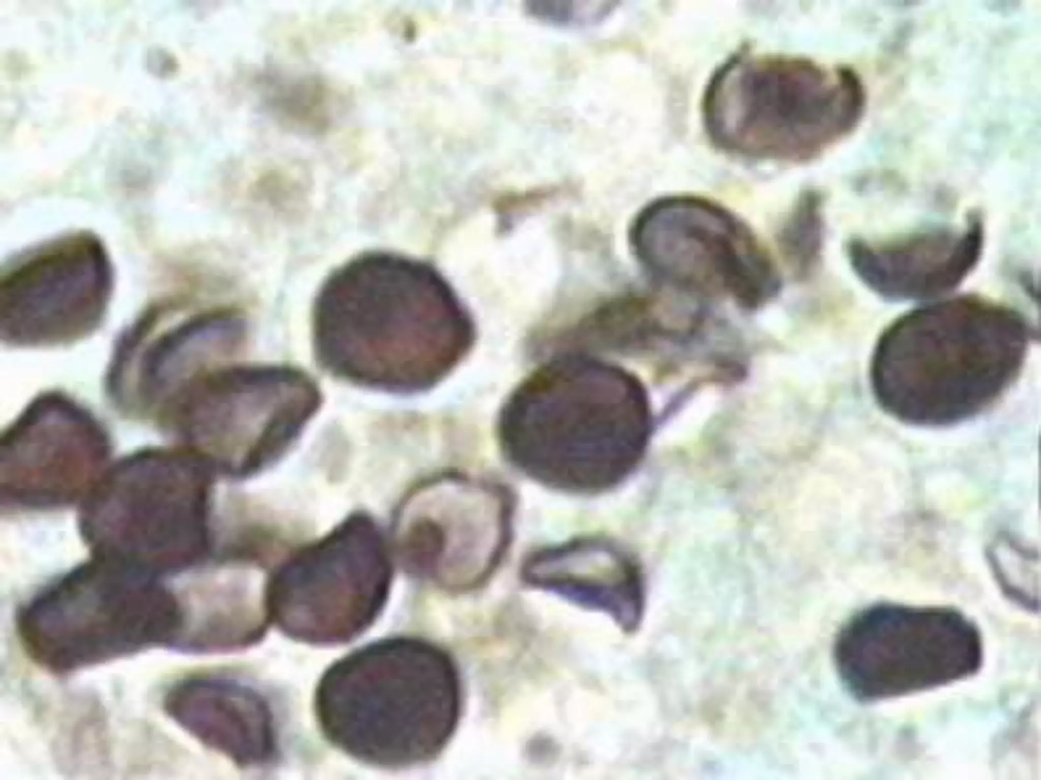
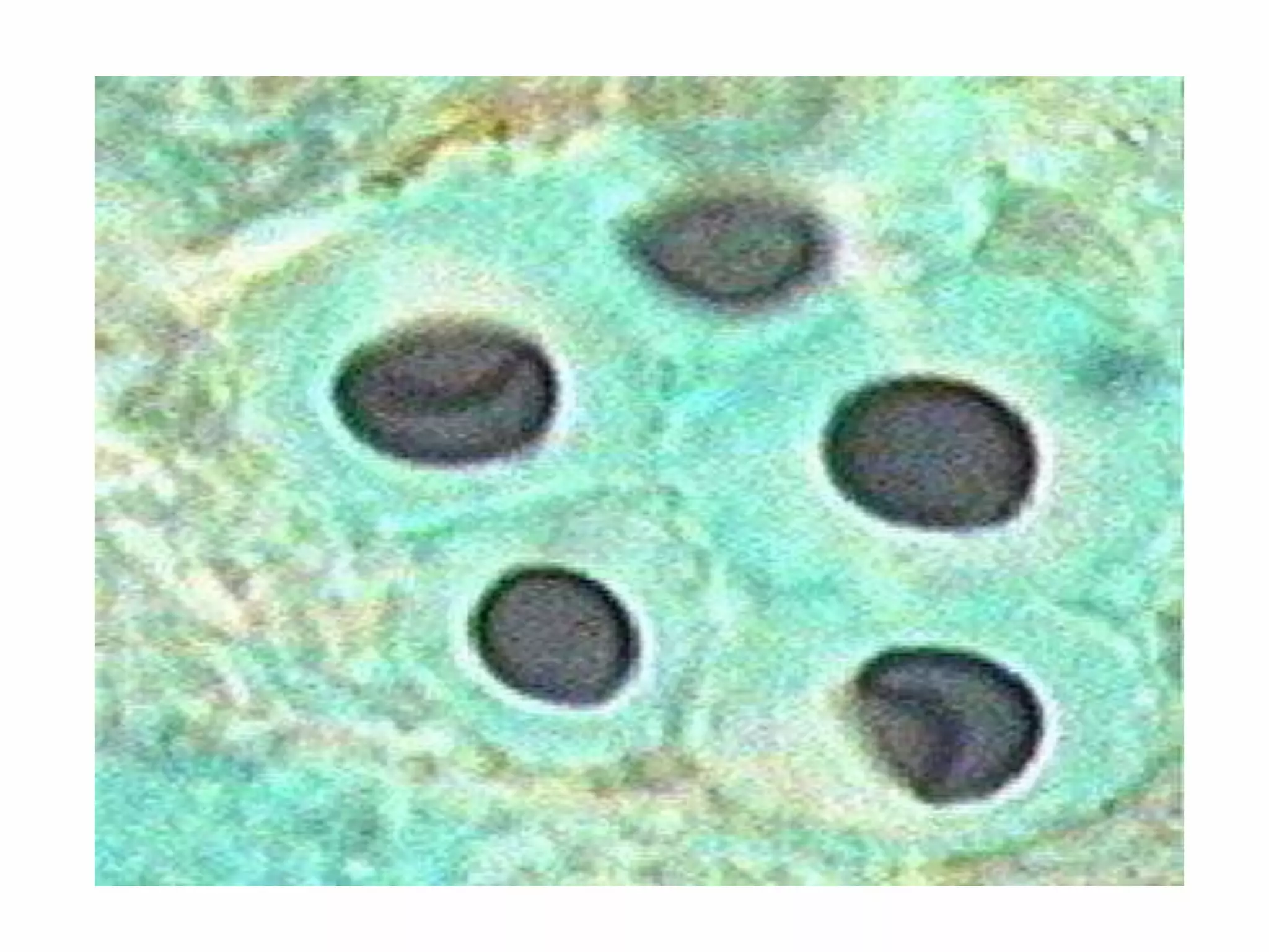
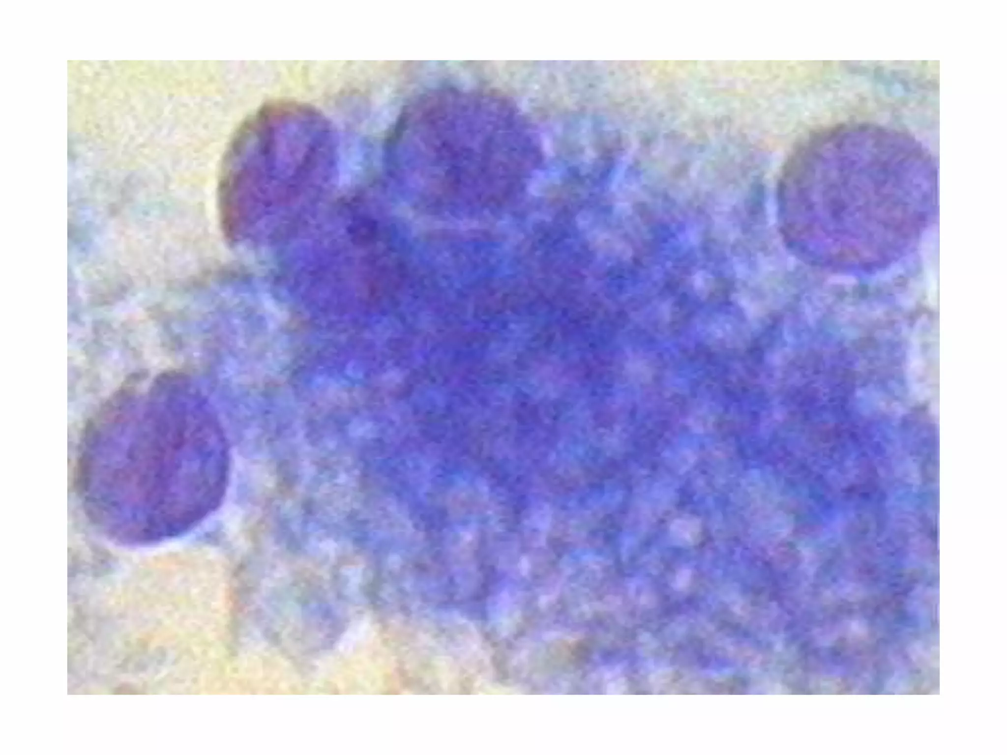
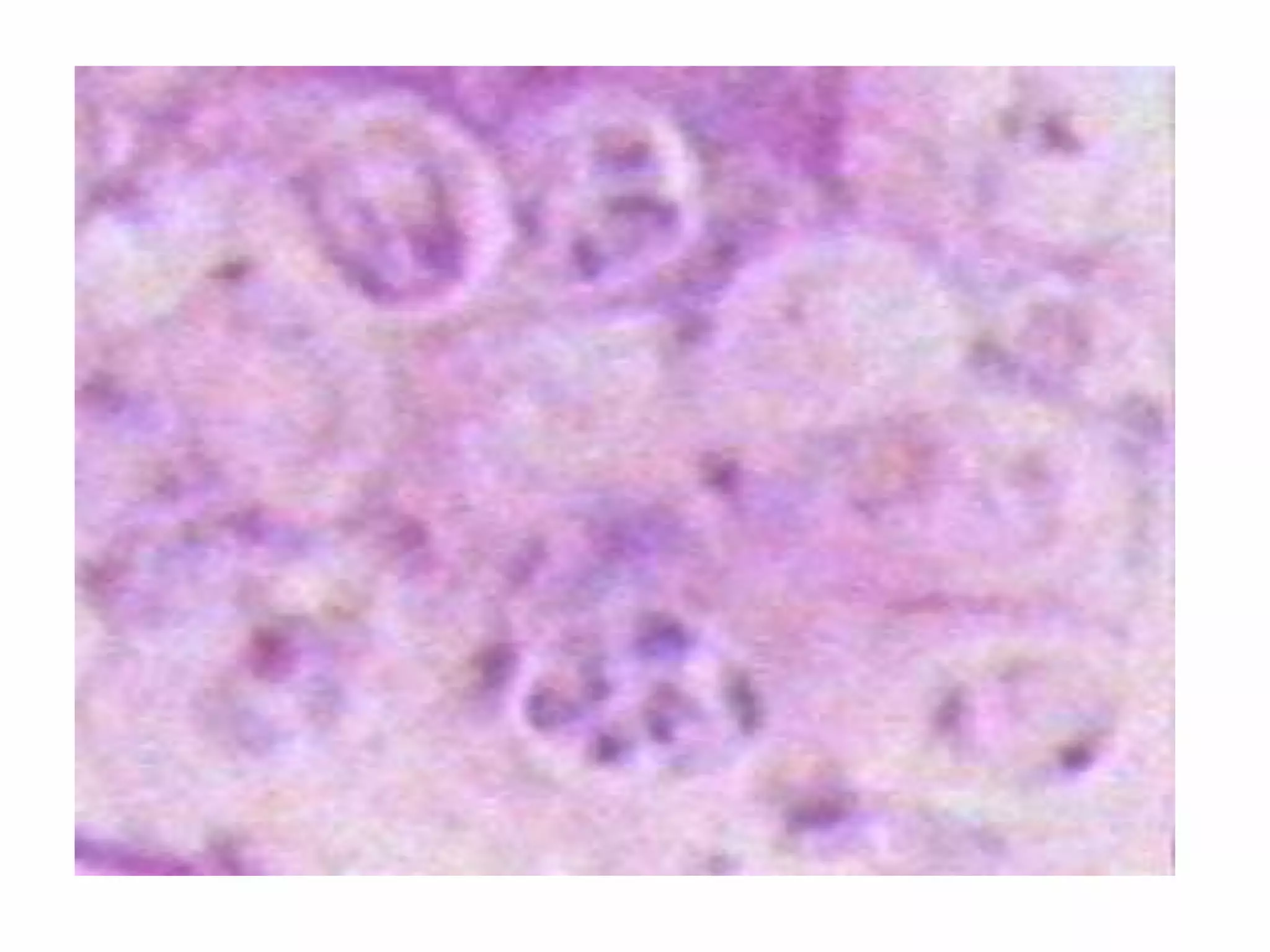
 Pneumocystis carinii pneumonia (PCP)
is an opportunistic infection that occurs in
immunosuppressed populations, primarily
patients with advanced human
immunodeficiency virus infection.
The classic presentation of
nonproductive cough, shortness of
breath, fever, bilateral interstitial
infiltrates and hypoxemia does
not always appear
 Before the epidemic of acquired
immunodeficiency syndrome (AIDS)
in the early 1980s,
Pneumocystis carinii pneumonia
(PCP) was a rare infection that
occurred in immunosuppressed
patients with protein malnutrition or
acute lymphocytic leukemia, or in
patients receiving corticosteroid
therapy.
Risk Factors and Clinical
Presentation
Defective T-cell immunity is the
primary risk factor for PCP.
 Associated clinical signs are well
defined in HIV infection and
reflect the degree of CD4+ cell
depletion
Physical findings are also
nonspecific.
 fine dry rales or may be
unremarkable.
In 2 to 6 percent of cases, PCP
may present with a spontaneous
pneumothorax.
Extrapulmonary pneumocystosis
occurs rarely, involving organs
such as the heart, skin, spleen,
thyroid or eyes.
The chest film typically shows
diffuse interstitial infiltrates but
can be normal in at least one
third of cases.
Less commonly, lobar infiltrates,
effusions or cavitary lesions mimic
other pulmonary processes.
Many other diseases may have a
similar presentation, including
mycobacterial, fungal, viral or
bacterial pneumonias, heart
failure, Kaposi's sarcoma and
pulmonary emboli.
Diagnosis
 P. carinii cannot be routinely cultured
and is identified by stains
demonstrating the cyst wall or the
trophozoite.
 Before the emergence of AIDS, PCP
was diagnosed by open lung biopsy.
Less invasive procedures-sputum
induction and bronchoalveolar
lavageare now the methods of
choice
Periodic acid-Schiff and
Papanicolou stains have been
used to display the characteristic
honeycombed froth associated
with P. carinii
Papanicolou staining
 It uses a combination of
haematoxylin, Orange G, eosin Y,
Light Green SF yellowish, and
sometimes Bismarck Brown Y.
PAS staining
 is a staining method used to etect
glycogen in tissues
The reaction of periodic acid
selectively oxidizes the glucose
residues, creates aldehydes that react
with the Schiff reagent and creates a
purple-magenta color
Solutions and Reagents:
0.5% Periodic Acid Solution:
 Periodic acid 0.5 g
 Distilled water 100 ml
14998019.ppt
14998019.ppt
14998019.ppt
14998019.ppt
14998019.ppt
14998019.ppt
14998019.ppt
14998019.ppt
14998019.ppt
14998019.ppt
14998019.ppt

14998019.ppt

  • 1.
    Pneumocystis carinii Pneumonia  Pneumocystiscarinii pneumonia (PCP) is an opportunistic infection that occurs in immunosuppressed populations, primarily patients with advanced human immunodeficiency virus infection.
  • 2.
    The classic presentationof nonproductive cough, shortness of breath, fever, bilateral interstitial infiltrates and hypoxemia does not always appear
  • 3.
     Before theepidemic of acquired immunodeficiency syndrome (AIDS) in the early 1980s,
  • 4.
    Pneumocystis carinii pneumonia (PCP)was a rare infection that occurred in immunosuppressed patients with protein malnutrition or acute lymphocytic leukemia, or in patients receiving corticosteroid therapy.
  • 6.
    Risk Factors andClinical Presentation Defective T-cell immunity is the primary risk factor for PCP.  Associated clinical signs are well defined in HIV infection and reflect the degree of CD4+ cell depletion
  • 7.
    Physical findings arealso nonspecific.  fine dry rales or may be unremarkable. In 2 to 6 percent of cases, PCP may present with a spontaneous pneumothorax.
  • 8.
    Extrapulmonary pneumocystosis occurs rarely,involving organs such as the heart, skin, spleen, thyroid or eyes.
  • 9.
    The chest filmtypically shows diffuse interstitial infiltrates but can be normal in at least one third of cases.
  • 10.
    Less commonly, lobarinfiltrates, effusions or cavitary lesions mimic other pulmonary processes.
  • 11.
    Many other diseasesmay have a similar presentation, including mycobacterial, fungal, viral or bacterial pneumonias, heart failure, Kaposi's sarcoma and pulmonary emboli.
  • 12.
    Diagnosis  P. cariniicannot be routinely cultured and is identified by stains demonstrating the cyst wall or the trophozoite.  Before the emergence of AIDS, PCP was diagnosed by open lung biopsy.
  • 13.
    Less invasive procedures-sputum inductionand bronchoalveolar lavageare now the methods of choice
  • 14.
    Periodic acid-Schiff and Papanicoloustains have been used to display the characteristic honeycombed froth associated with P. carinii
  • 15.
    Papanicolou staining  Ituses a combination of haematoxylin, Orange G, eosin Y, Light Green SF yellowish, and sometimes Bismarck Brown Y.
  • 16.
    PAS staining  isa staining method used to etect glycogen in tissues
  • 17.
    The reaction ofperiodic acid selectively oxidizes the glucose residues, creates aldehydes that react with the Schiff reagent and creates a purple-magenta color
  • 18.
    Solutions and Reagents: 0.5%Periodic Acid Solution:  Periodic acid 0.5 g  Distilled water 100 ml