1. ORIGINAL ARTICLE
Journal of Evolution of Medical and Dental Sciences/ Volume 2/ Issue 25/ June 24, 2013 Page 4599
PTERION ITS LOCATION AND CLINICAL IMPLICATIONS- A STUDY
COMPARED
Suchit Kumar1, Anurag2, Shashi Munjal3, Puja Chauhan4, Alok Chaudhary 5, Sanjeev Kumar Jain6.
1. Post Graduate, Department of Anatomy, Shri Guru Ram Rai Institute of Medical and Health Sciences, Dehradun.
2. Professor, Department of Anatomy, Shri Guru Ram Rai Institute of Medical and Health Sciences, Dehradun.
3. Associate Professor, Department of Anatomy, Shri Guru Ram Rai Institute of Medical and Health Sciences,
Dehradun.
4. Associate Professor, Department of Anatomy, Shri Guru Ram Rai Institute of Medical and Health Sciences,
Dehradun
5. Lecturer, Department of Anatomy, Shri Guru Ram Rai Institute of Medical and Health Sciences, Dehradun
6. Professor & HOD, Department of Anatomy, Shri Guru Ram Rai Institute of Medical and Health Sciences,
Dehradun
CORRESPONDING AUTHOR:
Dr. Suchit Kumar,
Shri Guru Ram Rai Institute of Medical and Health Sciences.
Patel nagar ,Dehradun
Uttarakhand.
E-mail: skumar1422@yahoo.com
HOW TO CITE THIS ARTICLE:
Suchit Kumar, Anurag, Shashi Munjal, Puja Chauhan, Alok Chaudhary, Sanjeev Kumar Jain. “Pterion its location
and clinical implications- A Study Compared”. Journal of Evolution of Medical and Dental Sciences 2013; Vol2,
Issue 25, June 24; Page: 4599-4608.
ABSTRACT: Pterion a cranio-metric point has been described according to its location, type and
relationship with the surrounding bony landmarks. Approach through pterion is mostly used to treat
lesions of anterior and middle cranial fossa. Pterion ossicle or Epipteric bone are sometimes
mistaken as a fracture at this point. AIM & OBJECTIVE: The study was set to explore the
morphometry of Pterion in the human dry skulls of Uttarakhand region. The data may be useful for
the anthropologists, forensic pathologists, neurosurgeons and maxillo-facial surgeons. MATERIAL
AND METHOD: 40 dry human skulls of unknown sex collected from the department of anatomy of
SGRR medical college. Instrument used for linear measurements - Sliding Caliper. ANALYTICAL
TEST: Students ‘t’ test. RESULT: Sphenoparietal type accounted 86.25%, frontotemporal 11.25% and
stellate 2.5% collectively on the both sides of skull. The pterion is located 3.25 ±1.05cm behind
frontozygomatic suture and 3.76 ±6.62cm above the temporozygomatic suture. CONCLUSION:
Pterion is less likely to be diagnosed as a fracture site due to nonoccurrence of epipteric type of
pterion in human skull of Uttarakhand. Pterion can be easily located with its relation to bony
landmarks, and is most preferable approach in neurosurgery.
KEY WORDS: pterion, skull, sutural pattern, pterional approach.
INTRODUCTION: Pterion is a significant region which is marked by the junction of frontal bone,
parietal bone, squama temporalis and the greater wing of sphenoid bone and forms the floor of
2. ORIGINAL ARTICLE
Journal of Evolution of Medical and Dental Sciences/ Volume 2/ Issue 25/ June 24, 2013 Page 4600
temporal fossa. This cranio-metric point on the lateral side of skull is used by neurosurgeons and
maxillo-facial surgeons due to its structural and anatomical importance.
It is an area of bone junction in the anterior part of the temporal fossa. It is usually indicated by an
H-shaped formation of sutures that unite the frontal, parietal, sphenoid (greater wing), and temporal
bones. Less commonly, the frontal and temporal bone articulate, sometimes all bones meet at a point
(1).
The pterion corresponds to the site of anterolateral fontanelle of neonatal skull which closes
in the third month after birth (2). The joints of the cranial vault are sutural joints which ossify in
membranes. As the bones are growing, the unossified sutural membranes connect the periosteum
covering the outer and inner surfaces of the bone, which helps in growth as well as binding the
bones together to their apposed margins(3). A sutural bone is sometimes present at the pterion (4).
This bone is called pterion ossicle or Epipteric bone or flower’s bone.
It is the region mostly used as a guiding point where the position of deeper structures and
their relations to the surface of the head are explained. This point is an important clinical landmark
because the calvarium is thin and gets fractured easily. It overlies anterior branch of the middle
meningeal artery which is the most common artery to be damaged producing extradural
haematoma, requiring burr hole surgery to evacuate haematoma(5).
The pattern of bone articulation at pterion however can be varied and small epipteric bones
may be present. Its center is approximately 4.0 cm superior to the zygomatic arch and 3.0-3.5 cm
posterior to the frontozygomatic suture.(6)
The Pterion was first classified into three types (sphenoparietal, frontotemporal and stellate)
by Broca in 1875.Four types of Pterion (sphenoparietal, frontotemporal, stellate, and epipteric) were
defined by Murphy in 1956. Lastly Wang et al. proposed six types of pterion (sphenoparietal,
frontotemporal, stellate, epipteric zygomatico-parietal and zygomatico-temporal ).
Pterion lies two fingers superior to the zygomatic arch and a thumb’s breadth posterior to the
frontal process of the zygomatic bone (1). It is the point where the greater wing of the sphenoid
meets the anterio-inferior angle of the parietal bone and is not marked by an eminence or a
depression(7). In neurosurgery, it is important to have the most suitable bony aperture in order to be
minimally invasive (8).
In present study 40 dry skulls were examined for morphology & variation of the pterion with
relation to the 2 bony landmarks. The distance between the pterion with respect to landmarks is
measured by the stainless steel sliding caliper and the data obtained was analyzed statistically. This
knowledge is mandatory for the surgeons in the pterional approach used for various microsurgeries
and surgeries. Due to scarcity of the data on the morphology & location of the Pterion in the
Uttarakhand dry skulls this study was undertaken.
MATERIAL AND METHODS: Present study is based on observation of 40 skulls of unknown sex. The
study is conducted in the department of Anatomy, Shri Guru Ram Rai Institute of Medical & Health
Sciences (SGRRIM & MS), Patel nagar, Dehradun.
The dry skull belongs to the department of Anatomy, SGRRIM & MS.
Morphology of the pterion on both the sides of each skull and the sutural pattern of the pterion was
determined.
The type of Pterion was identified in all 40 skulls on both the sides.
3. ORIGINAL ARTICLE
Journal of Evolution of Medical and Dental Sciences/ Volume 2/ Issue 25/ June 24, 2013 Page 4601
The center of pterion was determined by drawing a circle of smallest radius connecting all the four
bones taking part in formation of pterion, a center of which was taken as a center of pterion. This
method is also used in the previous studies done on Pterion.
Stainless steel sliding caliper with an accuracy of 0.1 cm was used to measure linear
distances between the pterion and specific identifiable bony landmarks. All the Measurements were
taken twice and then averaged so as to minimize bias errors.
Parameters used in the present study are as follows:
PF: distance from the center of the pterion to the anterior aspect of the frontozygomatic
suture.
PT: distance from the center of the pterion to the superior aspect of the temporozygomatic
suture.
The linear measurement were taken for the PF with the help sliding caliper, one pointed jaw was
kept at anterior aspect of the frontozygomatic suture and other at the center of Pterion. Similarly all
the skulls were measured for the particular variable bilaterally. Data collected is summarized in table
I.
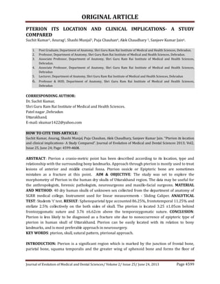
Fig 1: Measurement taken with the help of sliding caliper for the parameter PF.
Table I
Parameter No: of skulls Mean Median Minimum Maximum
PF ( r ) 40 3.44875 3.47500 2.20000 4.30000
PF (l) 40 3.46250 3.50000 2.20000 4.30000
PT variable was also measured by keeping the pointed jaws of sliding caliper at the center of the
pterion and at the superior aspect of the temporozygomatic suture. All skulls were measured for this
particular variable bilaterally and data collected is tabulated in table II.
5. ORIGINAL ARTICLE
Journal of Evolution of Medical and Dental Sciences/ Volume 2/ Issue 25/ June 24, 2013 Page 4603
Keys:
PF-linear distance between pterion and frontozygomatic arch.
PT-linear distance between pterion and temporozygomatic arch.
(r)- right side of skull.
(l)- left side of skull.
Fig 3 shows that the sphenoparietal type is a sutural pattern in which the sphenoid and parietal
bones are in direct contact, preventing the frontal and temporal bones making contact with one
another.
Fig 3.
Fig 4 shows that frontotemporal type is a sutural pattern in which the frontal and temporal bones
are in direct contact, preventing the sphenoid and parietal bones making contact with one another.
Fig 4
6. ORIGINAL ARTICLE
Journal of Evolution of Medical and Dental Sciences/ Volume 2/ Issue 25/ June 24, 2013 Page 4604
Fig 5 shows that stellate type is characterized by articulation of four bones (frontal, parietal, temporal
and sphenoid) at a point.
Fig 5
Table IV: Percentage of pterion observed on right and left side of skulls
Type of Pterion Right side Left side Both sides
n =40 n =40 n =80
Sphenoparietal 90% 82.50% 86.25%
Frontotemporal 12.5% 10% 11.25%
Stellate 5% 0% 2.5%
Keys: n- number of skulls
RESULT: Sphenoparietal type of pterion accounted 86.25% (90% on the right side and 82.5% on the
left side), frontotemporal 11.25% (12.5% on the right side and 10% on the left side) and stellate
2.5% (5% on the right side) in all 40 skulls.
The Student’s t test was employed in the assessment of each variable on either side of the
skull. A p value ≤ 0.05 was considered significant.
The pterion is located 3.25 ±1.05cm behind the anterior aspect of the fronto-zygomatic
suture on right side and 3.25 ±1.05cm on left side of the skulls. This is comparatively same on either
side. The p value for this variable is 0.43824 which is higher than the p = 0.05, this is statistically
insignificant.
The Pterion is located 3.825 ±.625cm above the superior aspect of the temporo-zygomatic
suture on the right side and 3.7±.7cm on the left side of skulls. Pterion on the left side is slightly
higher than the right side. The p value for this is 0.27527 which is statistically insignificant.
7. ORIGINAL ARTICLE
Journal of Evolution of Medical and Dental Sciences/ Volume 2/ Issue 25/ June 24, 2013 Page 4605
TABLE: V Analysis of linear distances of Pterion from bony landmarks.
Variables
Descriptive Statistics (STAmaster.sta)
Valid N Mean Median Minimum Maximum Variance S.D. S.E
PF( r ) 40 3.5 3.47500 2.20000 4.30000 0.164176 ±0.44922 0.0710
PF (l) 40 3.41 3.50000 2.20000 4.30000 0.186551 ±0.48287 0.07635
PT (r) 40 3.7775 3.70000 3.20000 4.45000 0.116505 ±0.35752 0.05653
PT (l) 40 3.69375 3.65000 3.00000 4.40000 0.088430 ±0.30153 0.04768
Keys:
S.D- standard deviation
S.E- standard error
(r)- right side
(l)- left side
N- number of skulls.
DISCUSSION: In primate evolution, the anterosuperior segment of the squamous part of the
temporal bone of lower primates became detached from its parent and incorporated into the
posterosuperior angle of the greater wing of the sphenoid bone of humans, thereby changing the
pterion pattern from the frontotemporal type of nonhuman primates to the sphenoparietal type of
humans(9).
Sphenoparietal type of pterion is most common in all the regions. It’s occurrence is higher in
Indians (95.1%)(10), Northern Indians (87.72%)(11), Southern Indians (93.55%)(12), western
Indian (91.7%)(9) and Nigerians (87.79% and 82.1%)(10,13), while it was significantly lower in
Korean (76.5%)(14) and Kenyan (66%)(15) populations as compared to this study.
The occurrence of frontotemporal type of Pterion also shows dissimilarity among different
groups, it accounts 10%–23.6% in Nigerians (10,13) and 15% of Kenyans(15), which are
significantly higher than present study in which the pterion is 11.25% being closest to that reported
in other populations of India.
Stellate variety was least present in the skulls of Uttarakhand region, study revealed it’s
occurrence nearly 2.5% (both sides). In the study done in Gujrat it’s extremely as low as 0.02% but in
the other study done in the region of Awadh area around Lucknow it accounts 5.17% which is higher
than the present study (11). It can be concluded that the occurrence of stellate variety increases from
east to west in India.
Epipteric type of pterion was not encountered in the present study, this is significant as in
Nigerians it accounts 23.6% (13), Australian Aborigines 18.5% (16), and in other Indian studies it
ranges from 6.74% to 11.79% which was mostly associated with sphenoparietal type(10,17).
The pterion lie 4.0cm above the arcus zygomaticus and 3.5 cm behind the sutura
Frontozygomatica, this is in agreement with the present study where the distance from the arcus
zygomaticus ranges from 3.0cm to 4.45cm and the distance from the sutura frontozygomatica ranges
from 2.2cm to 4.3cm.
8. ORIGINAL ARTICLE
Journal of Evolution of Medical and Dental Sciences/ Volume 2/ Issue 25/ June 24, 2013 Page 4606
In the 1970s Yasargil laid to the foundation of the pterional approach. There are many variants for
the pterional approach. Mainly it is a trepanation which permits access to the frontal and to the
temporal lobe as well as the Sylvian fissure (18, 19). Pterion is a keyhole approach to such kind of
intracranial surgeries (20,21).The combination of both a vital artery in this area and the relatively
thin bone structure has lent itself to the name "God's little joke" by some physicians and
clinicians(22). The study done on Korean population also adds up that the bone thickness at pterion
of the left side was significantly thicker than the right side of Korean adult skulls (23).
The variation in type and location of the pterion from different bony landmarks have been studied
and explained in different populations, the findings of this study might be useful in providing
information and data to the anthropologists, forensic pathologist neurosurgeons and maxilla-facial
surgeons.
Table VI: Types of Pterion in different populations (in percentage)
Study, year Population, n (skulls),
sex
Type of pterion
Sphenoparietal Frontotemporal Stellate Epipteric
In % In % In % In %
Saxena et al,
1988
Nigerian, n=40,
unknown sex
87.79 10.11 5.06 3.79
Saxena et al,
1988
Indian, n=72, unknown
sex
95.3 3.46 1.38 11.79
Manjunath et al,
1993
Southern Indian, n=172,
unknown sex
93.55 3.52 2.93 17.3
Asala et al, 1996 Nigerian, n=212,
unknown sex
82.1 23.6 - 5.7
Lee et al, 2001 Korean, n=149,
unknown sex
76.5 - - 40.3
Saxena et al,
2003
Northern Indian, n=203,
both sex
87.72 10.01 5.17 0
Mwachaka et al, Kenyan, n=50, both sex 66 15 7 12
Prabha et al,
2012
Southern Indian, n=50,
both sex
74 3 9 14
Present
study,2013
Uttarakhand, n=40,
unknown sex
86.25 11.25 2.5 0
Keys: n-number of skull
ACKNOWLEDGEMENT: First of all, I am grateful to the almighty god for establishing me to complete
this article. I take this opportunity to record my sincere thanks to all the faculty members of the
department of anatomy for their help, guidance and encouragement in completing this article. I am
extremely grateful and indebted to them for their expert, sincere and valuable guidance extended to
me.
I would like to thank statistician (Raman Nautiyal) for helping me in statistical analysis.
9. ORIGINAL ARTICLE
Journal of Evolution of Medical and Dental Sciences/ Volume 2/ Issue 25/ June 24, 2013 Page 4607
I also place on record, my sincere gratitude to one and all who, directly or indirectly, have lent their
helping hand in this article.
These acknowledgments would not be complete without thanking my family especially my wife
(Gunjan) and my daughter (Sugun) for their constant support and care.
REFERENCES:
1. Moore KL, Dalley AF. Clinically oriented anatomy, 4th edn. Lippincott Williams & Wilkins,
Baltimore, 1999;836–840
2. Williams PL, Bannister LH, Berry MM, Collins P, Dyson M, Dussek JE, et al. The skull. In.
Gray’s Anatomy, 38thed.London: Churchill Livingstone; 1995. p. 583-606.
3. Natekar PE, DeSouza FM, Natekar SP. Pterion: An anatomical variation and surgical
landmark. Indian J Otol 2011; 17:83-5.
4. Hussain SS, Haseena S, Prasanna LC. Unusual Wormian bones at Pterion – Three case
reports: JBiomed Sci and Res. 2010; 2 (2):116-18
5. Snell Richard S. Clinical Anatomy by Regions, 8th edi. Lippincott. Williams and Wilkins,
2008; p.673, 686
6. Apinhasmit W, Chompoopong S, Chaisuksunt V, Thiraphatthanavong P, Phasukdee N.
Anatomical consideration of pterion and its related references in Thai dry skulls for
pterional surgical approach. Journal Of The Medical Association Of Thailand Chotmaihet
Thangphaet.2011; 94(2): 205-214.
7. Richard S. Snell. Clinical Anatomy.7th ed. Lippincott Williams & Wilkins, Baltimore, 2004;
896
8. Ersoy M, Evliyaoglu C, Bozkurt M, Konuskan B, Tekdemir I. Epipteric bone in the pterion may
be a surgical pitfall. Minim. Invas. Neurosurg.2003; 46:363-5.
9. Zalawadia DA, Vadgama DJ, Ruparelia DS, Patel DS, Patel DSV. Morphometric Study Of Pterion
In Dry Skull Of Gujarat Region. NJIRM. 2010; 1(4): 25-29.
10. Saxena SK, Jain SP, Chowdhary DS. A comparative study of pterion formation and its
variations in the skulls of Nigerians and Indians. Anthropol Anz 1988;46:75-82
11. Saxena R, Bilodi A, Mane S, Kumar A. Study of pterion in skulls of Awadh area-in and around
Lucknow. Kathmandu University Medical Journal (KUMJ) .2003; 1(1): 32-33.
12. Manjunath KY, Thomas IM. Pterion variants and epipteric ossicles in South Indian skulls. J
Anat Soc India. 1993; 42:85-94.
13. Asala SA, Mbajiorgu FE. Epigenetic variation in the Nigerian skull: sutural pattern at the
pterion. East Afr Med J 1996;73:484-6
14. Lee UY, Park DK, Kwon SO, Paik DJ, Han SH. Morphological analysis of the pterion in Korean.
Korean J Phys Anthropol. 2001; 14:281-9.
15. Mwachaka PM, Hassanali J, Odula P. Sutural morphology of the pterion and asterion among
adult Kenyans. Braz J Morphol Sci .2009; 26:4-7
16. Murphy, T. The pterion in the Australian aborigine. Am. J. Phys. Anthropol., 14:225-44, 1956
17. Gopinathan K., Dhall U., Chhabra S. Sutural bones in the North Indian population, J. Anat. Soc.
India. 1998; 47(2):91-96.
18. Scholz M, Parvin R, Thissen J, Löhnert C, Harders A, Blaeser K. Skull base approaches in
neurosurgery. Head & Neck Oncology. 2010; 216.
10. ORIGINAL ARTICLE
Journal of Evolution of Medical and Dental Sciences/ Volume 2/ Issue 25/ June 24, 2013 Page 4608
19. Chao S, Shen C, Cheng W. Microsurgical removal of sylvian fissure lipoma with pterion
keyhole approach-case report and review of the literature. Surgical Neurology.2008; 70
Suppl 1S1:85-90.
20. Cheng W, Lee H, Sun M, Shen C. A pterion keyhole approach for the treatment of anterior
circulation aneurysms. Minimally Invasive Neurosurgery: MIN .2006; 49(5): 257-262.
21. Samson D, Hodosh R, Clark W. Microsurgical evaluation of the pterional approach to
aneurysms of the distal basilar circulation. Neurosurgery .1978; 3(2): 135-141.
22. Praba AMA, Venkatramaniah C. Morphometric Study of different types of Pterion and it’s
relation with middle meningeal artery in dry skulls of Tamil Nadu. JPBMS. 2012; 21 (04):1-4.
23. Hwang K, Kim JH, Baik SH. The thickness of the skull in Korean adults. J Craniofac Surg. 1999;
10: 395-9.