Absence of the Palmaris Longus Tendon in Mid Eastern Population..pdf
SKJAIN
1. J. Morphol. Sci., 2011, vol. 28, no. 3, p. 184-188184
Original
article
Relationship between palmar skin creases and osseous anatomy -
a radiological study identification
Chauhan, P.1
*, Kalra, S.2
, Jain, SK.1
, Munjal, S.1
and Anurag, A.1
1
Department of Anatomy, Shri Guru Ram Rai Institute of Medical and Health Sciences Patel Nagar,
Dehradun, Uttarakhand, 248001, India
2
Department of Anatomy, University College of Medical Sciences, and Associated GTB Hospital,
Delhi, 110095, India
*E-mail: drpujachauhan@yahoo.co.in
Abstract
Functional association of the Palmar creases overlying the various osseous structure of the hand have proved as
an important anatomical landmark. This fact can be of use in various orthopaedic and surgical procedures on
the hand. These lines upon the skin may not exactly mark the line of underlying joint, therefore it is important
to know the relationship between the two. The study was performed on the right hands of 50 subjects.
Most consistent creases were marked using a 25 gauge copper wire on the palm and radiographs were taken
in AP view. Various measurements were taken from the crease to the associated joint/osseous structure.
Statistical analysis proved significant positive correlation in the distance between distal transverse crease(DTC)
and metacarpophalangeal (MCP) joint and proximal digital crease (PDC) and MCP joints in the index
finger and middle finger with p value of 0.03 and 0.008 respectively, whereas, highly significant negative
correlation was found in the distance between proximal transverse crease (PTC)-MCP joint and proximal
digital crease(PDC)‑MCP joint of the index finger(p = 0.038). PTC was consistently proximal to MCP joint
in the study group. It was observed that in majority of cases the associated joints do not lie under the flexion
creases, therefore if any joint exposure is essential, then for better field of surgery incision can be placed parallel
to the crease so that it exactly overlaps the joint if the position of joint with respect to the crease is known.
Keywords: flexion crease, palmar crease, hand reconstruction.
1 Introduction
The study of palmar skin creases has evoked substantial
interest among forensic science experts, astrologers, clinicians
and geneticists. These are exclusive to any human being and
no two people can have exactly identical creases in hand. Both
primary genetic determination and development secondary
to flexion function have been suggested as the mechanisms
underlying the crease development. The evidence offered has
been mostly indirect, related to the timing of the onset of the
fetal hand movement and the crease aberrations in malformed
hands and fingers (KIMURA, 1991). Flexion lines are of
significance and concern as they are the surface registration
of the mobility of the parts. They indicate the folding points
of the skin and subcutaneous tissue and mark the site of skin
joint, brought into action by the underlying bony joint. The
major markings are found in the vicinity of synovial joints,
where the skin is strongly attached to the underlying deep
fascia .These flexion lines develop during early fetal life and
are unique features of the hand. Documentation of pre-
natal stage of development of the flexion creases on human
palm revealed that most flexion creases develop concurrently
with the appearance of fetal volar pads and the rest develop
independently of them. A study on development of human
palmar and digital flexion creases documented that the
palmar creases are consistently seen by 13 weeks of gestation
(STEVENS, CAREY, SHAH et al., 1970). The volar pads
are present from 8 to 14 fetal weeks. A hand malformation
or specific insult that occurs before the time of crease
development and that alters the form or function of the fetal
hand can cause secondary alterations in the crease patterns
of the hand. However, there are also studies to indicate that
digital and palmar hand creases are secondary features, being
the consequence of flexional folding of palmar skin in early
fetal life (KIMURA and KITAGAWA, 1986; POPICH and
SMITH, 1970). But contrarily, a research in the inheritance
of liability estimated from the incidence among relatives
suggested that these palmar creases are inherited in polygenic
manner and genetic factors and environmental both interplay
to produce these creases (TAY, 1979).
Because of the functional association of the various
osseous structure of the hand with the palmar creases,
these can be used as an important anatomical landmark to
the underlying structures and can help in inspection and
palpation of the hand and prediction of structures, which are
liable to be damaged in relation to a specific surface wound.
Palmar skin creases are considered to be ideal lines for giving
incision in hand surgeries and the design of every skin
incision must take into account the structure and mobility
of area it crosses, its relationship to deeper structures, the
blood supply to skin, the sensory nerve distribution, the
previous wounds and cosmetic considerations (TUBIANA,
THOMINA and EVELYN, 1984). A mislaid incision can
often lead to the damage of underlying important structures
and unsightly tender contracted scar. The correlation can also
be of practical therapeutic value in orthopaedic rehabilitation
of hand malformations. The most important objective of this
study was to measure the distance between Palmar/digital
2. Palmar creases and osseous anatomy
J. Morphol. Sci., 2011, vol. 28, no. 3, p. 184-188 185
of Vernier Calipers. Perpendicular line was measured which
was drawn from the mid-point of metacarpophalangeal joint
of various fingers to the distal/ proximal palmar creases.
Distance between thenar crease and hook of hammate was
also measured. The position of thenar crease was also noted
on X-Ray with respect to second metacarpal, second inter-
metacarpal space, third metacarpal and third intermetacarpal
space. A horizontal line was measured which was passing
from the thenar crease to the hook of hamate medially and
from hook of hamate to first carpometacarpal joint laterally.
The effect of potential radiograph magnification was
determined by measuring skin crease to skin crease distances
directly in 5 hands and obtaining same measurements
from their respective radiographs. Comparison was done
statistically by applying paired t-test. The test statistics for
T = D - mD ÷ SD/n where,
• D = Sample average difference between each pair of
observation
• SD = Sample standard deviation
• m = sample size, i.e. the number of pairs of observation
• mD = Population mean difference under the null
hypothesis
When the null hypothesis is true and the population
mean difference is mD, the statistic has a T-distribution
with n-1 degree of freedom. The error due to hand size was
determined by measuring the length of the 3rd
metacarpal
and size ranking the hands by this measurement followed by
grouping them in 3 categories consisting of: Subjects within
±2SD of the mean value Lying below –2SD Lying above
+2SD, and the subject falling within ±2SD were chosen for
study. To eliminate the gender specific differences, the study
group comprised exclusively of the female volunteers.
All the measurements taken were expressed as mean
values and Standard Deviation. Various distances of the
same finger were correlated statistically for any positive/
negative/no correlation by using Karl Pearson’s test of
correlation. Differences in the mean distances which were
proximal and those, which were distal, were compared for
significance for the same parameter by applying Student’s
t-test. Normal curve distribution of various parameters in
sample study group was observed by expressing the results
with the help of graphs and confirmed by using Kolmogorov
Smirnov’s statistical test of normality. Quantification of these
relationships would aid in hand examination and placement
of surgical incisions and provide additional insight into
anatomic and functional associations of the hand with these
creases.
3 Results
In the present study, distal transverse crease (DTC)
was proximal to MCP joint of all the fingers in majority of
volunteers (95-100%). In index finger, the DTC reached
the level of MCP joint in only 18% volunteers. Distance
gradually increased from index to ring finger and there was
abrupt fall in the mean distance of the little finger (Table 1).
In index and little finger the crease was almost equidistant
from MCP joint.
Distance between the DTC and MCP joint in the study
group was:
• Index finger:Proximaltothejoint- 3observationswere
between 20-25 mm and 5 were between 5 -9.9 mm.
skin creases and the related joints (joint causing deepening
of the skin crease), quantification of these measurements,
their statistical analysis to find out the relationship between
the creases and osseous anatomy and to study its clinical
implications.
2 Materials and methods
The study was conducted on 50 adult female volunteers.
Exclusion criteria included any known medical history of
congenital anomaly, surgery or trauma of the hand. Twenty
five Gauge copper wire was used to mark 8 consistently
present skin creases on the right hands of all the volunteers
(Figure 1). The creases marked were thenar crease
(TC), proximal transverse crease (PTC), distal transverse
crease (DTC), palmar digital crease (PDC), proximal
interphalangeal crease (PIP) and distal interphalangeal crease
(DIP). Radiographs were taken in an anteroposterior (AP)
view with fingers extended (0° flexion) and wrist in neutral
position. In the neutral position the wrist is in 0° flexion,
extension, ulnar deviation and radial deviation. Although in
this study the hands were placed in a standardized position
to obtain radiographs, it is rational to believe, that, because
of the relative immobility of the creases the relationships
described here would retain validity regardless of the hand
or fingers and this position was selected for the study
because many palmar hand procedures are performed in this
position. Markers placed along palmar skin creases of 50
hands radiographically demonstrated creases superimposed
on osseous anatomy.
Various measurements from each skin crease to its nearest
and functionally associated joints/osseous structure were
taken directly from the radiographs of the hand with the help
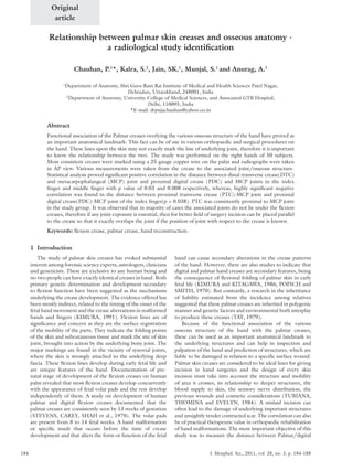
Figure 1. Radiograph of right hand with copper wire placed on
the digital creases, transverse creases, wrist creases and thenar
crease.
3. Chauhan, P., Kalra, S., Jain, SK. et al.
J. Morphol. Sci., 2011, vol. 28, no. 3, p. 184-188186
of reconstructive surgery are paramount but perfectly
executed reconstruction can return this very important part
of the body to near perfect condition.
In compliance with our observations, Bugbee and Botte
(1993), also reported DTC to be proximal in middle, ring
and little fingers. However, there was no mention about the
index finger. In the present study, the crease was found to be
farthest (distance = 13.4 mm) from the MCP joint of ring
fingers which is in conformity with (distance = 10.3 mm) as
reported by Bugbee and Botte (1993) and was nearest to the
joint in the little fingers (present study: distance = 8.8 mm,
study by Bugbee and Botte (1993) distance = 7.9 mm). In
index finger, test of correlation was found to be significantly
positive when distance between PDC & MCP joint and
DTC & MCP joint were compared (Pearson’s correlation
Coefficient = -.699, P = .036). Middle finger also showed
highly significant negative correlation when the distance
between the DTC & MCP joint between PDC & MCP joint
were compared (Pearson’s correlation Coefficient = -.374,
P = .008).
PTC was found to be proximal to the MCP joint of all
the digits in maximum number of volunteers (96%) except
the little finger). There was a gradual increase in the distance
of the crease to the MCP joint from index to little finger.
This crease reached the level of MCP joint of little finger
in 48% cases, whereas Bugbee and Botte (1993) did not
mention about its presence in relation to little finger. This
crease was farthest from the joint in little finger in our
study in contrast to that of proximal digital crease. Both the
studies (present study: D = 25.1 mm, Bugbee and Botte
(1993): D = 22.1 mm) reported this crease to be nearest
to the joint in index finger (present study: D = 11.6 mm,
Bugbee and Botte (1993): D = 9.1 mm). Difference in the
observations was in the range of 2-3 mm. Other researchers
(BUGBEE and BOTTE, 1993), also reported that the line
joining the radial border of proximal transverse crease and
ulnar border of distal transverse crease accurately overlapped
the metacarpal neck in majority of volunteers (73%), where
as in our study it was passing through the metacarpal neck
Distal to the joint- 2 (4%) observations were recorded
in the range of 1-5 mm and 1 (2%) in the range
of 15.1-20 mm. In one case (2%), DTC was found
to be overlapping with the joint, in the rest of the
volunteers, DTC didn’t reach till the measurable
point.
• Middle finger: Proximal to the joint- 12 cases (24%)
were between 15-19.9 mm, 18 (36%) fell in the range
of 10-14.9, 10 (20%) between 5-9.9 mm and 6 (12%)
were between 0.1-4.9 mm. Distal to the joint- 2 (4%)
observations were recorded in the range of 1-5 mm,
1 (2%) was in the range of 5.1-10 mm and 1 (2%) case
was in the range of 15.1-20 mm.
• Ring finger: All the observations were proximal to the
joint. 2 (4%) observation were in 20-25 mm, 21 (42%)
were between 15-19.9 mm, 15 (30%) in 10-14.9 mm
range, 11 (22%) were between 5-9.9 mm and only
1 (2%) was in the range of 1-4.9 mm.
• Little finger: All the cases were proximal to
the joint. 1 (2%) observation fell between
20‑25 mm, 1 (2%) between 15-19.9 mm, 16 (32%)
between 10‑14.1 mm, 28 (56%) between 5-9.9 mm
and 2 (4%) were found to be in the range of
0.1‑4.9 mm, 2 were overlapping the joint.
In middle finger, maximum observations, n = 46; were
proximal to the joint and were in the range of 5-20 mm.
In ring finger, the crease was proximal to the joint with the
most frequent range of 5-20 mm in 100%. In little finger
also, all the observations were proximal with the most
frequent range of 5-15 mm.
4 Discussion
Studies of palmar and digital creases in the normal hand
may be of diagnostic value in particular syndromes, as well
as being of functional significance in serious malformations
of the hand .The importance of this study involves not
only diagnostic purposes but more important for various
reconstructive hand surgeries. Not only the aesthetic benefits
Table 1. Mean distance of palmar creases from metacarpo-phalangeal joints.
Sl.No. Distance Position No. of volunteers Mean ± Standard Deviation
1. Index dist. tr.-mcp jt.
P 5 8.7 ± 7.02
D 3 12 ± 6.08
2. Middle dist. tr.-mcp jt.
P 46 11.78 ± 4.55
D 4 6.75 ± 7.02
3. Ring dist. tr.-mcp jt.
P 49 13.45 ± 4.01
D 1 14.5
4. Little dist. tr.-mcp jt.
P 50 8.8 ± 3.48
D 0 0
5. Index px. tr.-mcp jt.
P 50 11.64 ± 4.97
D 0 0
6. Middle px. tr.-mcp jt.
P 50 21.68 ± 6.16
D 0 0
7. Ring px. tr.-mcp jt.
P 48 25.12 ± 7.26
D 0 0
8. Little px. tr.-mcp jt.
P 23 26.86 ± 12.93
D 0 0
4. Palmar creases and osseous anatomy
J. Morphol. Sci., 2011, vol. 28, no. 3, p. 184-188 187
5 Conclusion
The hand serves important functions can be severely
hinder anyone with a hand defect or injury from doing the
job due to physical trauma such as due to burns, accidents or
a birth defect. Reconstructive hand not only does improve
the appearance of the hand, but its primary utility is that
of restoring the hand’s functionality, allowing the long-
suffering individual to live a nearly perfectly normal life.
The knowledge of these creases in relation to the osseous
anatomy and the joints of the hand are extremely essential
in hand surgeries.
References
BUGBEE, WD. and BOTTE, MJ. Surface anatomy of hand: The
relation between palmar skin creases and osseous anatomy. Clinical
Orthopaedics and Related Research,1993, vol. 296, p. 122-126.
PMid:8222413.
in maximum cases (46%) and was overlapping the head of
metacarpal in 38% volunteers. Correlation of distances was
seen only in the readings of index finger. Significant negative
correlation was seen in the distance between PTC and MCP
joint; and PDC and MCP joint. (Pearson’s correlation
Coefficient = -.294). In the present study, thenar crease
in the palm, which originated from its radial border in
common with the proximal transverse crease, was located in
2nd
intermetacarpal space in majority of cases (76%), contrary
to another study by Bugbee and Botte (1993), which
reported it to be crossing the 3rd
metacarpal in maximum
cases (54%). The position of the crease was least common on
the third intermetacarpal space. In the proximal palm, thenar
crease crossed scaphoid in maximum number of volunteers
(26%) (Figure 2), but Bugbee and Botte (1993) reported
it to be crossing the capitate in the majority (47%). In the
current study the thenar crease was 3 to 4 mm nearer to
the first carpometacarpal joint and the similar was the case
with the hook of hamate.Thenar crease was passing through
second intermetacarpal space in 38 (76%) volunteers. It was
passing through the third metacarpal in 10 (20%) and the
third intermetacarpal space in 2 (4%) volunteers (Figure 3).
In the present study distance of thenar crease from the first
carpo metacarpal joint and the hook of hamate was observed
to be 7.2 mm and 15.6 mm respectively. Whereas Bugbee
and Botte (1993), reported this distance to be 22.6 mm
and 18.7 mm respectively which probably indicates the racial
differences and shows that North Americans have wider
carpal region as compared to Indians. In another research
(KOMATZ, DAIJO and YOSHIDA, 1978) digital flexion
creases on the little fingers of 283 male and 268 female
individuals were studied. Two males and two females were
found to have an extra interphalangeal transverse crease
situated between the metacarpophalangeal and proximal
interphalangeal creases. The extra crease of the little finger
was unilateral or bilateral. None of the little fingers with
extra crease had radiographic anomalies of the bones or
joints, or evidence of dyskinesia. In the present study none
of the fingers or palm had an extra crease.
The knowledge of these creases in relation to the
osseous anatomy and the joints of the hand can be of
great use to the operating surgeon in various procedures
on hand such as for tendon repairs, corrective procedures
for webbed fingers, carpal tunnel hand surgery, trigger
finger surgery, ganglion cyst removal etc. In Dupuytren’s
contracture accepted indications (TOWNLEY, BAKER,
SHEPPARD et al., 2006) for surgical intervention include
metacarpophalangeal joint contracture of 30° and any
degree of proximal interphalangeal joint contracture
(TOWNLEY, BAKER, SHEPPARD et al., 2006). For
contracture release fasciotomy/fasciectomy, is done via
zigzag incisions along natural palmar and digital creases to
prevent volar contractures and allow adequate exposure.
Resurfacing after a total degloving injury to the hand is one
of the most difficult management problems in hand surgery.
In a report the authors resurfaced a totally degloved hand
using extremely thin and broad perforator-based cutaneous
free flaps, and the donor defects were covered with split-
thickness skin grafts (KIM, KIM, KIM et al., 2003). Several
procedures, including the formation of palmar and digital
creases and interdigitation, defatting were performed to
obtain the final appearance and function of the hand.
Figure 2. Comparative analysis of thenar crease in proximal
palm.
Figure 3. Comparative analysis of of thenar crease in mid palm.
5. Chauhan, P., Kalra, S., Jain, SK. et al.
J. Morphol. Sci., 2011, vol. 28, no. 3, p. 184-188188
STEVENS, CA., CAREY, JC., SHAH, M. and BAGLEY, GP.
Development of human palmar and digital flexion creases. Journal
of Pediatrics, 1970, vol. 113, n. 1, p. 128-132.
TAY, JSH . The genetics of plamar creases: A study in the inheritance
of liability estimated from the incidence among relatives. Annals of
Human Genetics, 1979, vol. 42, n. 3, p. 327-332. PMid:434775.
http://dx.doi.org/10.1111/j.1469-1809.1979.tb00666.x
TOWNLEY, WA., BAKER, R., SHEPPARD, N. and
GROBBELAAR, A. Dupuytren’s contracture unfolded.
British Medical Journal, 2006, vol. 332, n. 7538, p. 397-400.
PMid:16484265. PMCid:1370973. http://dx.doi.org/10.1136/
bmj.332.7538.397
TUBIANA, R., THOMINA, JM. and EVELYN, M. Examination of
hand and upper limb: The skin of hand. Philadelphia: WB Saunders
Company, 1984.
Received February 15, 2011
Accepted June 21, 2011
KIM,KS.,KIM,ES.,KIM,DY.,LEE,SY.andCHO,BH.Resurfacing
of a totally degloved hand using thin perforator-based cutaneous
free flaps. Annals of Plastic Surgery, 2003, vol. 50, n. 1, p. 77‑81.
PMid:12545113. http://dx.doi.org/10.1097/00000637-
200301000-00013
KIMURA, S . Embryologic development of flexion creases. Birth
defects: original article series, 1991, vol. 27, n. 2, p. 113-129.
PMid:1786348.
KIMURA, S. and KITAGAWA, T. Embryological development
of human palmar, plantar and digital flexion creases. Anatomical
Record, 1986, vol. 216, p. 1199391-197. PMid:3777451.
http://dx.doi.org/10.1002/ar.1092160211
KOMATZ, Y., DAIJO, K. and YOSHIDA, O. Extra interphalangeal
transverse creases of the little finger. Japanese Journal of Human
Genetics, 1978, vol. 23, n. 1, p. 31-37. http://dx.doi.org/10.1007/
BF01871380
POPICH, GA. and SMITH, DW. The genesis and significance
of digital and palmar band creases: Preliminary report. Journal
of Pediatrics, 1970, vol. 77, n. 6, p. 1017-1023. http://dx.doi.
org/10.1016/S0022-3476(70)80086-5