2. 30
Status of Magnesium and Manganase in Selected Anti-Diabetic Medicinal Plants
used in Adamawa State, Nigeria
and food supplements for revitalizing body system, (Junaid, et al., 2006, Odukoya 2007, Stef et
al.,2010). According to world health organization report about 80% of the world population
is taking interest in indigenous medicinal plants remedies. These medicinal plants have
usually been used in the form of fruit and vegetables, drugs or their extract for the treatment
of different diseases in both developed and developing countries (Sofowora, 2008, Sahito et
al., 2005). Studies have shown that medicinal plants contain both organic and inorganic
constituents, and many medicinal plants are found to be rich in one or more individual
elements, thereby providing a possible link to the therapeutic action of the medicine (Singh et
al., 1997). Studies on the organic constituents of the medicinal plants have been going on long
time while little has been done on the inorganic aspect in the medicinal use of these plants
(Singh and Garg, 2006). It is important to know the elemental concentration in medicinal
plants from the point of view of nutritional requirement and intoxication risk associated with
their consumption. The effects and influence of trace elements on administration of medicinal
plants is also essential to understand the pharmacological action of the medicinal plants and
to decide the dosage of the herbal drugs prepared from these plant materials (Naga Raju et
al., 2013). The human body needs a number of minerals in order to maintain good health
(Balaji et al., 2000, Ajasa et al., 2004 Yagi et al., 2013,Magili,et al.,2014,). Macro and trace
elements influence biochemical processes in the human organism. Active constituents of
medicinal plants that is the secondary metabolic and a number of mineral elements play an
important role in the metabolism (Kolasani,et al., 2011).
Deficiency or excess of elements may cause a number of disorders. For example, Iron
deficiency causes anemia which has been reported to affects one third of the world
population (Kumari,et al.,2004,Leterme,et al.,2006). Low levels of Zn can induce the
pathogenesis of lung cancer ( Cobanoglu,et al., 2010). Breast cancer patients had low levels of
Ca, Mg, Fe, Cu, Mn and Zn in their hair (Joo,et al., 2009). Diabetes mellitus is one of the
metabolic disorders that have gravely troubled the human health and quality of life.
Conventional agents are being used to control diabetes along with lifestyle management.
However, they are not entirely effective and no one has ever been reported to have fully
recovered from this disease. Numerous medicinal plants have been used for the management
of diabetes mellitus in various traditional systems of medicine worldwide as they are a great
source of primary and secondary metabolites and many of them are known to be effective
against diabetes mellitus (Kayode,et al.,2012). Medicinal plants with anti-hyperglycemic
activities are being more desired, owing to lesser side-effects and low cost. Naga Raju et al.,
(2013) reported that anti-diabetic medicinal plants undoubtedly have significant effect on
lowering blood sugar. Numerous medicinal plants have been reported to be effective in
diabetes management, yet plenty of research is still needed to be carried out in this area with
3. 31
S.T.Magili, H.M.Maina, J.T.Barminas, O.N.
Maiteraand Y.K. Musa
a view of finding cheap alternative ways for the treatment of diabetes mellitus (Piero et al.,
2013).Trace elements have been identified for long time as potential candidates for improving
metabolic disorders such as, insulin resistance, obesity, metabolic syndrome or diabetes
(Mertz, 1993,)..Trace element plays a vital part in the metabolism of plants and animals
(Stitch, 1957). In the human body, the trace element is made of up to 0.01% of the body’s mass
(Nason and Schroeder, 1971). Trace elements, for example the metals selenium, zinc,
magnesium, manganase and copper, are essential to maintain the metabolism of the human
body. Studies have demonstrated that some trace elements are involved in potentiating
insulin action (Anderson et al., 1997, underwood, 1997). Read marked alterations in trace
elemental concentrations in human body are therefore associated with occurrence of diabetes
mellitus. Therefore, regulation of trace elemental concentrations has been proposed as a
potential prevention and management of diabetes mellitus. A good example to illustrate their
important contribution is magnesium; low magnesium levels have been associated with
increased type 2 diabetes (Chaudhary et al., 2010, Wells, 2008). Considering the importance of
trace elements in various human metabolic processes and also their curative properties, this
Study is designed to determine the bioavailability of Mg and Mn in selected anti-diabetic
medicinal plants which have been utilized in the study area. In the present investigation one
of the sensitive analytical techniques was applied like Instrumental neutron activation
analysis (INAA) to analyze the content of Mg and Mn in some anti-diabetic medicinal plants
parts.
MATERIALS AND METHODS
Sample Collection and Treatment
The plant samples were obtained from Mubi North, Mubi South and Maiha Local
Government, Areas of Adamawa State, from October to December 2011. The medicinal plants
were identified by Mr.Jarafu Ulam Mamza of the Department of Biological Sciences
Adamawa State University, Mubi, the scientific names, local name and traditional uses of the
studied medicinal plants are presented in Table 1. The collected plant material was washed
thoroughly with running tap water to remove the dust particles. They were shade dried,
powdered and stored in closed air tight polythene bag and kept away from moisture until
needed for analysis.
4. 32
Status of Magnesium and Manganase in Selected Anti-Diabetic Medicinal Plants
used in Adamawa State, Nigeria
Table 1: List of the Selected Antidiabetic Medicinal Plant Analysed in this Study.
S/No. Botanical Name Family Name Common Name Local Name (Hausa) Traditional Uses.
1.
Terminalia
avicennioides
Combretaceae
Terminalia
dictyonuna
diels.
Baushe
Skin, diseases,
headaches, bronchitis,
sore throat, high, blood
pressure, diabetes,
antibacterial properties,
vitamin deficiency,
diarrhoea.
2.
Hymenocardia
acida
Hymenocardiaceae Red onion Janyaaro
Malaria, fever, bronchitis,
dysentery, jaundice,
diabetes, wounds, skin
diseases.
3 Leptadenia hastata Asclepiadaceae
Cyandum
hastatum
Dan barawo
Fever, cough, diabetes,
gonorrhoea, wounds,
colds, diarrhoea.
4.
Balamites
aegyptiacae
Balanitiaceae Soapberry tree Aduwaa
Diarrhoea, wounds,
constipation, diabetes, etc
5.
Ageratum
conyzoides
Asteraceae
white weed,
Billy-Goat
weeds
Gwiwan Jimina
fever, diarrhea, wounds,
bactriocide, headache,
pneumonia, Diabetes
6. Sclerocarya birrea Anacaardiaceae Spondias birrea Daniya/Lule/ Nunu
Tooth, decay, malaria,
Diarrhea sore throats,
diabetes, anti-venom.
7
Anogeissus
leiocarpus
Combretaceae African birch Markee
Antibacterial properties,
high blood pressure,
diarrhoea, dysentery,
skin, disease, wounds,
diabetes, fever coughs
rheumatism.
8
Jatropha
gossypiifolia
Euphorbiaceae Wild cassada Zugu
Leprosy, coughs, fever,
high blood pressure,
diabetes, skin disease,
wounds, ulcers, scabies
etc.
9 Daniellia oliveri Caesalpinioideae
Paradaniellia
oliveri
maje
Headache, wounds,
ulcers, skin disease,
fever, jaundice, tooth
decay, menstrual
disorders, diabetes, burns
etc.
10
Sarcocephalus
latifolius
Rubiaceae Nauclea latifolia tafashiya
Tooth decay, jaundice,
indigestion, hernia,
wounds, fever ,malaria,
kidney failure, diabetes,
leprosy, syphilis,
swellings etc.
5. 33
S.T.Magili, H.M.Maina, J.T.Barminas, O.N.
Maiteraand Y.K. Musa
Sample Preparation for NAA Analysis
The method described by Funtua et al. (2012) and Kogo et al.,(2009) with some modifications
was adopted. The sealed sample in the polyethylene bags were put in a vial for irradiation.
The plant samples were in the ranges of 250- 300 mg as adopted for NIRR-1at Centre for
Energy Research and Training ABU, Zaria.
Analysis of Samples
Standard reference material SRM 1547 (NIST PEACH LEAVES) was analyzed along with the
samples for method substantiation and quality control purposes. From results obtained, it
was observed that most of the elemental concentrations are comparable to the certified
values. The Instrumental neutron activation analysis (INAA) technique has been widely
employed for the determination of major, minor and trace elements in medicinal plants,
water, clays, pottery, ceramics and other allied materials (Oladipo, 1992, Oladipo, 2003, Kogo
et al., 2009).The samples were irradiated using Nigeria Research Reactor-1 (NIRR-1) at a
neutron flux of 2.5 x 10¹¹
n/cm2
s in the outer irradiation channels for short lived irradiations.
Long-lived irradiations involved neutron irradiation of a batch of reference samples and
standards for 6 h at 5.0 x 10¹¹
n/cm 2
sec in the inner irradiation channels using the same
facility (Debrah et al., 2011). Each sample or standard underwent two irradiations procedure
as described in a work performed by Jonah et al. (2005) for short and long irradiations
respectively. For the short irradiation, each sample or standard were irradiated for one
minute, allowed to decay for a few minutes, followed by 10 min counting on a HPGe detector
coupled to its associated electronics. For the long-lived irradiations, first counting exercise
began four days after irradiation, each sample or standard were counted for 30 min to
analyze those nuclides with half-lives mainly in the order of hours or few days (Jonah et
al.,2006). The same batch of samples were recounted for one hour each after nine to ten days
decay in order to analyze those nuclides with half-lives in the order of days and years.
Finally, the identification of gamma ray of product radio-nuclides through their energies and
quantitative analysis of their concentration were obtained by using the gamma-ray spectrum
analysis software, WINSPAM, 2004.
Statistical Analysis
The obtained results are presented as mean ± SD (standard deviation). All differences are
considered significant at 5% level, therefore P-values less than 0.05 (P<0.05) were considered
statistically significant at p<0.05 using Analyse-it version 2.3 statistical software for Microsoft
Excel. Significant elemental concentration differences in plants samples were determined by
analysis of variance (ANOVA).
6. 34
Status of Magnesium and Manganase in Selected Anti-Diabetic Medicinal Plants
used in Adamawa State, Nigeria
RESULTS AND DISCUSSIONS
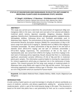
Table-2 and figures 1 and 2 shows the mean concentrations and distribution levels of Mg and
Mn in the leaves, stem bark and root bark of the selected anti-diabetic medicinal plants. The
results for the distribution of Mg in leaves stem bark and root bark of anti-diabetic medicinal
plants studied is shown on Figure 1. Read the concentrations Mg in leaves varied from
Terminalia avicennioides (1980±178 mg/kg) to Daniellia oliveri (6012±331 mg/kg) with a
variability coefficient of 40%. Mg was not detected in the leaves of Hymenocardia acida. More
than 60% of all pair wise concentration variation differences were statistically significant
(P<.05).
Table 2 Magnesium and Manganese Contents (mg/kg) in selected Antidiabetic Medicinal
Plants Parts Species
Plant Species Plant Parts
Element Concentrations (mean±SD)
Mg Mn
Ageratum conyzoides Leaves 5402.0±265.0 226.1±0.5
Root Bark 2729.0±306.0 220.4±0.4
Stem Bark 3463.0±208.0 80.9±0.3
Anogeissus leiocarpus Leaves 2307.0±164.0 32.9±0.2
Root Bark 1693.0±198.0 37.5±0.2
Stem Bark 2647.0±185.0 38.9±0.2
Balamites aegytiacae Leaves 3216.0±232.0 61.1±0.3
Root Bark 2597.0±255.0 52.6±0.3
Stem Bark 1706.0±229.0 39.1±0.2
Daniellia oliveri Leaves 6012.0±331.0 339.2±0.7
Root Bark 739.0±177.0 46.1±0.2
Stem Bark 593.0±147.0 43.8±0.3
Hymenocardia acida Leaves BDL 1969.0±14.0
Root Bark 3522.0±320.0 317.6±0.6
Stem Bark 1190.0±163.0 139.0±0.4
Jathropha gossypiifolia Leaves 4785.0±321.0 88.7±0.4
Root Bark 4386.0±307.0 103.1±0.4
Stem Bark 3029.0±233.0 48.4±0.2
Leptadenia hastata Leaves 5641.0±299.0 128.9±0.4
Root Bark 3080.0±243.0 165.0±1.0
Stem Bark 2375.0±221.0 44.9±0.3
Sarcocephalus latifolius Leaves 2403.0±147.0 49.3±0.3
Root Bark 3618.0±242.0 45.9±0.3
Stem Bark 1551.0±158.0 35.4±0.2
Sclerocarya birrea Leaves 4212.0±211.0 63.1±0.3
Root Bark 4298.0±232.0 114.2±0.5
Stem Bark 2951.0±177.0 25.9±0.2
Terminalia avicennioides Leaves 1980.0±178.0 173.0±1.0
Root Bark 591.0±140.0 44.4±0.3
Stem Bark BDL 44.2±0.3
BDL: Below detection limit. SD: Standard deviation
7. 35
S.T.Magili, H.M.Maina, J.T.Barminas, O.N.
Maiteraand Y.K. Musa
The concentration of Mg in the Stem bark of Daniellia oliveri ranged from (593±147 mg/kg) to
Ageratum conyzoides (3463±208 mg/kg) with a variability coefficient of 57%. Mg was not
detected in the stem bark of Terminalia avicennioides. Similarly, more than 60% of all pairwise
concentration variation differences were statistically significant (P<.05).Root bark Mg
concentration ranged from Terminalia avicennioides (591±140 mg/kg) to Jathropha gossypiifolia
(4386±307 mg/kg) with a variability coefficient of 48%. Mg was present in all root bark
samples analyzed. Again, more than 60% of all pairwise concentration variation differences
in root bark samples were statistically significant (P<.05).The order of Mg concentrations
distribution level is leaves > root bark > stem bark, on average. Summation of Mg
concentration in plants parts revealed that Terminalia avicennioides (2571±318mg/kg) was
lowest while Jathropha gossypiifolia (12200±861mg/kg) was highest.
Figure 1: Distribution of Mg in leaves, stem bark and root bark of anti-diabetic medicinal
plant
The result of this study indicated that the overall order of elemental contents of plants tissues
analyzed for Mg in the leaves, stem bark and root bark samples, generally suggests that the
most potent source of Mg is the leaves samples as the order of preference revealed Daniellia
8. 36
Status of Magnesium and Manganase in Selected Anti-Diabetic Medicinal Plants
used in Adamawa State, Nigeria
oliveri (6012±333.1 mg/kg) followed by Leptadenia hastata (5641±299.0 mg/kg), Ageratum
conyzoides (5402±265.0 mg/kg), and the leaves of Jathropha gossypiifolia (4785±321.0 mg/kg).
The results further suggest that root bark alternative for a potent source of Mg is Jathropha
gossypiifolia (4386±307.0 mg/kg), while stem bark alternative is Ageratum conyzoides
(3463±208.0 mg/kg). Mg status is associated with insulin sensitivity, and a low magnesium
intake predicts the development of type II diabetes. Mg supplements largely potentiate
insulin (Yagi et al., 2013).This element was proved to have significant biological function and
is hence essential to the human body. Moreover, there is evidence that Mg could be
implicated in the production of insulin and in the regulation of blood glucose levels. Mg
plays a significant role in the release of insulin and the maintenance of the pancreatic Beta-
cells (Durlach and Altura, 1983). The element was also reported to have anti-oxidant
activities that would help in the management of diabetics mellitus. Therefore, the
determination of Mg in medicinal plants may justify the use in Diabetes Therapy.
Magnesium is an important cofactor for enzymes and is involved in the carbohydrate
metabolism. A strong relationship between magnesium and insulin action has been reported
(Magili et al., 2014).The hypoglycemic activity of the plants studied can justifiably be
attributed to the presence of Mg in these plants. The element plays a vital role in potentiating
insulin (Piero et al., 2012).
The distribution of Mn in leaves, stem bark and root bark of antidiabetic medicinal plants
studied is also presented on Table 2 and Fig.2. The results revealed that Mn was present in all
plants samples analyzed. The concentrations of Mn in leave samples varied from Anogeissus
leiocarpus (32.9±0.2 mg/kg) to Hymenocardia acida (1969.25±14.0 mg/kg) with a variability
coefficient of 182%. Excepting Balanites aegytiacae v Sclerocarya birrea, Sclerocarya birrea v
Sarcocephalus latifolius and Anogeissus leiocarpus v Sarcocephalus latifolius all other pair wise
concentration variation differences of Mn in leaves samples are statistically significant
(P<.05). The concentration of Mn in the Stem bark ranged from Sclerocarya birrea (25.9±0.2
mg/kg) to Hymenocardia acida (139±0.4 mg/kg) with a variability coefficient of 59%. With
exception of Terminalia avicennioides v Leptademia hastata, Terminalia avicennioides v Daniellia
oliveri and Balanites aegytiacae v Anogeissus leiocarpus all other pair wise concentration
variation differences of Mn in stem bark samples are statistically significant (P<.05). The
concentration of Mn in Root bark ranged from Anogeissus leiocarpus (37.54±0.23 mg/kg) to
Hymenocardia acida (317.6±0.6 mg/kg) with a variability coefficient of 79%. Excepting
Terminalia avicennioides v Daniellia oliveri, Terminalia avicennioides v Sarcocephalus latifolius and
Daniellia oliveri v Sarcocephalus latifoliusall other pairwise concentration variation differences
of Mn in root bark samples were statistically significant (P<.05). The order of Mn
concentrations distribution level is leaves > root bark > stem bark, on average. Summation of
9. 37
S.T.Magili, H.M.Maina, J.T.Barminas, O.N.
Maiteraand Y.K. Musa
Mn concentration in plants parts revealed that Anogeissus leiocarpus (109.34±0.63mg/kg) is
lowest while Hymenocardia acida (2425.6±15mg/kg) is highest Fig.2.
Figure 2.: Distribution of Mn in leaves, stem bark and root bark of anti-diabetic medicinal
plant
The concentration of Mn in leaves, stem bark and root bark samples of the medicinal plants
samples studied, generally indicated potent plant sources of Mn. These are the leaves of
Hymenocardia acida (1969.25±14.0mg/kg, root bark (317.6±0.6) and stem bark (139.0±0.4 mg/kg)
samples and the leaves of Daniellia oliveri (339.2±0.7 mg/kg) respectively. However, the
results suggest Ageratum conyzoides’s leaves (226.1±0.5 mg/kg) and root bark (220.4±0.4
mg/kg) as suitable alternative source of Mn Table 2. Manganese deficiency can impair
glucose utilization. It is a key component of enzyme systems. In humans, the range between
deficiency and toxicity of Mn is narrow. The recommended FAO/WHO (1984) values for
adults range from 2 to 5 mg Mn/day (Merian, et al; 2004). These plants parts contain
appreciable concentration level of Mn and this element is important in the regulation of
insulin and control of the blood sugar levels in the human body. Manganese deficiency has
10. 38
Status of Magnesium and Manganase in Selected Anti-Diabetic Medicinal Plants
used in Adamawa State, Nigeria
been observed in various species of animals with the signs of impaired glucose tolerance and
alterations in carbohydrates and lipid metabolism. It has been established that Mn deficiency
interferes with normal skeletal development in various animal species (Freeland- Graves et al,
1987). Mn is known to be an enzyme activator of the insulin metabolism (Keen, et al., 1984).
Mn occurs naturally in foods and plants material and the human body can benefit highly
from it. Among the many benefits of Mn, it helps with natural insulin production. According
to Bailey and Day (1989) Mn supplementation is effective in maintaining normal blood
glucose. These plants parts contain appreciable amount of Mn. This shows that the plants can
be used for the management of diabetes mellitus and as a source of Mn supplement. The
concentration of this element in plants parts justifies its usage in the management of diabetes
mellitus in the study area.
CONCLUSION
The results of the present study provide justification for the usage of these medicinal plants
in the management of diabetes mellitus since they are found to contain appreciable contents
of Mg and Mn which play vital roles in blood glucose reduction, thereby aiding in
management of diabetes mellitus. This suggest that the analyzed medicinal plants can be
considered as potential sources for providing a reasonable amount of the required elements
other than diet to the patients of diabetes mellitus. The anti-diabetic potential of these plants
can be attributed to the presence of these elements in them. This investigation showed that
the leaves, stem bark and the root bark are good source of Mn and Mg. This property can be
exploited by the use of these plants for medicinal purposes. In spite of these interesting
findings, efforts should be made to quantify the antinutrients in these plants parts so as to
actually determine their safety consumption as medicinal plants. This work has further
demonstrated that Instrumental Neutron Activation Analysis is a useful technique in the
multi elemental analysis over a wide range of concentration since its free of matrix
interference hence reduced possibility of contamination due to extensive sample preparation
and treatment.
REFERENCE
Anderson R.A, Cheng N.& Bryden N.A, (1997) Elevated intakes of Supplemental Chromium
Improve Glucose and Insulin Variables with type 2 Diabetes. Diabetes;( 46): 1786–91
Ajasa, M.A.; Bello, O.M.; Ibrahim, O.M.; Ogunwande, A.I.& Olawore, O.N (2004). Heavy
Metals and Macronutrients Status in Herbal Plants of Nigeria. Food Chem. 85: 67–71.
Balaji, T., Acharya, R. N., Nair, A. G. C., Reddy, A. V. R., Rao, K.S., Naidu, G. R. K. &
Manohar, S. B.( 2000) Determination of Essential Elements in Ayurvedic Medicinal
11. 39
S.T.Magili, H.M.Maina, J.T.Barminas, O.N.
Maiteraand Y.K. Musa
leaves by k0 Standardized Instrumental Neutron Activation Analysis. J Radioanal Nucl
Chem, 243: 783-788.
Bailey C.J,& Day, C. (1989) Traditional Plant Medicines as Treatments for Diabetes. Diabetes
Care.( 12):553–564
Chaudhary D.P, Sharma R.& Bansal D.D ( 2010): Implications of Magnesium Deficiency in
Type 2 Diabetes: A Review. Biol Trace Elem Res 134 :119-129.
Cobanoglu, U., Demir, H., Sayir, F., Duran, M. & Mergan, D.( 2010). Some Mineral, Trace
Element and Heavy Metal Concentrations in Lung Cancer. Asian Pacific J. Cancer Prev.
11: 1383-1388.
Debrah S.K, Ayivor J.E D,enutsui D, Buah – Kwofie A, Forson A.& Nuviadenu C.(2011)
Elemental Evaluation of some herbal plants used in Ghana using INAA Der Pharma
Chemica, (http://derpharmachemica.com/archive.html) 3 (5):202-207
Durlach J.& Altura B.M (1983). Magnesium, Diabetes and Carbohydrate Metabolism.
Magnesium (2):173-336.
FAO/WHO, (1984). Contaminants in Codex Alimentarius XVII.( Ed..I.). Contaminants in
Codex Alimentarius Commission, Rome.
Funtua, I. I., Oladipo, M.O.A., Njinga, R. L., Jonah ,S.A., Yusuf, I.& Ahmed ,Y.A,(2012)
Evaluation for Accuracy and Applicability of Instrumental Neutron Activation Analysis
of Geological Materials on Nigeria Nuclear Research Reactor-1(NIRR-1) International
Journal of Applied Science and Technology 2 (1) : 286-292
Freeland-Graves J.H. Bales, C.W. & Behmardi F.(1987). Manganese Requirements of Humans,
In: Kies, C.L (Ed). Nutritional Bio-availability of Manganese. American Chemical Society,
Washington D.C. : 90-104.
Junaid, S. A., Agina, S. E, Mgbojikwe, L. O & Obobode, A. O. (2006): Anticardidas Properties
of Aqueous and Methanolic Extracts of Bridelia Ferruginea Stem Bark. Nigeria Journal
Biotech, 17(1 – 2), :1 – 8.
Jonah, S.A., Balogun, G.I., Umar, M. I.& Mayaki, M.C., (2005). Neutron spectrum parameters
in irradiation channels of the Nigeria Research Reactor-1 (NIRR-1) for k0-NAA
standardization. Journal of Radioanalytical and nuclear chemistry, .266 (1) P. 83-88.
12. 40
Status of Magnesium and Manganase in Selected Anti-Diabetic Medicinal Plants
used in Adamawa State, Nigeria
Jonah, S.A., Umar, I.M. Oladipo M.O.A., Balogun G.I.& Adeyemo, D. J.( 2006)
Standardization of NIRR-1 irradiation and Counting Facilities for Instrumental Neutron
Activation Analysis. Centre for Energy and Research Training, Ahmadu Bello
University, Zaria. Science Direct, pp: 818-822
Joo, N., Kim, S., Jung, Y. & Kim, K.( 2009). Hair iron and other Minerals’ level in Breast
Cancer Patients. Bio Trace Elem Res, 129: 28-35.
Kolasani, A., XU, H. & Millikan, M.( 2011). Evaluation of Mineral Content of Chinese
Medicinal herbs used to improve Kidney function with chemometrics. Food Chem. 127:
1465-1471.
Kayode, F.F. Danckwerts, , M. P. & Crowther, N (2012) Oral Glucose Tolerance of Traditional
Medicines in a Diabetes Induced Rat Model. International Journal of Pharmacognosy and
Phytochemical Research 4(2);41-48
Kumari, M., Gupta, S., Lakshmi, A.&Prakash, J. (2004) Iron Bioavailability in green leafy
Vegetables Cooked in different Utensils. Food Chem. 86: 217-222.
Keen, C. L. Zidenberg-cherr S.& Lonnerdal B (1984) Nutritional and Toxicological Aspects of
Managanese intake. An Overview In: Djama, A.A.D, Kouassi, Goffri M.C, Koua, A.A.
Ofosu, F.G. and Aboh, I.J.K. (2012). Heavy Metal Analysis of Some Anti-Diabetic
Medicinal Plants in Cote-D’Ivoire. Current research Journal of Biological Science 4 (5) : 633-
637
Kogo,B.E, Gajere, E.N, Ogunmola ,J.K.& Ogbole, J.O ( 2009) Neutron Activation Analysis of
Soil Samples from Different Parts of Abuja Metropolis Middle-East Journal of Scientific
Research 4 (4): 254-262
Kumar, A. Nair, A. G. C. Reddy A. V. R.& Garg, A. N. (2003), Analysis of Essential Elements
in Pragya – peya, A Herbal drink and its constituents be NAA. J. Pharma, Biomed. Anal.
(37): 631-638.
Leterme, P., Buldgen, A., Estrada, F. & London, A. M. (2006). Mineral Content of Tropical
Fruits and Unconventional Foods of the Andes and the Rain Forest of Colombia. Food
Chem. 95: 644-652.
Magili S. T., Maina H. M. Barminas J. T. Maitera O. N. & Onen A. I (2014) Study of some
Trace and Macro Elements in selected Anti-diabetic Medicinal Plants used in Adamawa
13. 41
S.T.Magili, H.M.Maina, J.T.Barminas, O.N.
Maiteraand Y.K. Musa
State, Nigeria by Neutron Activation Analysis (NAA) Peak Journal of Medicinal Plant
Research .2 (2), pp 13-22
Merian, E. Anke, W., Ihnat M.& Stoepher M., (2004). Elementts and their Compounds in the
Environment, 2nd
Edn, Wiley- Vctt, Weinheim : 901-930.
Mertz W. (1993). Chromium in Human Nutritional: A Review J. Nutr. (123) pp 626 -633. In
Djama, A.A. Kouassi Goffri M.C., Koua A.A., Ofosu, F.G. and Aboh, I.J.K. (2012).Heavy
Metal Analysis of Antidiabetes Medicinal Plants in CotedIvoire: Current Research
Journal of Biology Sciences 4(5) : 633-637.
Naga Raju G.N. Sarita, P, Chandra, S. R. J. Rao K.C. B.& Bhuloka R.S.(2013) Correlation of
Trace Elemental Content in selected Anticancer Medicinal Plants with their curative
Ability using Particle Induced X-Ray Emission (PIXE) Journal of Medicinal Plants
Research 7(16), pp: 1081-1086.
Nason, A.P.& Schroeder, H.A.(1971) Trace-Element Analysis in Clinical Chemistry. Clin.
Chem. (17) :461–474.
Odukoya, O. (2007): Herbal Medicine is Original and Real Medicine while Orthodox
Medicine is Alternative. Daily Sun, Newspaper, Tuesday, August 7, 2007: 34.
Oladipo, M.O.A., (1992). Neutron Activation Analysis of Clay and their Classification for
mineral prospecting. Centre for Energy Research and Training CERT Zaria. University
Press Zaria.
Oladipo, M.O.A., (2003). Establishment of Geological References Materials from Clay
Sources: Comparism of Results obtained from Collaborating Laboratories, Centre for
Energy Research and Training CERT Zaria. University Press Zaria.
Piero N, Njagi M J, Kibiti M.C, Maina D, Ngeranwa J.N.J, Njagi N.M.E, Njue M.W, Gathumbi
K.P (2012). Trace elements content of selected Kenyan Antidiabetic Medicinal Plants. Int.
J. Curr. Pharm. Res. 4(3):39-42.
Sofowora, A.(2008) Medicinal plants and Traditional Medicine in Africa. Polygraphics
Ventures Ltd .Ibadan. 3rd
Ed .
Sahito, S. R, Kazi, G.T Kazi, G.H M. Jakhrani, M.A.& Shaikh, M.S (2005) Trace Elements in
two Varieties of Indigenous Medicinal Plants, Catharanthus Roseus (Vinca Rosea).
National Centre of Excellence in Analytical Chemistry: 74 – 77
14. 42
Status of Magnesium and Manganase in Selected Anti-Diabetic Medicinal Plants
used in Adamawa State, Nigeria
.Stef, D.S. Gergen, I Ioan,T T. Hărmănescu,M. Stef, L. Drugă,M. Biron, R.&Hegheduş-
Mîndru, G (2010) Screening of 33 Medicinal Plants for the Microelements Content
Scientific Papers: Animal Science and Biotechnologies, 43 (1)PP 127-132
Singh, R.P. Tripathi, R.D. Sinha, S.K. Maheshwari, R.& Srivastava, H.S.( 1997).Response of
Higher Plants to Lead Contaminated Environment. Chemosphere.34:2467-2493.
Stitch, S.R.( 1957) Trace Elements in Human Tissue. A Semi-quantitative Spectrographic
Survey.Biochem. J., 67:97–103.
Singh, V.& Garg A.N.(2006) Availability of Essential Trace Elements in Indian Cereals,
Vegetables and Spices using INAA and the Contribution of Spices to Daily Dietary
Intake. Food Chem ; 94: 81-89.
Underwood, E.J.(1997) Trace Element in Human and Animals Nutrition. 4th
Edition,
Academic Press Inc. New York.
Wells I .C (2008): Evidence that the Etiology of the Syndrome Containing type 2 Diabetes
Mellitus Results from Abnormal Magnesium Metabolism. J. Physiol Pharmacol, 86:16-24.
Yagi S. , Rahman, A.E.A ELhassan, G.O.M. & Mohammed,A.M.A(2013) Elemental Analysis
of Ten Sudanese Medicinal Plants Using X-ray Luorescence Journal of Applied and
Industrial Sciences, 1 (1): 49-53
.