
Recommended
More Related Content
What's hot
What's hot (19)
Similar to Ectopic p puster
Similar to Ectopic p puster (20)
Recently uploaded
Recently uploaded (20)
Ectopic p puster
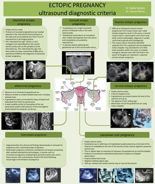
- 1. ECTOPIC PREGNANCY ultrasound diagnostic criteria Dr: Nadia Rahaim Dr: Hanaa Mossa • Visualizations of a single interstitial portion of fallopian tube in the main uterine body. • Gestational sac/products of conception seen mobile and separate from the uterus and completely surrounded by myometrium. • A vascular pedicle adjoining the gestational sac to the unicornuate uterus. • difficult to distinguish ovarian ectopic pregnancies from corpus luteal cysts, tubal ectopic pregnancy stuck to the ovary, a second corpus luteum, ovarian germ cell tumor's and other ovarian pathologies, diagnosis is usually confirmed surgically and histologically. • The corpus luteum should be identified separate from the suspected ovarian pregnancy. Colour Doppler may aid detection of a fetal heart pulsation within the ovary. A complex echogenic adnexal mass with free fluid in the pouch of Douglas can represent a ruptured ovarian ectopic pregnancy. • Empty uterine cavity. • A barrel-shaped cervix. • A gestational sac present below the level of the internal cervical os. • The absence of the ‘sliding sign’. • Blood flow around the gestational sac using Colour Doppler. • Empty uterine cavity. • Gestational sac or solid mass of trophoblast located anteriorly at the level of the internal os embedded at the site of the previous lower uterine segment caesarean section scar. • Thin or absent layer of myometrium between the gestational sac and the bladder. • Evidence of prominent trophoblastic/placental circulation on Doppler examination. • Empty endocervical canal. • Negative (sliding organs) sign. • Yolk sac, embryo and cardiac activity may or may not be present. caesarean scar pregnancy • Absence of an intrauterine gestational sac. • Absence of both an evident dilated tube and a complex adnexal mass. • A gestational cavity surrounded by loops of bowel and separated from them by peritoneum. • A wide mobility similar to fluctuation of the sac, particularly evident with pressure of the transvaginal probe toward the posterior cul-de-sac. • Empty uterine cavity. • Products of conception/gestational sac located laterally in the interstitial (intramural) part of the tube and surrounded by less than 5 mm of myometrium in all imaging planes. • The ‘interstitial line sign’, which is a thin echogenic line extending from the central uterine cavity echo to the periphery of the interstitial sac. The ‘interstitial line sign’ has been shown to have a sensitivity of 80% and a specificity of 98% for the diagnosis of interstitial ectopic pregnancy. • diagnosed when the ultrasound findings demonstrate an intrauterine pregnancy and a coexisting ectopic pregnancy. • Heterotopic pregnancy should be considered in all women presenting after assisted reproductive technologies, in women with an intrauterine pregnancy complaining of persistent pelvic pain and in those women with a persistently raised b-hCG level following miscarriage or termination of pregnancy abdominal pregnancy Ovarian ectopic pregnancy Cornual ectopic pregnancy Interstitial ectopic pregnancy caesarean scar pregnancy cervical ectopic pregnancy heterotopic pregnancy