1. Proc. Natl. Acad. Sci. USA
Vol. 93, pp. 11907–11912, October 1996
Microbiology
A novel clade of protistan parasites near the animal–
fungal divergence
(Dermocystidium͞Ichthyophonus͞Psorospermium͞small-subunit ribosomal RNA͞molecular phylogenetics)
MARK A. RAGAN*†‡, C. LOUISE GOGGIN§¶ʈ, RICHARD J. CAWTHORN§, LAGE CERENIUS**,
ANGELA V. C. JAMIESON††, SUSAN M. PLOURDE§, THOMAS G. RAND††, KENNETH SO¨DERHA¨LL**,
AND ROBIN R. GUTELL*‡‡
*Canadian Institute for Advanced Research, Program in Evolutionary Biology, and †Institute for Marine Biosciences, National Research Council of Canada,
Halifax, NS Canada B3H 3Z1; §Atlantic Veterinary College, University of Prince Edward Island, Charlottetown, PEI Canada C1A 4P3; ¶Department of
Parasitology, University of Queensland, Brisbane 4072, Queensland, Australia; **Department of Physiological Botany, University of Uppsala, S-752 36 Uppsala,
Sweden; ††Department of Biology, Saint Mary’s University, Halifax, NS Canada B3H 3C3; and ‡‡Departments of Molecular, Cell, and Developmental Biology,
and Chemistry and Biochemistry, University of Colorado, Boulder, CO 80309
Communicated by David M. Prescott, University of Colorado, Boulder, CO, July 11, 1996 (received for review September 22, 1995)
ABSTRACT Sequences of nuclear-encoded small-subunit
rRNA genes have been determined for representatives of the
enigmatic genera Dermocystidium, Ichthyophonus, and Pso-
rospermium, protistan parasites of fish and crustaceans. The
small-subunit rRNA genes from these parasites and from the
‘‘rosette agent’’ (also a parasite of fish) together form a novel,
statistically supported clade. Phylogenetic analyses demon-
strate this clade to diverge near the animal–fungal dichotomy,
although more precise resolution is problematic. In the most
parsimonious and maximally likely phylogenetic frameworks
inferred from the most stably aligned sequence regions, the
clade constitutes the most basal branch of the metazoa; but
within a limited range of model parameters, and in some
analyses that incorporate less well-aligned sequence regions,
an alternative topology in which it diverges immediately
before the animal–fungal dichotomy was recovered. Mitochon-
drial cristae of Dermocystidium spp. are flat, whereas those of
Ichthyophonus hoferi appear tubulovesiculate. These results
extend our understanding of the types of organisms from
which metazoa and fungi may have evolved.
Identifying the group of protists from among which the animal
lineage arose stands as one of the oldest and least tractable
problems in zoology (1–3). Recent phylogenetic analyses based
on protein (4) and rDNA (5, 6) sequences have identified the
fungi (Eumycota) as the major group of multicellular organ-
isms most closely related to animals. However, certain pro-
tistan groups appear to have arisen from the animal and fungal
lineages subsequent to the animal–fungal dichotomy. Chytrid-
iomycetes, which some authorities include among the protists
(7), are widely considered to constitute the most basal branch
within the fungal lineage. Choanoflagellates, likewise consid-
ered protists (8, 9), have been proposed on structural and
biochemical grounds to constitute the most basal clade in the
animal lineage (10); this proposal has received support from
some analyses of rDNA sequence data (5), although in other
rDNA trees the choanoflagellates diverge among the radiate
animals (9) or even before green plants (11).
The enigmatic ‘‘rosette agent,’’ an intracellular parasite of
salmonids (12, 13), was recently shown by rDNA sequence
analysis to be closely related to choanoflagellates and, thus, to
animals (14). Investigations in our laboratories have now
revealed that aquatic parasites of the enigmatic genera Der-
mocystidium, Ichthyophonus, and Psorospermium are specifi-
cally related to rosette agent.
More than 20 species of Dermocystidium occur either as cysts
in skin or gill cysts, or as systemic infections in carp, goldfish,
salmonids, eels, newts, and frogs (15). The genus has been a
depository for organisms that have a spherical stage with a
large, refractile-inclusion-bearing vacuole that restricts the
cytoplasm and nucleus to the cell periphery; at least some
species have septate hyphae (15), uniflagellate zoospores (16),
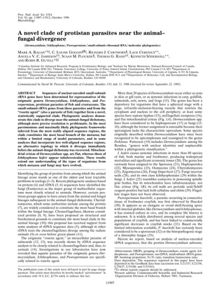
and flat mitochondrial cristae (Fig. 1A). Dermocystidium spp.
have been considered to be haplosporeans (17) or fungi (15,
18), although the former assignment is untenable because their
sporangium lacks the characteristic operculum. Some species
originally described within Dermocystidium have since been
recognized to be apicomplexans and reassigned to the genus
Perkinsus. Patterson (19) includes Dermocystidium among his
Residua, ‘‘genera with unclear identities and unplaceable
within a phylogenetic classification.’’
I. hoferi causes systemic infections in more than 80 species
of fish, both marine and freshwater, producing widespread
mortalities and significant economic losses (20). The genus has
variously been assigned to the Sporozoa (21), Haplosporidea
(22), Myxosporidea (23), Oomycetes (24), Chytridiomycetes
(25), Zygomycetes (26), Fungi Imperfecti (27), Fungi incertae
sedis (28), and its own class Ichthyophonales (29) within the
fungi. I. hoferi (25) resembles eumycota in possessing spindle
pole bodies, but its mitochondria have tubulovesiculate, not
flat, cristae (Fig. 1B); its cell walls are periodic acid͞Schiff
reagent-positive but lack both cellulose and chitin (29). Flagel-
late stages have not been observed.
Psorospermium haeckelii, a parasite occurring in connective
tissue of freshwater crayfish, was first observed by Haeckel
(30). It appears as an elongate or ovoid shell-bearing spore
with internal globules; like Dermocystidium and Ichthyophonus,
it has resisted culture in vivo, and its complete life history is
unknown. It is widely distributed among several species and
populations of crayfish, and has been linked to commercially
significant decreases in crayfish stocks (31). Based on the
limited information available, P. haeckelii has variously been
considered to be a sporozoan (32) or the histopathogenic stage
of a dimorphic fungus (33).
Herein we report, based on analysis of nuclear-encoded
rDNA sequences, that the protists Dermocystidium salmonis,
The publication costs of this article were defrayed in part by page charge
payment. This article must therefore be hereby marked ‘‘advertisement’’ in
accordance with 18 U.S.C. §1734 solely to indicate this fact.
Abbreviations: DRIPs, grouping of Dermocystidium, rosette agent, Ich-
thyophonus, and Psorospermium; ssu-rDNA, small-subunit rRNA genes;
BP, bootstrap proportion; Ts͞Tv ratio, transition͞transversion ratio.
Data deposition: The sequences reported in this paper have been
deposited in the GenBank data base (accession nos. U21337, U21336,
U25637, U33180, and U33181).
‡To whom reprint requests should be addressed.
ʈ
Present address: Commonwealth Scientific and Industrial Research
Organization Division of Fisheries, Hobart 7001, Tasmania.
11907
2. Dermocystidium sp., I. hoferi, and P. haeckelii are specifically
related to rosette agent to the exclusion of choanoflagellates.
This unanticipated and as-yet-unnamed grouping of Dermo-
cystidium, rosette agent, Ichthyophonus, and Psorospermium,
which we provisionally call the DRIPs clade, is part of the
‘‘eukaryotic crown assemblage,’’ and in our best phylogenetic
inferences appears (although without definitive statistical sup-
port) as the deepest branch within the animal kingdom.
MATERIALS AND METHODS
Cysts of D. salmonis were obtained by microsurgery from gills
of the chinook salmon Onchorhynchus tschawytscha (Trask
State Fish Hatchery, OR), and cysts of Dermocystidium spp.
were obtained from gills of the brook trout Salvelinus fontinalis
(Fraser’s Mills Hatchery, St. Andrews, NS Canada). Cysts were
crushed by vigorous grinding in microfuge tubes at Ϫ196ЊC and
incubated with Proteinase K and 4% SDS. I. hoferi was isolated
from liver lesions of the yellowtail flounder Limada ferruginea,
collected from Sable Island Bank and Brown’s Bank, northwest
Atlantic Ocean. Inocula were plated onto Petri dishes con-
taining EFS medium (34), and sporangiophores growing onto
the surface of the medium were excised. P. haeckelii was
isolated, by centrifugation through a Percoll cushion, from
subcarapacial connective tissue of the crayfish Astacus astacus
(35), collected from Lake Aspen, So¨dermanland, Sweden by
Erland Ma¨hlstro¨m. DNA was purified by phenol-chloroform
extraction and ethanol precipitation. Nuclear small-subunit
rRNA genes (ssu-rDNAs) were amplified by PCR and either
sequenced directly (Dermocystidium spp.) or cloned in pAmp1
(GIBCO͞BRL) for sequencing (36, 37).
The rDNAs of D. salmonis, Dermocystidium sp., I. hoferi, and
P. haeckelii were aligned with those of 40 other higher eukaryotes
selected to represent all major eukaryotic ‘‘crown’’ taxa, avoid
ssu-rDNAs highly divergent in branch length or nucleotide com-
position, and minimize (so far as possible) lengths of internal
edges in the inferred trees, and thereby to avoid topological
artifacts that can arise from unequal apparent rates of acceptance
of mutations (38). Alignment was based, to the extent possible, on
conservation of secondary- and higher-order structures as re-
vealed by covariation of nucleotides (39, 40). Every position was
identified as unpaired, involved in a short-range base pair (e.g.,
stem position), or long-range base paired in the folded rRNA
(Fig. 2), and in some analyses these assignments were used either
directly or to aid Hidden Markov Model-based assignment of rate
categories.
Columns of data corresponding to PCR primers (47 nucle-
otide positions) and the most sparsely populated alignment
positions were removed to yield ‘‘full’’ matrix A (44 species ϫ
1983 positions). Three additional matrices were produced and
differed in the degree to which ambiguously aligned positions
FIG. 2. Maskings for phylogenetic analyses of nucleotide positions,
displayed on a folded ssu-rRNA. ●, Most conservative positions, included
in all analyses; ⅐, positions excluded from matrix C; E, positions excluded
from matrices C and E; solid lines, positions excluded from matrices C,
E, and B; ϩ, PCR primers, excluded from all analyses.
FIG. 1. Electron micrographs of mitochondria of (A) schizont of Dermocystidium sp. from brook trout and (B) Ichthyophonus hoferi, showing
morphology of cristae. Widths of the fields shown are 0.65 mm (A) and 2.2 mm (B).
11908 Microbiology: Ragan et al. Proc. Natl. Acad. Sci. USA 93 (1996)
3. were removed (Fig. 2). Removal of the more ambiguously
aligned positions yielded matrix B (44 ϫ 1622 positions);
rigorous removal gave matrix E (44 ϫ 1461 positions); and
deletion of all positions for which a secondary structure could
not be unambiguously assigned yielded the ‘‘most conserva-
tive’’ matrix C (44 ϫ 1370 positions). Third and subsequent gap
positions were coded as unknown characters for parsimony
analyses.
Phylogenetic trees were inferred by maximum likelihood
using FASTDNAML version 1.0.6 (41) and an alpha version of
DNAML from PHYLIP (version 4.0) (42) with random addition
order and global (42-level) optimization. In some analyses,
base paired positions were assigned higher relative rates of
evolution to effect downweightings similar to those sometimes
employed in parsimony analysis (43–45). Parsimony trees were
inferred using DNAPARS in PHYLIP (version 3.53c) (42); mul-
tiple (n ϭ 500 or 1000) iterations were made to increase the
likelihood of finding maximally parsimonious solutions, and a
majority-rule consensus tree was calculated from the equally
parsimonious solutions using CONSENSE (42). Distance matri-
ces corrected for superposed substitutions were calculated
under a generalized Kimura two-parameter model using
DNADIST, and neighbor-joining trees were derived using
NEIGHBOR (42). Analyses were bootstrapped (n ϭ 500 or 1000)
by sequential use of the PHYLIP programs SEQBOOT, DNAPARS
(parsimony) or DNADIST and NEIGHBOR (neighbor-joining),
and CONSENSE (46). Alternative topologies were subjected to
the nonparametric Templeton–Felsenstein test under parsi-
mony (47) and the Kishino–Hasegawa test under likelihood
(48) using DNAPARS (version 3.53c) and DNAML (version 4.0),
respectively. The decay index (49) and tree statistics were
computed using PAUP (version 3.1) (50) on a Macintosh IIci;
most other computation was done on a Sun 10͞61 Unix
workstation.
RESULTS
Single products corresponding to intron-free nuclear rDNAs
were amplified and sequenced from D. salmonis, Dermocys-
tidium sp., I. hoferi, and P. haeckelii (Table 1). Alignment with
ssu-rDNAs of 40 other selected eukaryote nuclear rDNAs was
straightforward, yielding (upon progressive removal of PCR-
primer regions, sparsely populated sites and ambiguously
alignable regions) a series of matrices. Among these, only
matrices C and E are unambiguously aligned (i.e., provide
unambiguous homology statements), the former fully sup-
ported by secondary and higher-order structure, the additional
91 positions of the latter based on extensive primary-sequence
identity.
Maximum-likelihood (Fig. 3), parsimony, and neighbor-
joining analyses revealed an unanticipated grouping of ssu-
rDNAs of the two Dermocystidium spp., I. hoferi and P.
haeckelii, and rosette agent. The consistency index of this
DRIPs clade ranged from 0.893 (full matrix A) to 0.957 (most
conservative matrix C). With the more conservative matrices
C and E, bootstrap proportions (BPs) for integrity of the
DRIPs clade were 60–79% under parsimony and 76–99%
under distance. Simulations indicate that BPs are highly
conservative estimates of accuracy (51, 52); although extrap-
olation to real-life trees remains problematic, here, where
internodal distances are short (51) and the number of species
is large (52), the probability that the DRIPs clade does not
appear in the true tree can be estimated to range from much
less than 15% (for BPs ϭ 60%) to essentially 0 (for BPs Ն
80%). In contrast, the greatest BP for any individual grouping
inconsistent with holophyly of the DRIPs clade [the flagellate
Apusomonas proboscidea (53, 54) grouping with one or more
DRIPs ssu-rDNAs; see below] was 10%, and BPs for alterna-
tive topologies in which one or more DRIPs group specifically
with the two choanoflagellates ranged from 10% (matrix C,
parsimony) to 0.9% (matrix E, parsimony); in simulations, BPs
of 10% correspond to near-zero probabilities (51, 52) and are
similar to the support observed for polyphyly of choanoflagel-
lates, i.e., background noise. The decay index (49) for the
DRIPs clade is Ͼ6 steps in heuristic analysis of matrix C,
likewise indicating good support. Within the DRIPs clade, the
two Dermocystidium spp. group stably together (BP ϭ 100%),
as do I. hoferi and P. haeckelii (BP ϭ 92–99%), and the two
Dermocystidium spp. with rosette (BP ϭ 81–100%).
Phylogenetic analysis in this region of the eukaryotic ssu-
rDNA tree is complicated by the volatility of ssu-rDNAs of
Cyanophora paradoxa, Acanthamoeba spp., and especially A.
proboscidea. These sequences have a tendency to migrate (with
poor bootstrap support) within the tree in response to minor
changes in model parameters, choice of alignment regions, or
presence of attractor sequences. In parsimony and maximum-
likelihood analyses of the two least stably aligned matrices, A
and B, A. proboscidea tends to intrude into the DRIPs clade to
group with Dermocystidium spp. The attraction between A.
proboscidea and Dermocystidium ssu-rDNAs involves transver-
sions as well as transitions, and does not appear to be purely
a compositional (GϩC) effect (data not shown). As analyses
of matrix A yielded conflicting, often biologically unreason-
able trees with low BPs even for well-established clades (i.e.,
21% for metazoa), we interpret the appearance of A. pro-
boscidea within the DRIPs clade (above) as an artifact of, or
exacerbated by, the alignment of nonhomologous or highly
diverged sites in regions that show no common secondary-
structural helices or obvious primary-structural similarity
among these ssu-rRNAs.
All analyses of all matrices (except some parsimony analyses
of the least conservative matrices, A and B) showed the DRIPs
clade (sometimes with A. proboscidea) to diverge either im-
mediately after or immediately basal to the animal–fungal
dichotomy. Maximum-likelihood and parsimony analyses of
the best-supported matrices, C and E, resolved the DRIPs
clade as the deepest branch within the animal lineage (Figs. 3
and 4, tree I). With matrix C, tree I was recovered under
maximum likelihood at all eight investigated Ts͞Tv ratios
between 0.50 and 1.70 and all seven between 2.05 and 10.00,
but tree II (Fig. 4), in which the DRIPs clade is the sister group
to animalsϩfungi, was most likely when the Ts͞Tv ratio was
between 1.75 and 2.00. Tree I, however, showed the greatest
overall likelihood (Ϫ16846.07632 at a Ts͞Tv ratio of 1.50),
more than 40 log-likelihood units greater than the best value
(Ϫ16886.90976 at a Ts͞Tv ratio of 1.75) observed for tree II.
BPs for positioning the DRIPs clade as the deepest branch
within animals, however, were only 37–53%.
The robustness of this result was examined in extensive
maximum-likelihood analyses of matrices C and E. In some
series, unpaired, stem, and long-range-paired positions were
assigned different relative rates of change (1, 0.5–5.0, and
0.6–3.0, respectively), hence, in effect, different weights; in
other series, positions were assigned to rate categories (values
as above, sometimes with a fourth, zero-rate category for
unvarying positions) using a Hidden Markov Model (55). The
Table 1. Nuclear-encoded ssu-rDNAs of D. salmonis,
Dermocystidium spp. from brook trout, I. hoferi, and P. haeckelii
Organism Length*
GϩC,
%
GenBank
accession no.
D. salmonis 1780 43.8 U21337
Dermocystidium sp. 1821 43.6 U21336
I. hoferi 1808 43.8 U25637
P. haeckelii† 1792 44.1 U33180
*Length in nucleotides of PCR-amplified ssu-rDNAs, including PCR
primer regions.
†The ssu-rDNA sequence of the crayfish host, Astacus astacus, has
been deposited in the GenBank data base (accession no. U33181).
Microbiology: Ragan et al. Proc. Natl. Acad. Sci. USA 93 (1996) 11909
4. greatest overall likelihood was always associated with tree I,
although in each series, tree II was locally more likely within
a narrow (although variable) range of Ts͞Tv ratios. Thus, these
two topologies (tree I and, less favorably, tree II) are robustly
inferred from the most stably aligned nucleotide positions
under a wide range of biologically reasonable models.
To examine further the relative support for these two (and
other possible) topologies, alternatives were tested by the
method of Templeton and Felsenstein under parsimony, and
by the method of Kishino and Hasegawa under maximum
likelihood (Fig. 4). Under these nonparametric tests, alterna-
tive topologies are rejectable at 95% confidence if they require
Ͼ1.96 SD more steps than the most parsimonious tree, or are
Ͼ1.96 SD less likely than the most likely tree; the correspond-
ing value for 90% confidence is Ͼ1.645 SD. Groups stable in
the above-mentioned analyses were maintained intact, but
their relative branching positions were permuted, exhaustively
for the DRIPs clade and nearby lineages, for more remote
FIG. 3. Maximum-likelihood tree inferred from ssu-rDNAs of Dermocystidium sp., D. salmonis, I. hoferi, P. haeckelii, and 40 other eukaryotes,
based on matrix C (1370 positions) at a transition͞transversion (Ts͞Tv) ratio of 1.50. In the topologically identical, most parsimonious tree inferred
from matrix C, L ϭ 2838 steps, ci ϭ 0.400, rc ϭ 0.222, and ri ϭ 0.555; 521 positions are ‘‘informative.’’ The upper and lower numbers give the
bootstrap support (percent of 500 replicates) in parsimony and neighbor-joining analyses, respectively, of matrices C and E. GenBank accession
numbers, from top to bottom: animals: X04025, D14358, U29235, X70210, L24489, X79878, X79872, X01723, X13457, L10829, L10826, D15068,
L10825, L10823, and L10824; DRIPs: U33180, U25637, L29455, U21336, and U21337; fungi: M59761, M59759, M59758, M60300, X62396, and
X54863; protists: L37037, U07411, U07413, and X68483; chlorophytes: M20017 and X74002; rhodophytes: Z14142 and L26177; heterokonts:
X54266, M87329, M87336, M55286, L27634, and U21338; apicomplexa: Z15106, M64244, L07375, and X75762.
11910 Microbiology: Ragan et al. Proc. Natl. Acad. Sci. USA 93 (1996)
5. lineages only if initial tests showed a significant influence upon
the position or length of the edge joining the DRIPs ssu-
rDNAs to the rest of the tree. Of the hundreds of alternative
topologies screened, 59 were selected in this way for more
rigorous analysis.
With the most conservative matrix, matrix C, variances were
relatively large for both tests, and about 40% of these alter-
native topologies could not be ruled out. Most of the accept-
able alternatives involved rearrangements among lower inver-
tebrates (Acanthocoepsis unguiculata, Diaphanoeca grandis,
Microciona prolifera, Mnemiopsis leidyi, and Beroe¨ cucumis),
and͞or migrations of the three volatile lineages mentioned
above; but in a minority, the DRIPs clade was repositioned as
the deepest clade within fungi, or immediately basal to the
animal ϩ fungal clade. Tests on matrix E were much more
discriminatory; all but three alternatives to tree I could be
rejected at 95% under parsimony, all but two at 90% under
likelihood, and none was acceptable under both tests. With
matrix B, all alternatives to tree I (but with A. proboscidea
joining the DRIPs clade as a sister group to Dermocystidium
spp.) were rejected at 95% under both tests.
DISCUSSION
The analyses presented herein clearly indicate the existence of
an unanticipated and as-yet-unnamed clade of eukaryotic
protists, all recognized members of which are parasites of
aquatic animals, including fish (Dermocystidium, Ichthyopho-
nus, rosette), amphibians (Dermocystidium), and crustaceans
(Psorospermium). No complete life-history is known for any of
these organisms, and none has been successfully cultured in
vivo; thus, it is premature to propose a concept, archetype, or
bauplan for this clade, or to speculate upon its likely phyletic
delimitation. We provisionally refer to this group as the DRIPs
clade after its presently known members.
Based on known life-history forms, Dermocystidium (15, 18),
Ichthyophonus (25–29), Psorospermium (33), and the rosette
agent (12) have all been proposed to have fungal affinities,
although without satisfactory assignment to any existing fungal
taxon. ‘‘Remarkable’’ similarities in gross pathology, intracellular
location, and ultrastructure between the rosette agent and sys-
temic Dermocystidium infections have already been noted (56).
Moreover, the encysted resting stages of P. haeckelii and Ichthyo-
phonus appear to be very similar in gross morphology, and an
amoeboid stage in P. haeckelii (57) may correspond to the
amoeboid (infective?) stage of Ichthyophonus.
Our analyses indicate that the DRIPs organisms diverged
near the animal–fungal dichotomy, although the precise posi-
tion could not be conclusively resolved. In the most likely and
most parsimonious trees inferred from the most stably aligned
ssu-rDNA regions, the DRIPs clade diverges subsequent to the
animal–fungal dichotomy, as the most basal branch within the
animal lineage. In their initial description of the ssu-rDNA
sequence of rosette agent, Kerk et al. (14) reported that
ssu-rDNAs of rosette agent and choanoflagellates form a
monophyletic group with a BP of 81–94%. As described above,
addition of ssu-rDNAs from Dermocystidium, Ichthyophonus,
and Psorospermium dissolves this association and resolves
choanoflagellates on the second-deepest branch within the
animal lineage, thereby further strengthening molecular sup-
port for the hypothesis that metazoa arose from a flagellated
protozoan (58) similar to modern choanoflagellates (5, 10).
As detailed above, one alternative hypothesis for the posi-
tion of the DRIPs ssu-rDNAs within the eukaryote tree
cannot, based on our data, be ruled out statistically, although
it is less likely and less parsimonious; in this hypothesis, the
origin of the DRIPs clade is immediately basal to the animal–
fungal dichotomy (tree II). This alternative is compatible with
the ssu-rDNA data and, like the most parsimonious and most
likely tree I, is consistent with monophyly of animals and of
true fungi. The relative strengths of these two inferences
depend to some degree on which ssu-rDNAs are included in
the analysis; for example, removal of sequences diverging
immediately basal to the animal–fungal dichotomy (Acanth-
amoeba spp., A. proboscidea, and C. paradoxa) diminishes the
acceptability of tree II, whereas removal of choanoflagellate
and sponge ssu-rDNAs resolves the DRIPs clade onto the
fungal branch (results not shown). We have minimized arbi-
trariness by including representatives of all major eukaryotic
crown groups meeting the branch-length and compositional
criteria described above, retaining even volatile lineages such
as A. proboscidea. However, ssu-rDNA sequence comparisons
(5, 6, 9, 11, 14) have not yet converged on a stable phylogeny
of the crown groups, and the relationships derived herein are
unlikely to constitute the final word on the origin of animals.
Phylogenetic analyses of protein sequences and comparison of
intron positions in protein-coding genes (4, 5) should help
select among these competing hypotheses.
Like almost all animals and eumycota, the Dermocystidium
sp. from brook trout has flattened mitochondrial cristae.
However, the mitochondrial cristae of I. hoferi appear tubu-
lovesiculate under a variety of fixation and embedding proto-
cols (Fig. 1). For technical reasons (e.g., thick spore walls),
cristal morphology is unknown in rosette (D. Kerk, personal
communication), D. salmonis (R. E. Olson, personal commu-
nication), and P. haeckelii (unpublished data). If (as in our best
inferences) the DRIPs clade diverged after the animal–fungal
dichotomy, the tubulovesiculate appearance of cristae in I.
FIG. 4. Alternative relationships among the DRIPs clade and other eukaryote lineages not rejectable under the Templeton–Felsenstein and
Kishino–Hasegawa tests. Tree I, most likely and most parsimonious topology based on matrices C and E (from Fig. 3); tree II, alternative topology
inferred from matrix C when the Ts͞Tv ratio was 1.75–2.00. Dashed line to Apusomonas ssu-rDNA denotes instability.
Microbiology: Ragan et al. Proc. Natl. Acad. Sci. USA 93 (1996) 11911
6. hoferi must, like the tubular cristae of mesozoa, represent a
reversion from the flat form typical of animals and true fungi.
Shape of mitochondrial cristae is in general an excellent guide
to phylogenetic position (19), but exceptions are known (59,
60).
It was recently suggested (61) that animal phyla do not
contain protist species; the almost simultaneous discovery,
based on analysis of ssu-rDNAs, that myxozoa are closely
related to bilateral animals (11) does not provide a firm
counterexample, as the classification of myxozoa among pro-
tists had long been controversial. The results presented herein
strongly suggest that the two earliest branches within the
animal kingdom include protists, as do early branches within
the other two crown kingdoms (62), Fungi and Plantae. The
appearance of an additional group of protists near the point of
divergence of the animal and fungal lineages (4–6) casts new
light on the nature of the ancestral animal, points to the need
for additional study of these enigmatic organisms, and reminds
us again that protist refers at best to a grade, not a clade, of
eukaryotic organisms.
We thank R. E. Olson for the D. salmonis cysts, D. Kerk for access
to ref. 14 before publication, T. Cavalier-Smith for communicating the
sequence of A. proboscidea ssu-rDNA before publication and for
helpful discussions, D. R. Stothard for advice on selection of Acanth-
amoeba ssu-rDNAs, and J. M. Munholland and C. A. Murphy for
expert technical assistance. C.L.G., T.G.R., and R.J.C. thank the
Natural Sciences and Engineering Research Council (Canada). L.C.
and K.S. thank the Swedish Natural Science Research Council and the
Swedish Research Council for Forestry and Agricultural Research.
R.R.G. acknowledges support from National Institutes of Health
Grant GM 48207 and the W. M. Keck Foundation. Issued as publi-
cation no. NRCC 39713.
1. Hyman, L. H. (1940) The Invertebrates (McGraw–Hill, New
York), Vol. 1, pp. 248–253.
2. Cue´not, L. (1952) in Traite´ de Zoologie, ed. Grasse´, P.-P. (Masson,
Paris), Tome I, Fasc. I, pp. 1–33.
3. Brusca, R. C. & Brusca, G. J. (1990) Invertebrates (Sinauer,
Sunderland, MA), pp. 112–116.
4. Baldauf, S. L. & Palmer, J. D. (1993) Proc. Natl. Acad. Sci. USA
90, 11558–11562.
5. Wainright, P. O., Hinkle, G., Sogin, M. L. & Stickel, S. K. (1993)
Science 260, 340–342.
6. Ragan, M. A. & Gutell, R. R. (1995) Bot. J. Linn. Soc. 118,
81–105.
7. Margulis, L., Corliss, J. O., Melkonian, M. & Chapman, D. J.,
eds. (1990) Handbook of Protoctista (Jones & Bartlett, Boston).
8. Leadbeater, B. S. C. (1977) J. Mar. Biol. Assoc. U.K. 57, 285–301.
9. Cavalier-Smith, T. (1993) Microbiol. Rev. 57, 953–994.
10. Cavalier-Smith, T. (1987) in Evolutionary Biology of the Fungi,
eds. Rayner, A. D. M., Brasier, C. M. & Moore, D. (Cambridge
Univ. Press, Cambridge, U.K.), pp. 339–353.
11. Smothers, J. F., von Dohlen, C. D., Smith, L. H., Jr., & Spall,
R. D. (1994) Science 265, 1719–1721.
12. Harrell, L. W., Elston, R. A., Scott, T. M. & Wilkinson, M. T.
(1986) Aquaculture 55, 249–262.
13. Elston, R. A., Harrell, L. & Wilkinson, M. T. (1986) Aquaculture
56, 1–21.
14. Kerk, D., Gee, A., Standish, M., Wainright, P. O., Drum, A. S.,
Elston, R. A. & Sogin, M. L. (1995) Mar. Biol. 122, 187–192.
15. Dykova´, I. & Lom, J. (1992) J. Appl. Ichthyol. 8, 180–185.
16. Olson, R. E., Dungan, C. F. & Holt, R. A. (1991) Dis. Aquat. Org.
12, 41–48.
17. Caullery, M. (1953) in Traite´ de Zoologie, ed. Grasse´, P.-P.
(Masson, Paris), Tome I, Fasc. II, pp. 922–934.
18. Allen, R. L., Meekin, T. K., Pauley, G. B. & Fujihara, M. P.
(1968) J. Fish. Res. Board Can. 25, 2467–2475.
19. Patterson, D. J. (1994) in Progress in Protozoology, eds. Haus-
mann, K. & Hu¨lsmann, N. (Fischer, Stuttgart), pp. 1–14.
20. Sindermann, C. J. (1970) Principal Diseases of Marine Fish and
Shellfish (Academic, New York), pp. 22–25.
21. Hofer, B. (1893) Allg. Fischwirtschaftsztg. 18, 168–171.
22. Caullery, M. & Mesnil, F. (1905) Arch. Zool. Exp. Ge´n. Ser. 4 4,
101–180.
23. Cox, P. (1916) Contrib. Can. Biol. Fish. 1914–1915, 81–85.
24. Sproston, N. G. (1944) J. Mar. Biol. Assoc. U.K. 26, 72–98.
25. Plehn, M. & Mulsow, K. (1911) Zentralbl. Bakteriol. Parasitenkd.
Orig. 59, 63–68.
26. Johnstone, J. (1913) Proc. Trans. Liverpool Biol. Soc. 27, 196–218.
27. Chauvier, G. (1979) Ann. Parasitol. (Paris) 54, 105–111.
28. Alderman, D. J. (1982) in Microbial Diseases of Fish, ed. Roberts,
R. J. (Academic, London), pp. 189–242.
29. Rand, T. G. (1994) Dis. Aquat. Org. 18, 21–28.
30. Haeckel, E. (1857) Arch. Anat. Physiol. Med. (Mu¨ller’s Arch.) 24,
469–568.
31. So¨derha¨ll, K. & Cerenius, L. (1992) Annu. Rev. Fish Dis. 2, 3–23.
32. Alderman, D. J. & Polglase, J. L. (1988) in Freshwater Crayfish
Biology, Management and Exploitation, eds. Holdich, D. M. &
Lowery, R. S. (Croom Helm, London), pp. 167–212.
33. Nylund, V., Westman, K. & Lounatmaa, K. (1983) Freshwater
Crayfish Pap. Int. Symp. 5, 307–314.
34. Rand, T. G. & Cone, D. (1990) J. Wildl. Dis. 26, 323–328.
35. Cerenius, L., Henttonen, P., Lindqvist O. V. & So¨derha¨ll, K.
(1991) Aquaculture 99, 225–233.
36. Sogin, M. L. (1990) in PCR Protocols, eds. Innis, M. A., Gelfand,
D. H., Sninsky, J. J. & White, T. J. (Academic, San Diego), pp.
307–314.
37. Bird, C. J., Rice, E. L., Murphy, C. A. & Ragan, M. A. (1992)
Phycologia 31, 510–522.
38. Hendy, M. D. & Penny, D. (1989) Syst. Zool. 38, 297–309.
39. Gutell, R. R. (1993) in The Translational Apparatus, eds. Nier-
haus, K. H., Franchschi, F., Subramanian, A. R. & Erdmann,
V. A. (Plenum, New York), pp. 477–488.
40. Gutell, R. R., Larsen, N. & Woese, C. R. (1994) Microbiol. Rev.
58, 10–26.
41. Olsen, G. J., Matsuda, H., Hagstrom, R. & Overbeek, R. (1994)
Comput. Appl. Biosci. 19, 41–48.
42. Felsenstein, J. (1989) Cladistics 5, 164–166.
43. Wheeler, W. C. & Honeycutt, R. L. (1988) Mol. Biol. Evol. 5,
90–96.
44. Dixon, M. T. & Hillis, D. M. (1993) Mol. Biol. Evol. 10, 256–267.
45. Bakker, F. T., Olsen, J. L., Stam, W. T. & van den Hoek, C.
(1994) Mol. Phylogenet. Evol. 3, 365–382.
46. Felsenstein, J. (1985) Evolution 39, 783–791.
47. Templeton, A. R. (1983) Evolution 37, 221–244.
48. Kishino, H. & Hasegawa, M. (1989) J. Mol. Evol. 29, 170–179.
49. Donoghue, M. J., Olmstead, R. G., Smith, J. F. & Palmer, J. D.
(1992) Ann. Mo. Bot. Gard. 79, 333–345.
50. Swofford, D. L. (1993) PAUP, Phylogenetic Analysis Using Par-
simony (Illinois Natural History Survey, Champaign, IL), Ver-
sion 3.1.
51. Hillis, D. M. & Bull, J. J. (1993) Syst. Biol. 42, 182–192.
52. Sitnikova, T., Rzhetsky, A. & Nei, M. (1995) Mol. Biol. Evol. 12,
319–333.
53. Vickerman, K., Darbyshire, J. F. & Ogden, C. G. (1974) Arch.
Protistenkd. 116, 254–269.
54. Cavalier-Smith, T. & Chao, E. E. (1995) Proc. R. Soc. London B
261, 1–6.
55. Felsenstein, J. & Churchill, G. A. (1996) Mol. Biol. Evol. 13,
93–104.
56. Hedrick, R. P., Friedman, C. S. & Modin, J. (1989) Dis. Aquat.
Org. 7, 171–177.
57. Grabda, E. (1934) Me´m. Acad. Pol. Sci. Cracovie B 6, 123–142.
58. Haeckel, E. (1874) Jena. Z. Naturwiss. 8, 1–55.
59. Patterson, D. J. (1990) J. Mar. Biol. Assoc. U.K. 70, 381–393.
60. O’Kelly, C. J. (1993) J. Eukaryotic Microbiol. 40, 627–636.
61. Corliss, J. O. (1994) Acta Protozool. 33, 1–51.
62. Knoll, A. H. (1992) Science 256, 622–627.
11912 Microbiology: Ragan et al. Proc. Natl. Acad. Sci. USA 93 (1996)