

Recommended
More Related Content
What's hot
Similar to Iontophoresis perimed
Similar to Iontophoresis perimed (20)
Recently uploaded
Recently uploaded (20)
Iontophoresis perimed
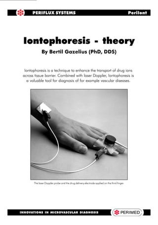
- 1. Iontophoresis - Theory 1 Iontophoresis is a technique to enhance the transport of drug ions across tissue barrier. Combined with laser Doppler, Iontophoresis is a valuable tool for diagnosis of for example vascular diseases. Iontophoresis - theory By Bertil Gazelius (PhD, DDS) PERIFLUX SYSTEMS PeriIont INNOVATIONS IN MICROVASCULAR DIAGNOSIS The laser Doppler probe and the drug delivery electrode applied on the third finger.
- 2. 2 Iontophoresis - Theory Introduction The method of iontophoresis was first described by Pivati in 1747. Galvani and Volta, two well- known scientists working in the 18th century, combined the knowledge that electricity can move different metal ions, and that movements of ions produce electricity. The method of administering pharmacological drugs by iontophoresis became popular at the beginning of the 20th century due to the work of Leduc (1900) who introduced the term iontotherapy and formulated the laws for this process. Iontophoresis is defined as the introduction, by means of a direct electrical current, of ions of solu- ble salts into the tissues of the body for therapeutic purposes (Singh and Maibach, 1994). It is a technique used to enhance the absorption of drugs across biological tissues, such as the skin. Another method for drug delivery through the skin, called phonophoresis, uses ultrasound instead of an electric current. Both of these techniques are complicated because of other processes that occur simultaneously with the delivery of the drug. With the present knowledge about these proc- esses, it is easier to select and prepare appropriate drugs and vehicles for iontophoresis than for phono- phoresis. In clinical practice, iontophoresis devices are used primarily for the treatment of inflammatory conditions in skin, muscles, tendons and joints, such as in temporomandibular joint dysfunctions. More recently, iontophoresis has been used in combination with laser Doppler technology as a diagnostic tool in diseases compromising the vascular bed.
- 3. Iontophoresis - Theory 3 Factors affecting iontophoretic transport Many factors have been shown to affect the results of iontophoresis. These include the physio- chemical properties of the compound (molecular size, charge, concentration), drug formulation (type of vehicle, buffer, pH, viscosity, presence of other ions), equipment used (available current range, constant vs. pulsed current, type of electrode), biological variations (skin site, regional blood flow, age, sex), skin temperature and duration of iontophoresis. Iontophoresis has mainly been used for therapeutic purposes, but in combination with the laser Doppler technique, it is possible to study the influence of the drugs used on the vascular bed. Until now, the combination of LDPM (Laser Doppler Perfusion Monitoring) and iontophoresis has been used mostly as a diagnostic tool for diseases affecting macro- and microcirculation and the control- ling regulatory nerves. When using iontophoresis as a diagnostic instrument, the following factors have to be considered. A) Influence of pH The pH is of importance for the iontophoretic delivery of drugs. The optimum is a compound that exists predominantly in an ionised form. When the pH decreases, the concentration of hydrogen Principle of iontophoresis By definition, iontophoresis is the increased movement of ions in an applied electric field. Ionto- phoresis is based on the general principle that like charges repel each other, and unlike charges attract each other. An external energy source can be used to increase the rate of penetration of drugs through the membrane. When a negatively charged drug is to be delivered across an epithelial barrier, it is placed under the negatively charged delivery electrode (cathode) from which it is repelled, to be attracted towards the positive electrode placed elsewhere on the body. In anodal iontophoresis (positively charged ions), the electrode orientation is reversed (see fig. 1). The choice of drug is of importance, depending on whether the compound is unionised or ionised. Non-ionised compounds are generally better absorbed through the skin than ionised substances. The penetration across the skin or other epithelial surfaces is usually slow due to their excellent barrier properties. Many drug candidates for local applications only exist in an ionised form, which makes effective membrane penetration impossible. Fig. 1. Electrode system applied to the skin surface. a) Negatively charged dispersive elec- trode (PF 384) placed at a minimum distance of 15 cm from b. b) Probe (PF 481), with an attached drug delivery electrode (PF 383) containing positively charged ions. c) By applying a positive current to the delivery electrode, the positively charged drug ions are repelled from the electrode and forced through the stratum corneum.
- 4. 4 Iontophoresis - Theory ions increases, and a vascular reaction (vasodilatation) is initiated because of C-fibre activation (fig. 2). Thus, it is important to keep the pH as close as possible to 7, at least when working with vasodilators. At pH 5.5 and below, there is an increasing risk for vascular reactions due to the high concentration of hydrogen ions rather than the compound used. Since hydronium ions are small, they penetrate the skin more easily than larger drug ions. B) Current Strength There is a linear relationship between the observed flux of a number of compounds and the applied current. With the present electrode area of 1 cm2 (PF 383), the current is limited to 1 mA due to patient comfort considerations. This current should not be applied for more than three minutes because of local skin irritation and burns. With increasing current, the risk of non-specific vascular reactions (vasodilatations) increases. At a current of 0.4 to 0.5 mA/cm2 , such a vascular reaction is initiated after a few seconds of iontophoresis with deionised or tap water. This latter effect is prob- ably due to the current density being high enough within a small area to stimulate the sensory nerve endings, causing reactions such as the release of Substance P from C-fibre terminals (fig. 2). C) Ionic Competition In a solution of sodium chloride, there is an equal quantity of negative (Cl- ) and positive (Na+ ) ions. Migration of a sodium ion requires that an ion of the opposite charge is in close vicinity. The latter ion of opposite charge is referred to as a counter-ion. An ion of equal charge but of a different type is referred to as a co-ion. When using iontophoresis, it is important to know that pH adjustment is performed by adding buffering agents. The use of buffering agents adds co-ions which are usually smaller and more mobile than the ion to be delivered. This results in a reduction of the number of drug ions to be delivered through the tissue barrier by the applied current. In our example, this means that when a POLYMODAL NOCICEPTIVE C-AFFERENTS LOW pH X<5.5 Fig. 2. When using drugs at low pH (5.5), the concentration of hydrogen ions is sufficient to activate the polymodal nociceptive C-fibres. The result is a local axon-reflex mediated vasodilatation beneath the delivery electrode which is not related to the drug per se. SP=Substance P CGRP=Calcitonin Gene Related Peptide MC=Mast Cells Hi= Histamine 5-HT=Serotonins
- 5. Iontophoresis - Theory 5 positively charged drug is diluted in saline, the sodium ions will compete with the amount of drug ions to be delivered. Ideally, the use of a buffer system should be avoided in iontophoresis, but if this is not possible, alternative buffers consisting of ions with low mobility or conductivity are pre- ferred. D) Drug Concentration Depending on the drug used, the steady-state flux (ion movement) has been shown to increase with increasing concentration of the solute in the donor compartment, i.e. in the delivery electrode. A limiting factor to be considered is the strength of the current used. At higher drug concentrations, the transport may become independent of concentration, probably because of the saturation of the boundary layer relative to the donor bulk solution (Phipps et al, 1989). E) Molecular Size It has been shown that the permeability coefficients in positively charged, negatively charged and uncharged solutes across excised human skin are a function of molecular size. When the molecular size increases, the permeability coefficient decreases (Yoshida et al., 1993). However, there are certain solutes with a relatively high molecular size (e.g. insulin, vasopressin and several growth hormones), which have also been shown to penetrate the skin barrier into the systemic circulation. F) Convective or Electro-osmotic Transport When performing iontophoresis with a specific current, the flow of ions across the membrane induces a flow of solvent called electro-osmosis. Compared to the ion transport, the electro-osmotic contribution is small. The penetration of uncharged substances (e.g. bovine serum albumin) has been shown to be facilitated by the volume flow effect induced by an applied potential difference across the membrane. Iontophoresis has also been observed to enhance the penetration of a number of dipolar ions (zwitterionic substances, such as phenylalanine). Most of these substances have been shown to be delivered in significantly higher amounts by anodic delivery than by ca- thodic delivery. In general, iontophoresis is more effective for charged compounds, especially monovalent ions. G) Current - Continuous vs. Pulsed mode Application of a continuous current over a long period of time can modulate iontophoretic delivery. Continuous DC current may result in skin polarisation, which can reduce the efficiency of ionto- phoretic delivery in proportion to the length of current application. This polarisation can be over- come by using pulsed DC, a direct current that is delivered periodically. During the “off time”, the skin becomes depolarised and returns to its initial unpolarised status. The enhanced skin depolari- sation using pulsed DC can, however, decrease the efficiency of pulsed transport if the frequency is too high (Bagniefski and Burnette, 1990). Enhanced iontophoretic transport has been reported for peptides and proteins by using pulsed DC compared to conventional DC (Chien et al, 1989). Most of the drug ions used for diagnostic purposes in combination with iontophoresis and LDPM are small in size. As a result, the time needed for an effect is relatively short (5-120 sec), compared to when iontophoresis is used for therapeutic purposes (20-40 min). H) Physiological Factors Iontophoresis reduces intra- and inter-subject variability in the delivery rate. This is an inherent disadvantage with the passive absorption technique. Experiments in vivo and in vitro give support for clinical findings that there are small differences in the flux rate following transdermal ionto- phoresis between males and females, as well as between hairy and hairless skin. The status of the vascular bed is also important; for instance, a pre-constricted vascular bed decreases the drug flux through the skin while a dilated vascular bed increases the yield of the drug through the skin.
- 6. 6 Iontophoresis - Theory Optimising Iontophoretic Transport 1. Iontophoretic transport can be regulated by varying the applied current density and area of application. A current density that is too high may be unpleasant for the patient. If possible, avoid using current settings that result in more than 500 mA/cm2 . At high current densities, there is a significant risk for unspecific electrically mediated vasodilatation that is not drug related. 2. The pH of the formulation should be optimised to ensure maximum ionisation of the com- pound. To prevent pH drifts during the iontophoresis, the choice of electrodes is of impor- tance. With a correct electrode material, decreased solubility and precipitation of the com- pound are avoided. 3. Before iontophoresis is carried out, carefully clean the skin area to be used with deionised water or preferably 70 per cent alcohol. Cleaning will decrease the current needed and mini- mise the risk for local spots of high current density, which could result in C-fibre activation, vasodilatation and local micro-burns. Disadvantages of Iontophoresis Major side-effects are very rare when using iontophoresis as a diagnostic tool. However, minor reactions such as itching, erythema and general irritation of the iontophoretic skin surface are common. There is an increased risk of minor reactions if the exposure time and/or current are increased, and with some drugs like histamine, capsaicin and acetylcholine. Some drugs induce long-lasting skin pigmentation after iontophoretic application, where the intensity of skin discolora- tion is proportional to the exposure time. The current density across the pores in the skin may be higher than the current per unit area applied, depending on the density of pores in a given area. These spots of high current density increase the possibility of current-induced skin damage. Burnette and Ongpipattanakul (1988) showed that the skin resistance was always less than the initial value when a current of 0.16 mA was applied for 10 minutes. This may result in a permanent skin damage. This phenomenon may explain the sudden vascular response with iontophoresis of deionised water, which seems not to be related to the dose. Under the microscope, small spots of skin damage within the pore area could be recognised. The vasodilatation initiated in this way may be caused by activation of nociceptive fibres terminating in the epidermis, which initiate an axon- reflex mediated vascular response. There are isolated reports of contact sensitisation to drugs (ketoprofen), components of electrodes and electrode gels (propylene glycol), serum albumin (in vehicles) and nickel (Zugerman, 1982 and Fisher, 1978). Contraindications for Iontophoresis Contraindications for iontophoresis are important in patients with higher susceptibility to applied currents. Such patients include those carrying electrically-sensitive implanted devices such as cardiac pacemakers, those who are hypersensitive to the drug to be applied, or those with broken or damaged skin surfaces.
- 7. Iontophoresis - Theory 7 References • Bagniefski T. and Burnette R.R. A comparison of pulsed and continuous current iontophoresis. J Cont Rel 1990;11:113. • Burnette R.R. and Ongpipattanakul B. Characterization of the pore transport properties and tissue alteration of excised human skin during iontophoresis. J Pharm Sci 1988;77:132. • Chien Y.W., Siddiqui O., Shi W., Lelawongs P. and Liu J.C. Direct current iontophoretic transdermal delivery of peptide and protein drugs. J Pharm Sci 1989;78:377. • Fisher A. A. Dermatitis associated with transcuteneous electrical nerve stimulation. Cutis 1978;21:24. • Leduc S. Introduction of medicinal substances into the depth in tissues by electric current. Ann d’electrobiol 1900;3:545. • Phipps J.B., Padmanabhan R.V. and Lattin G.A. Iontophoretic delivery of model inorganic and drug ions. J Pharm Sci 1989;78:365. • Singh P. and Maibach H.I. Iontophoresis in drug delivery: Basic principles and applications. Crit Rev Ther Drug Car Sys 1994;11:161. • Zugerman C. Dermatitis from transcutaneous electric nerve stimulation. J Am Acad Dermatol 1982;6:936. • NOTE: References to papers using the combination of Iontophoresis and laser Doppler can be found in the Perimed Literature Reference List (available on our website at: http://www.perimed.se)
- 8. 8 Iontophoresis - Theory Part.no.4.400.612.799.01.12 We have distributors in many countries, please contact Perimed AB for more information. HEADQUARTERS Perimed AB ˜ Box 564 ˜ SE-175 26 Järfälla, Stockholm ˜ Sweden Tel: +46-8-580 119 90 ˜ Fax: +46-8-580 100 28 E-mail: mail@perimed.se ˜ Website: http://www.perimed.se UNITED KINGDOM Perimed UK Ltd 15 Sheerwater Close Bury St. Edmunds SUFFOLK IP32 7HR UK Phone:+44-1284-700110 Fax: +44-1284-844330 E-mail: pbasham@perimeduk.softnet.co.uk CHINA (People´s Republic) Perimed China Ltd 10/F. Ya-Fei Mansion 13 Xiao-Ying Road Chao Yang District BEIJING 100101 PR China Phone:+86-10-64961700 +86-10-64975725 Fax: +86-10-64975726 ITALY Perimed Italia Srl V Privata dei Fiori 22 I-20012 CUGGIONO (MI) Italy Phone:+39-2-972 40534 Fax: +39-2-9746925 E-mail: moneta@est.it FRANCE Perimed France SARL 557 Avenue Pierre Auguste Roiret Z.A Les Tourrais F-69290 CRAPONNE France Phone:+33-47-8578200 Fax: +33-47-8578202 E-mail: perimed@imaginet.fr USA Perimed, Inc 4873 Princeton Dr. North Royalton OH 44133 USA Phone: +1-440-877 0537 Fax: +1-440-877 0534 E-mail: perimed@aol.com INNOVATIONS IN MICROVASCULAR DIAGNOSIS