Dr screening training for nurses interpretation of retinal images for dr screening
•
0 likes•21 views

Diabetic retinopathy-screening training program for nurses St John Eye Hospital - UNRWA

Recommended
Recommended
More Related Content
Similar to Dr screening training for nurses interpretation of retinal images for dr screening
Similar to Dr screening training for nurses interpretation of retinal images for dr screening (20)
More from Riyad Banayot
More from Riyad Banayot (20)
Recently uploaded
Recently uploaded (20)
Dr screening training for nurses interpretation of retinal images for dr screening
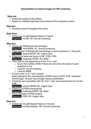
- 1. 1 Interpretation of retinal images for DR screening Step one Check the quality of the photos. Adjust for suitable brightness and contrast of the computer screen. Step two Carefully screen throughout the retina. Step three If there are no pathological lesions of retina Diagnosis: no DR. R0. Annual screening Step four If there are MA/dot-blot hemorrhages Diagnosis: mild NPDR. R1. Annual screening If the number of MA/dot-blot hemorrhage in each quadrant is > 20 points. Diagnosis: severe NPDR. R2. Urgent refer If there is at least one lesion of HE, CWS, FSH Diagnosis: moderate NPDR. R2. Refer Then look for the appearance of the "4-2-1 rule". o Count the number of MA if there are more than 20 points in each quadrant or not. o Look for venous beading. o Look for IRMA. If none of the "4-2-1 rule" present Keep looking for the characteristic of PDR such as NVD, NVE, preretinal hemorrhage, vitreous hemorrhage, fibrous proliferation If findings are compatible with the "4-2-1 rule" and ensured that it is not the PDR Diagnosis: severe NPDR. R2. Urgent refer If there is no PDR characteristic Diagnosis: moderate NPDR. R2. Refer If any character of PDR present Diagnosis: PDR. R3. Urgent refer Step five If there are no pathological lesions of macula Diagnosis: no Maculopathy. M0. Annual screening
- 2. 2 Step Six If there is any of the following: Exudate < or = 1DD of center of fovea Circinate or group of exudates within macula Microaneurysm or hemorrhage < or = 1DD of center of fovea Retinal thickening < or = 1DD of center of fovea Diagnosis: Diabetic Maculopathy. M1. Refer Step Seven If there is any of the following: Other lesion Ungradable image Diagnosis: Other lesion. OL. Refer for Assessment Diagnosis: Un-gradable. UG. Refer for Assessment