Obat Aborsi Makassar WA 085226114443 Jual Obat Aborsi Cytotec Asli Di Makassar
bjsm.2006.031237.pdf
1. ORIGINAL ARTICLE
The effect of cryotherapy on nerve conduction velocity, pain
threshold and pain tolerance
Amin A Algafly, Keith P George
. . . . . . . . . . . . . . . . . . . . . . . . . . . . . . . . . . . . . . . . . . . . . . . . . . . . . . . . . . . . . . . . . . . . . . . . . . . . . . . . . . . . . . . . . . . . . . . . . . . . . . . . . . . . . . . . . . . . . . . . . . . . . . . . . . .
See end of article for
authors’ affiliations
. . . . . . . . . . . . . . . . . . . . . . . .
Correspondence to:
K P George, Research
Institute for Sport and
Exercise Sciences, 15–21
Webster Street, Liverpool L3
2ET, UK; k.george@ljmu.
ac.uk
Accepted 4 December 2006
Published Online First
15 January 2007
. . . . . . . . . . . . . . . . . . . . . . . .
Br J Sports Med 2007;41:365–369. doi: 10.1136/bjsm.2006.031237
Objectives: To determine the impact of the application of cryotherapy on nerve conduction velocity (NCV),
pain threshold (PTH) and pain tolerance (PTO).
Design: A within-subject experimental design; treatment ankle (cryotherapy) and control ankle (no
cryotherapy).
Setting: Hospital-based physiotherapy laboratory.
Participants: A convenience sample of adult male sports players (n = 23).
Main outcome measures: NCV of the tibial nerve via electromyogram as well as PTH and PTO via pressure
algometer. All outcome measures were assessed at two sites served by the tibial nerve: one receiving
cryotherapy and one not receiving cryotherapy.
Results: In the control ankle, NCV, PTH and PTO did not alter when reassessed. In the ankle receiving
cryotherapy, NCV was significantly and progressively reduced as ankle skin temperature was reduced to
10˚C by a cumulative total of 32.8% (p,0.05). Cryotherapy led to an increased PTH and PTO at both
assessment sites (p,0.05). The changes in PTH (89% and 71%) and PTO (76% and 56%) were not different
between the iced and non-iced sites.
Conclusions: The data suggest that cryotherapy can increase PTH and PTO at the ankle and this was
associated with a significant decrease in NCV. Reduced NCV at the ankle may be a mechanism by which
cryotherapy achieves its clinical goals.
C
ryotherapy has been accepted for decades as an effective,
inexpensive and simple intervention for pain manage-
ment after many acute sport injuries.1 2
It is widely
believed that the therapeutic application of cryotherapy leads to
a reduction in pain and swelling, but the physiological basis for
this effect is still incompletely understood.3 4
Saeki5
and other
authors concluded that pain relief with cold application could
be due to many mechanisms including altered nerve conduc-
tion velocity (NCV), inhibition of nociceptors, a reduction in
muscle spasms and/or a reduction in metabolic enzyme activity
levels.6–8
NCV can be altered by gender, age and, more pertinently,
skin temperature.9
On this basis it is plausible to propose that
cryotherapy could reduce pain via an alteration in NCV.
Alternatively, cryotherapy could also be effective as a counter-
irritant to pain via diffused noxious inhibitory controls, pain
gate theory, suppressed nociceptive receptor sensitivity or via
the analgesic descending pathway of the central nervous
system such as endorphins.1 5 10 11
Evaluation of a counter-
irritant role is difficult to directly evaluate, but if cryotherapy
can reduce pain threshold (PTH) and pain tolerance (PTO)
independent of any effect on NCV then these processes may be
more important.
The aim of the current study, therefore, was to assess
changes in NCV, PTH and PTO concomitantly as ankle skin
temperature was reduced via cryotherapy. We hypothesise that
cryotherapy reduces skin temperature to a level that decreases
NCV, and that changes in NCV, are associated with an increase
in PTH and PTO.
METHODS
Subjects
A convenience sample of 23 volunteers from local sports clubs
were informed individually about the purpose, nature and risks
involved with the study. Written informed consent was
obtained, and ethical approval was granted through the
Manchester Metropolitan University, Manchester, UK. The
study conformed to the Declaration of Helsinki. Inclusion
criteria required the subjects to be young (20–29 years), male
and physically active. Exclusion criteria prevented the recruit-
ment of subjects who were having/had central and/or periph-
eral nervous system disorders, overweight (body mass index
.30), skin problems and/or allergic response on to exposure
cold conditions.
Research design
All subjects were fully familiarised before to testing, at which
time the experimental (EXP; cryotherapy) and control (CON)
ankles were determined randomly by the toss of a coin. All data
collection occurred in one visit to the laboratory (ca 3 h) where
the room temperature was held constant between 21˚C and
24˚C. Both ankles were subjected to the same measurement
protocols at the same time, but the order of testing between
EXP and CON ankle was again randomised by the toss of a coin.
For the EXP ankle this meant measures of NCV, PTH and PTO
at baseline before ice application and then at skin temperatures
of 15˚C and 10˚C with ice cooling and a skin temperature of
15˚C with re-warming after ice removal. Chesterton et al12
reported that to achieve a desirable physiological response
(reduction in pain) with cryotherapy requires that the skin
tissue is cooled to specific temperature levels (,13.2˚C). This
specifically drove the rationale for the choice of temperatures
assessed in this research design.
Abbreviations: ANOVA, analysis of variance; CON, control; EMG,
electromyogram; EXP, experimental; NCV, nerve conduction velocity; PTH,
pain threshold; PTO, pain tolerance
365
www.bjsportmed.com
group.bmj.com
on December 11, 2014 - Published by
http://bjsm.bmj.com/
Downloaded from
2. Protocols
Skin temperature was measured using a Digital Thermometer
(Chavin Arnoux, France). An electrode was placed on the iced
area of leg where NCV, PTH and PTO were measured. The
thermistor was applied directly to the skin so temperature
measures reflected skin changes with heat loss rather than the
direct effect of ice on the thermometer.
In this study, NCV measurement was acquired by using a
portable electromyogram (EMG) system (Medtronic Keypoint,
Copenhagen, Denmark). Anode and cathode electrodes were
fixed to the fifth toe and the two EMG electrodes were placed
over the tibial nerve. Proper placement of the EMG head would
result in the contraction of muscles of the fifth metatarsal once
stimulated. The active electrode was placed above the flexor
retinaculum and medial to the medial malleolus for both
medial and lateral plantar nerves. The ground electrode was
placed on the dorsum of the foot. The stimulus intensity needed
to obtain the maximal amplitudes is usually three times the
sensory threshold. Lateral plantar stimulation was applied
orthodromically by means of a ring electrode on the fifth toe.
The EMG was set to a frequency of 8 Hz or 1.6 kHz with a
sweep speed of 2 or 5 ms/div and a gain of 5–10 mV. NCV was
then calculated by software inherent to the EMG as the time
difference between the stimulation and the stimulation and
onset of EMG activity divided by the distance (assessed by
callipers) between the fifth-digit ring electrodes to the ankle
pick-up.
Both PTH and PTO were assessed by a pressure algometer
(Pain Diagnostic and Thermography, Great Neck, NY, USA) by
a single investigator at two different sites of reference for the
tibial nerve on the lateral aspect of both the treatment and
control ankles. The first assessment site (PTH1, PTO1: iced) was
located posterior to the lateral aspect of the lateral malleolus
1 cm to the lower tip of the lateral malleolus, where ice
application and skin temperature measurements were made.
The second assessment site (PTH2, PTO2: non-iced) was located
on the lateral aspect of the shaft of the fourth metatarsal bone
in close proximity to its head, distal to the first point. This was
not iced but still served by the tibial nerve. The algometer has a
force gauge (0–20 kg) fitted with a rubber tip with a 1 cm2
surface area13–15
that was mounted on a stand vertically above
each of the two ankles. A lever arm on the stand allowed the
circular probe head of the algometer to be lowered gradually to
make initial contact with the skin and once in place, the probe
head was further lowered at a steady rate until discomfort was
reported (PTH) and removed at the point the pain became
unbearable (PTO).
Ice application
After baseline measures, ice was applied to lower the tissue
temperature on the EXP ankle. Crushed ice has been found to
be most effective in reducing skin temperature compared with
other cold modalities,12 16
and was hence used in this study. At
the specific skin temperatures noted (15˚C and 10˚C), the ice
pack was removed and all variables were reassessed.
Data analysis
Descriptive statistics (mean (SD)) were used to summarise the
data for NCV, as well as for PTO and PTH, at both assessment
sites on the EXP and CON ankles. Data for NCV, PTO and PTH
at both iced and non-iced sites were analysed using repeated-
measure two-way analysis of variances (ANOVAs) with the
timeline (pre (baseline), 15˚C, 10˚C and 15˚C) and ankle (EXP
and CON) as repeated factors. For each significant F ratio, post
hoc comparisons of group means were made using the Tukey
honestly significant difference test. Pearson product–moment
correlations were then performed on the association between
changes (delta scores from baseline to 10˚C) in NCV with PTO
and PTH. For all statistical tests, differences were considered to
be significant if p,0.05. Data analysis was performed using the
SPSS V.10 software package.
RESULTS
Nerve conduction velocity
The average ice application time for skin temperature to reach
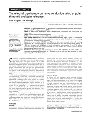
10˚C was 26 min (range 20–31 min). For NCV, significant main
effects for ankle (EXP vs CON: F = 5.4, p = 0.02) and timeline
(pre, 15˚C, 10˚C and 15˚C: F = 31.8, p,0.005), as well as the
ankle–timeline interaction, (F = 27.8, p,0.005) were demon-
strated. Figure 1 presents the pattern of response, and post hoc
analysis stated that NCV for the EXP ankle was significantly
depressed from baseline as ankle skin temperature decreased in
a progressive fashion. At both 15˚C and 10˚C NCV was also
significantly lower in the EXP ankle than in the CON ankle
(p,0.05). Data for NCV did not differ across the timeline in the
CON ankle (p.0.05; fig 1). The change in NCV with ice
application from baseline (pre) to 10˚C represented a 33%
reduction.
PTH and PTO at assessment site one (iced)
Both PTH1 and PTO1 were significantly altered by ice
application (figs 2 and 3). For PTH1, significant ANOVA F
ratios were reported for the main effects of ankle (F = 47.5,
p,0.005), timeline (F = 56.7, p,0.005) and their interaction
(F = 41.3, p,0.005). This was mirrored by statistical outcomes
for PTO1 (ankle: F = 34.5, p,0.005; timeline: F = 53.2,
45
40
35
30
25
20
NCV
(m/s)
15
10
5
0
PRE 15°C
* *
EXP CON
*
**
10°C
Timeline
15°C
Figure 1 The impact of ice application on nerve conduction velocity in
experimental (EXP) and control (CON) ankles (data are mean (SD)).
Timeline: pre, baseline; 15˚C, skin temperature cooling; 10˚C, skin
temperature cooling; 15˚C, skin temperature with passive warming.
*Significant difference between EXP and CON ankles as well as between
pre and 10/15˚C measurements in EXP ankle (p,0.05). **Significant
difference between 15 and 10˚C measurements in the EXP ankle (p,0.05).
PTH
(kg)
2
4
6
8
10
12
0
PRE 15°C
* *
EXP CON
*
**
10°C
Timeline
15°C
Figure 2 The impact of ice application upon pain threshold (PTH) at point
1 (iced) in the experimental (EXP) and control (CON) ankles (data are
represented as mean (SD)). PTO, pain tolerance. *Significant difference
between EXP and CON ankles as well as between baseline (pre) and 10/
15˚C measurements in the EXP ankle (p,0.05). **Significant difference
between 15 and 10˚C measurements in the EXP ankle (p,0.05).
366 Algafly, George
www.bjsportmed.com
group.bmj.com
on December 11, 2014 - Published by
http://bjsm.bmj.com/
Downloaded from
3. p,0.005; interaction: F = 43.2, p,0.005). Data for both PTH1
and PTO1 represent a significant and progressive increase in
threshold and tolerance for pain perception, with a decrease in
skin temperature. In both cases, these changes in the EXP ankle
were significantly different from the CON ankle, which
remained unchanged across the timeline of repeat assessments.
The relative percentage change in PTH1 from baseline (pre) to
10˚C was 89% in the EXP ankle. Likewise, the relative
percentage change in PTO1 from baseline (pre) to 10˚C was
76% in the EXP ankle.
PTH and PTO at assessment site 2 (non-iced)
Both PTH2 and PTO2 were significantly altered by ice
application (figs 4 and 5). For PTH2, significant ANOVA F
ratios were reported for the main effects of ankle (F = 19.1,
p,0.005), timeline (F = 46.5, p,0.005) and their interaction
(F = 30.0, p,0.005). This was mirrored by statistical outcomes
for PTO2 (ankle: F = 14.4, p,0.005; timeline: F = 35.6,
p,0.005; interaction: F = 21.7, p,0.005). Data for both PTH2
and PTO2 represent a significant and progressive increase in
threshold and tolerance for pain perception, with a decrease in
skin temperature. In both cases, these changes in the EXP ankle
were significantly different from the CON ankle, which
remained unchanged across the timeline of repeat assessments.
The relative percentage change in PTH2 from baseline (pre) to
10˚C was 71% in the EXP ankle. Likewise, the relative
percentage change in PTO2 from baseline (pre) to 10˚C was
56% in the EXP ankle.
Delta scores for NCV, PTH and PTO from baseline (pre) to 10˚C
were correlated and all relationships were significant (NCV and
PTO2 = 0.41, NCV and PTO1 = 0.55, NCV and PTH2 = 0.68, NCV
and PTH1 =0.71, all p,0.05). Figure 6 presents an exemplar
scatterplot for the relationship between the change in NCV and
PTH1 (r= 0.71). These data suggest a close association between
the ice-induced alteration in NCV and the consequent changes in
PTH and PTO irrespective of site assessed.
DISCUSSION
Our data support the contention that cryotherapy is an
effective, inexpensive and simple intervention for pain manage-
ment,1 2
and uniquely provides some insight into the potential
mechanisms by which this may be brought about. Specifically,
NCV is significantly and progressively reduced concomitantly
with skin temperature at the ankle during cryotherapy.
Associated with the changes in NCV, we observed significant
increases in PTH and PTO at both assessment sites on the ankle
served by the same nerve, even though only the first site
received direct ice application.
We reported an average reduction of 33% in NCV from
baseline (pre) to 10˚C, which equates to a 0.4 m/s decrease in
sensory NVC for each 1˚C fall in skin temperature (ca 31–10˚C).
This trend supports previous data.17
A drop in NCV with a
reduction in skin temperature was also reported by Chesterton
et al,12
although the magnitude of relative change in NCV was
smaller than that observed in the current study. Specifically,
Chesterton et al12
reported that a skin temperature of 13.5˚C was
required to reduce NCV by 10% compared with the 17%
reduction in NCV at 15˚C and the 33% reduction in NCV
reduction at 10˚C in the present study. The difference in
magnitude of these changes may be due to a number of
methodological differences between the studies, notably the
site of the acquired temperature measurements.
Any specific explanation of the decrease in NCV with
cryotherapy in the current study is purely speculative. However,
previous research has suggested that temperature can affect the
exchange between Ca2+
and Na+
in neural cells.17
Reid et al
reported that low temperature could increase the friction between
Ca2+
and its cellular ‘‘gate’’ during the exchange that could result
in the delay of action potential generation.
Associated with the drop in NCV was an increase in PTH and
PTO at both assessment sites. As with the changes in NCV, the
significant increases in PTH and PTO were progressive with the
decrease in skin temperature recorded at assessment site 1.
Previous studies have documented an increase in both PTH and
PTO with the use of cooling.5
The fact that PTH and PTO were
increased in a similar manner at both assessment site 1 (iced)
and site 2 (non-iced) as well as the fact that PTH and PTO were
PTO
(kg)
4
2
8
6
18
16
14
12
10
20
0
PRE 15°C
*
*
EXP CON
*
**
10°C
Timeline
15°C
Figure 3 The impact of ice application on pain tolerance at point 1 (iced)
in the experimental (EXP) and control (CON) ankles (data are represented
as mean (SD)). PTH, pain threshold. *Significant difference between EXP
and CON ankles as well as between baseline (pre) and 10/15˚C
measurements in the EXP ankle (p,0.05). **Significant difference between
15 and 10˚C measurements in the EXP ankle (p,0.05).
PTH
(kg)
2
1
4
5
3
9
8
7
6
10
0
PRE 15°C
*
*
EXP CON
*
**
10°C
Timeline
15°C
Figure 4 The impact of ice application on pain threshold (PTH) at point 2
(non-iced) in the experimental (EXP) and control (CON) ankles (data are
represented as mean (SD)). *Significant difference between EXP and CON
ankles as well as between baseline (pre) and 10/15˚C measurements in the
EXP ankle (p,0.05). **Significant difference between 15 and 10˚C
measurements in the EXP ankle (p,0.05).
PTO
(kg)
2
6
8
4
16
14
12
10
0
PRE 15°C
* *
EXP CON
*
**
10°C
Timeline
15°C
Figure 5 The impact of ice application on pain tolerance (PTO) at point 2
(non-iced) in the experimental (EXP) and control (CON) ankles (data are
represented as mean (SD)). *Significant difference between EXP and CON
ankles as well as between baseline (pre) and 10/15˚C measurements in the
EXP ankle (p,0.05). **Significant difference between 15 and 10˚C
measurements in the EXP ankle (p,0.05).
Cryotherapy and nerve conduction velocity 367
www.bjsportmed.com
group.bmj.com
on December 11, 2014 - Published by
http://bjsm.bmj.com/
Downloaded from
4. significantly different between the EXP and CON ankles, may
provide some mechanistic insight into the change in pain
perception with ice application. Interestingly, some investiga-
tors have reported that with exposure to a cold environment
(19˚C) for 30 min, plasma b-endorphin levels doubled.18 19
In
this case, b-endorphin should affect the organism as a whole
and increase PTH and PTO throughout the body. In the EXP
and CON ankles different PTH and PTO responses were noted,
suggesting no general b-endorphin effect. Although we did not
assess b-endorphin levels in this study, it does not seem to be a
plausible theory. For a similar reason, the role of any higher
nervous system activity in explaining the current data can also
be largely discounted.
The diffuse noxious inhibitory control theory, the pain gate
theory and the theory of suppression of nociceptive receptor
sensitivity can be largely discounted as these would predict a
different PTH and PTO response at the two assessment sites
(iced and non-iced). The diffuse noxious inhibitory control
theory suggests that responses to noxious stimuli applied in the
excitatory receptive field are inhibited by other noxious stimuli
applied to parts of the body distant from the excitatory
receptive field. Assessment site 2 was distal to the ice
application, but PTH and PTO responded similarly to changes
seen at site 1. In the pain gate theory, pain pulses transmitted
by A d fibres and C fibres are inhibited by various inputs
conducted by A d to dorsal horn cells, leading to the regulation
of pain perception by a gate that may be opened or closed.20
In
the present study, assessment sites 1 and 2 would have their
own neural pathways or gates (spinal cord level). When site 1
was iced, on the basis of the pain gate control theory, the pain
signals from the iced site should be blocked at the spinal cord
level but not at the non-iced site. This does not coincide with
the data obtained for PTO and PTH at both sites.
Changes in PTO and PTH at both assessment sites could,
therefore, plausibly be explained by the reduction in NCV of the
tibial nerve. At the site on the treatment ankle that was not iced
a decrease in NCV was still apparent, as were changes in PTO
and PTH, even though no local cooling had taken place. This
viewpoint is indirectly supported by the work of Walmesley et
al,21
who reported that the application of transcutaneous nerve
stimulation to relieve pain was associated with a significant
reduction in median NCV.
As with any study, it is pertinent to note some limitations
and suggest future research to address these and other issues.
The current study used cooling during a completely non-active
timeline and thus the generalisability of the influence of ice
application on pain perception within a sporting context may be
limited. Indeed, not all sports-injury related cryotherapy can be
applied for the same time period. In these circumstances, skin
cooling may be interspersed with periods of muscle activity
producing localised heating. Such interactions may be worthy
of further research. Future studies may examine the responses
of different sensory fibres to cooling. It is pertinent to note that
the tibial nerve is superficially located and, as such, the use of
cryotherapy at other sites/injuries would probably lead to a
different relationship between skin temperature change and an
alteration in NCV. This would result in different skin temperature
changes and thus mediate a range of responses in NCV.
In conclusion, the present study revealed that ice application
to the ankle resulted in an increase in PTH and PTO at the point
of ice application as well as at an assessment site distal to the
ice application but also served by the tibial nerve. A significant
decrease in NCV was also observed with a decrease in ankle
skin temperature. It is, therefore, plausible in the current study
to suggest that cryotherapy at the ankle exerts its clinical effect,
a reduction in pain, primarily via altered NCV in the tibial
nerve.
Authors’ affiliations
. . . . . . . . . . . . . . . . . . . . . . .
Amin A Algafly, Department of Physiotherapy, Qatif Central Hospital,
Qatif, Kingdom of Saudi Arabia
Keith P George, Research Institute for Sport and Exercise Sciences,
Liverpool John Moores University, Liverpool, UK
Competing interests: None.
This work was completed while Amin A Algafly was studying for an MSc in
Sports Injury and Therapy at Manchester Metropolitan University.
REFERENCES
1 Kottke FJ, Stillwell GK, Lehamann JF. Krusen’s handbook of physical medicine
and rehabilitation, 3rd edn. Philadelphia: WB Saunders, 1996.
2 Sauls J. Efficacy of cold for pain: fact or fallacy? Online J Knowl Synth Nurs
1999;22:6–8.
3 Edwards DJ, Rimmer M, Keene GC. The use of cold therapy in the postoperative
management of patients undergoing arthroscopic anterior cruciate ligament
reconstruction. Am J Sports Med 1996;24:193–5.
4 Rakel B, Barr JO. Physical modalities in chronic pain management. Nurs Clin
North Am 2003;38:477–94.
5 Saeki Y. Effect of local application of cold or heat for relief of pricking pain. Nurs
Health Sci 2002;4:97–105.
6 Wahren LK, Torebjork E, Jorum E. Central suppression of cold-induced C fiber
pain by myelinated fiber input. Pain 1989;38:313–19.
7 Konrath GA, Lock T, Goitz HT, et al. The use of cold therapy after anterior
cruciate ligament reconstruction: a prospective randomized study and literature
review. Am J Sports Med 1996;24:629–33.
8 Airaksinen OV, Kyrklund N, Latvala K, et al. Efficacy of cold gel for soft tissue
injuries. Am J Sports Med 2003;31:680–4.
9 Binnie CD, Cooper R, Fowler CJ, et al. Clinical neurophysiology, 1st edn. London:
Butterworth-Heinemann, 1995.
10 Low J, Reed A. Electrotherapy explained principles and practice, 2nd edn.
London: Butterworth-Heinemann, 1994.
11 Skinner K, Basbaum AI, Fields HL. Cholecystokinin and enkephalin in brain stem
pain modulating circuits. Neuroreport 1997;8:2995–8.
12 Chesterton LS, Foster NE, Ross L. Skin temperature response to cryotherapy. Arch
Phys Med Rehabil 2002;83:543–9.
13 McDowell BC, McCormack K, Walsh DM, et al. Comparative analgesic effects of
H-wave therapy and transcutaneous electrical nerve stimulation on pain
threshold in humans. Arch Phys Med Rehabil 1999;80:1001–4.
Delta
PTH1
(kg)
2
1
4
5
3
10
9
8
7
6
0
5
0 10
r=0.71, p⬍0.05
15 20 25 30 40
Timeline
35
Figure 6 The relationship between changes from baseline (pre) to 10˚C in
nerve conduction velocity and pain threshold (PTH) at assessment point 1
(iced).
What is already known on this topic
N Cryotherapy is routinely used in the frontline treatment of
a large number of sports injuries, although its mechanism
of action is unclear.
What this study adds
N We have shown that in a specific model of ankle cooling
the association between changes in pain sensation and
nerve conduction velocity (NCV) suggests a role for NCV
in mediating the clinical impact of cryotherapy.
368 Algafly, George
www.bjsportmed.com
group.bmj.com
on December 11, 2014 - Published by
http://bjsm.bmj.com/
Downloaded from
5. 14 Hou CR, Tsai LC, Cheng KF, et al. Immediate effects of various physical
therapeutic modalities on cervical myofascial pain and trigger-point sensitivity.
Arch Phys Med Rehabil 2002;83:1406–14.
15 Arendt-Nielsen L, Sumikura H. From pain research to pain treatment: role of
human pain models. J Nippon Med Sch 2002;69:514–24.
16 Mecomber SA, Herman RM. Effects of local hypothermia on reflex and voluntary
activity. Phys Ther 1971;51:271–81.
17 Reid G, Babes A, Pluteanu F. A cold and menthol-activated current in rate dorsal
root ganglion neurones: properties and role in cold transduction. J Physiol
2002;545:595–614.
18 Schoenfeld AD, Lox CD, Chen CH, et al. Pain threshold changes by acute
exposure to altered ambient temperatures. Peptides 1985;6:19–22.
19 Melzack S, Wall PD. Pain mechanisms: a new theory. Science
1965;150:971–9.
20 Walmesley RP, Monga TN, Prouix M. Effect of transcutaneous nerve stimulation
on sensory nerve conduction velocity: a pilot project. Physiother Pract
1986;2:117–20.
. . . . . . . . . . . . . . . COMMENTARY . . . . . . . . . . . . . . .
Cryotherapy is often used in the treatment of sports injuries.
The paper presented demonstrates that the mode of action of
cryotherapy is likely to be related to the diminishing of nerve
conductance velocity. This finding is of great significance to
those treating soft-tissue injuries and may also influence
decisions relating to return to play after the use of this
treatment modality.
Lee Herrington
Salford University, School of Healthcare Professionals, Manchester, UK;
l.c.herrington@salford.ac.uk
EDITORIAL BOARD MEMBER . . . . . . . . . . . . . . . . . . . . . . . . . . . . . . . . . . . . . . . . . . . . . . . . . . . . . . . . . . . . . . . . . . . . . . . . . . . .
Karl Fields
K
arl B Fields, MD, graduated from Yale University and the
University of Kentucky Medical School. He completed
family practice residency at Charlotte Memorial Hospital
in Charlotte, NC, and after a stint in private practice completed
a faculty development fellowship at UNC Chapel Hill. He is
residency and sports medicine fellowship director of the Moses
Cone Family Practice Residency in Greensboro, NC as well as
Professor of Family Medicine and Associate Chairman of the
Department of Family Medicine at the University of North
Carolina Medical School in Chapel Hill and an adjunct
professor of sports science at the University of North Carolina
in Greensboro. He developed the sports medicine fellowship
programme in Greensboro in 1992 and received his CAQ in
sports medicine in 1993. Currently he serves as team physician
for Guilford College and consults in the care of several
university and high school teams, running clubs and elite
athletes. He has been a visiting professor for the Australian
Institute of Sport and for the Norwegian Primary Care
Association. He is a past president of the American Medical
Society of Sports Medicine and a member of the American
College of Sports Medicine.
Figure 1 Karl Fields.
Cryotherapy and nerve conduction velocity 369
www.bjsportmed.com
group.bmj.com
on December 11, 2014 - Published by
http://bjsm.bmj.com/
Downloaded from
6. velocity, pain threshold and pain tolerance
The effect of cryotherapy on nerve conduction
Amin A Algafly and Keith P George
doi: 10.1136/bjsm.2006.031237
2007
2007 41: 365-369 originally published online January 15,
Br J Sports Med
http://bjsm.bmj.com/content/41/6/365
Updated information and services can be found at:
These include:
References
#BIBL
http://bjsm.bmj.com/content/41/6/365
This article cites 17 articles, 5 of which you can access for free at:
service
Email alerting
box at the top right corner of the online article.
Receive free email alerts when new articles cite this article. Sign up in the
Notes
http://group.bmj.com/group/rights-licensing/permissions
To request permissions go to:
http://journals.bmj.com/cgi/reprintform
To order reprints go to:
http://group.bmj.com/subscribe/
To subscribe to BMJ go to:
group.bmj.com
on December 11, 2014 - Published by
http://bjsm.bmj.com/
Downloaded from