
Recommended
More Related Content
Similar to SPINA BIFIDA PPT.
Similar to SPINA BIFIDA PPT. (20)
Recently uploaded
Recently uploaded (20)
SPINA BIFIDA PPT.
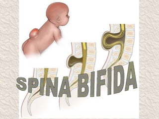
- 2. DEFINITION Spina bifida (Latin: "split spine") is a developmental congenital disorder caused by the incomplete closing of the embryonic neural tube. The closure of the neural tube occurs around the 23rd and 27th day after fertilization. Some vertebrae overlying the spinal cord are not fully formed and remain unfused and open.
- 3. CAUSES • Maternal diabetes • Family history • Obesity • Medications such as some anticonvulsants ex. Valproic acid have an increased risk of having children with spina bifida • Folic acid deficiency
- 6. SPINA BIFIDA OCCULTA • Occulta is Latin for "hidden". This is the mildest form of spina bifida. • In occulta, the outer part of some of the vertebrae is not completely closed. • The splits in the vertebrae are so small that the spinal cord does not protrude. The skin at the site of the lesion may be normal, or it may have some hair growing from it, there may be a dimple in the skin .
- 7. MENINGOCELE • The least common form of spina bifida in which cyst or fluid filled sac pokes through an open part of the spine . • The sac contain membranes that protect spinal cord ie CSF and meninges.
- 8. MYELOMENINGOCELE • This type of spina bifida often results in the most severe complications. • In which cyst or fluid filled sac pokes through an open part of the spine . • The sac contain spinal cord, CSF and meninges.
- 9. CLINICAL MANIFESTATIONS Physical Signs: • Bladder and bowel control problems, including incontinence, urinary tract infections, and poor renal function. • Pressure sores and skin irritations • Paralysis
- 10. CONT….. • Scoliosis • Back pain • Partial or complete lack of sensation • Weakness of the hips, legs, or feet of a newborn • Other symptoms may include: Dimpling of the sacral area • Below-average intelligence.
- 11. EXECUTIVE FUNCTION • Specific areas of difficulty in some individuals include planning, organizing, initiating, and working memory. Problem-solving, abstraction, and visual planning may also be impaired.
- 12. DIAGNOSTIC EVALUATION Neonatal examination Pregnancy screening: • Neural tube defects can usually be detected during pregnancy by testing the mothers blood (AFP screening) or a detailed fetal ultrasound. • Increased levels of maternal serum alpha- fetoprotein is seen in neural tube defects. • Ultrasonography, X ray , CT Scan, MRI
- 13. TREATMENT • PRENATAL SURGERY The surgeon opens the uterus and repair the spinal cord of the fetus, usually during week 19 to 25 of pregnancy. • POSTNATAL SURGERY Laminectomy and closure of defect within 24 to 48 hours of birth. The spinal cord and its nerve roots are put back inside the spine and covered with meninges.
- 14. PREVENTION • Dietary supplementation with folic acid in pregnancy has been shown to be helpful in reducing the incidence of spina bifida. Sources of folic acid include whole grains, cereals, dried beans, leaf vegetables and fruits. • It is recommended that any woman considering becoming pregnant should take 0.4 mg of folic acid a day.
- 15. NURSING MANAGEMENT Pre-Op • Position the child in prone with legs abducted. This reduces tension and risk of sac trauma. • Put the child in an incubator or warmer . This maintains normal body temperature. • Apply moist and sterile dressing to avoid drying of the area due to heat in the incubator. • Change dressing two-four hourly to avoid infection.
- 16. Post-Op • Position the child in prone to avoid pressure on suture, or side lying position alternatively. • Monitor the child`s vital signs every 30 minutes until stable. • Monitor input and output. • Encourage the mother to continue breastfeeding if the child is being breastfed. • Remove the dressings after 48hrs to check any signs of bleeding or bulging. Observe for leakage.
- 17. COMPLICATIONS • Frequent urinary tract infections • Hydrocephalus • Loss of bowel or bladder control • Meningitis • Permanent weakness or paralysis of legs
- 18. THANK YOU