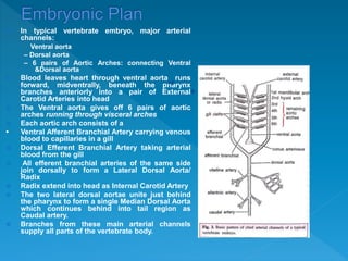
SlideShare a Scribd company logo
1 of 9
 In typical vertebrate embryo, major arterial
channels:
Ventral aorta
– Dorsal aorta
– 6 pairs of Aortic Arches: connecting Ventral
&Dorsal aorta
 Blood leaves heart through ventral aorta runs
forward, midventrally, beneath the pharynx
branches anteriorly into a pair of External
Carotid Arteries into head
 The Ventral aorta gives off 6 pairs of aortic
arches running through visceral arches
 Each aortic arch consists of a
• Ventral Afferent Branchial Artery carrying venous
blood to capillaries in a gill
 Dorsal Efferent Branchial Artery taking arterial
blood from the gill
 All efferent branchial arteries of the same side
join dorsally to form a Lateral Dorsal Aorta/
Radix
 Radix extend into head as Internal Carotid Artery
 The two lateral dorsal aortae unite just behind
the pharynx to form a single Median Dorsal Aorta
which continues behind into tail region as
Caudal artery.
 Branches from these main arterial channels
supply all parts of the vertebrate body.
 Although arterial system of different adult
vertebrates shows major differences, but it is
actually built according to the same basic
architectural plan as seen in the vertebrate
embryo
 The differences are due to increasing
complexity of heart on account of a shift from
gill respiration to lung respiration.
 The modifications mainly concern the aortic
arches which undergo a progressive reduction
in number from lower to higher vertebrates
 Branchiostoma (Amphioxus): nearly 60 pairs
of aortic arches are present, connecting the
ventral and dorsal aortae
 Petromyzon: 7 pairs of aortic arches are
found
 Other cyclostomes: varies from 6 (Myxine) to
15 pairs (Eptatretus)
FISHES
 Embryos = primitive plan with 6 or more
pairs of aortic arches, each passing through
a gill
 Adults = the number is reduced to 4 or 5
Elasmobranchs
 Sharks = only 5 pairs (II, III, IV, V, and VI) are
functional
 1st gill slit forms the spiracle (a non-
functional gill)
 1st arch (mandibular) is absent or
represented by an efferent pseudobranchial
artery
 Heptanchus = 7 pairs of aortic arches
 Each arch forms one afferent and two
Bony fishes
 Teleosts = I and II arches
disappear, so that only 4 pairs (III,
IV, V and VI) remain functional
 Each gill has one afferent and
one efferent artery
Lungfishes
 Polypterus and lungfishes
(Dipnoi) = gills are poorly
developed, so that a pulmonary
artery arises from the efferent
part of the VI arch on both sides
and supplies blood to the
developing air bladder or lung
 Protopterus = III and IV
embryonic arches are
uninterrupted by gill capillaries
 Each arch forms one afferent and
two efferent arteries (by splitting)
in each gill
 Transition from gills to lungs
 URODELES live in water and retain
external gills + lungs
 So, aortic system shows only partial
shift w.r.t. fishes
 4 pairs of arches (III to VI)
 Except Necturus, Siren, Amphiuma
 V arch = incomplete, reduced or
absent only 3 pairs of aortic arches
 III arch forms the carotid arches, IV the
systemic arches
 The radix or lateral aorta between III &
IV arches may persist as a vascular
nconnection: ductus caroticus
 VI arch on either side becomes the
pulmocutaneous artery or arch,
supplying blood to skin and lungs
 It also retains connection with radix
aorta called ductus Botaili or ductus
arteriosus
ANURANS
 Larvae- arrangement of aortic
arches = adult urodele (gill
respiration)
 At metamorphosis, with loss of
gills, aortic arches I, II and V
disappear
 Ductus caroticus disappears so
that the III or carotid arch takes
oxygenated blood only to head
region
 IV or systemic arch on each
side continues to dorsal aorta
to distribute blood elsewhere
except head and lungs
 Ductus arteriosus disappears
so that VI or pulmocutaneous
arch supplies venous blood
exclusively to lungs and skin
for purification
 Adults exhibit only 3 functional
arches, (III, IV and VI) similar to
amniotes
 Fully terrestrial vertebrates, gills disappear
altogether & are replaced by lungs
 Only 3 functional arches (III, IV and VI) present
 Elongation of neck, posterior shifting of heart and
partial division of ventricle brings about certain
innovations in the aortic system
1. Entire ventral aorta and Conus split: forming only
3 trunks-two aortic/systemic + one pulmonary
2. Right systemic arch (IV): arises from left ventricle
carrying oxygenated blood to the carotid arch (III) to
be sent into head
3. Left systemic arch (IV) leads from right ventricle
carrying deoxygenated or mixed blood to the body
through dorsal aorta
4. Pulmonary trunk (VI) also emerges from right
ventricle carrying deoxygenated blood to the lungs
for purification
5. Ductus caroticus and ductus arteriosus are
absent. But, ductus caroticus is present in certain
snakes and lizards (Uromasitx), ductus arteriosus in
some turtles, and both in Sphenodon.
 Reptiles also remain cold-blooded, like amphibians
and fishes, due to mixing of blood.
 Warm-blooded: ventricle is
completely divided, no mixing of
O2 and CO2bloods
 As usual, 6 arches develop in the
embryo, but only 3 (III, IV, VI)
persist in the adult
 Other modifications include
1. Ventral aorta is replaced by two
independent aortae or trunks-
systemic & pulmonary
2. IV arch is represented by a
single systemic aorta, right in birds
and left in mammals, emerging
from left ventricle and carrying
oxygenated blood. Uniting with the
radix aorta of its side it forms the
dorsal aorta
3. The only remaining part of the
other lost systemic arch is
represented by a subclavian artery,
on left side in birds and on right
side in mammals
4. Arch III with remnants of
lateral and ventral aortae
represents carotid arteries,
which arise from systemic
aorta
5. Arch VI forms a single
pulmonary trunk taking
deoxygenated blood from
right ventricle to the lungs
6. Embryonic ductus
caroticus and ductus
arteriosus also disappear.
The latter closes but
persists until hatching or
birth in some cases as a
thin ligament of Botalli or
ligamentum arteriosum

More Related Content

Similar to Evolution of arotic arches.pptx

Circulatory system
Circulatory systemCirculatory system
Circulatory system
Reyna Federo
 

Similar to Evolution of arotic arches.pptx (20)

Heart in vetrebrates
Heart in vetrebratesHeart in vetrebrates
Heart in vetrebrates
 
Major Arteries AS.pptx
Major Arteries AS.pptxMajor Arteries AS.pptx
Major Arteries AS.pptx
 
Blood-circulation.pdf
Blood-circulation.pdfBlood-circulation.pdf
Blood-circulation.pdf
 
Heart in vertebrates
Heart in vertebratesHeart in vertebrates
Heart in vertebrates
 
Evolution of Heart
Evolution of HeartEvolution of Heart
Evolution of Heart
 
Cvs dev
Cvs devCvs dev
Cvs dev
 
Heart .
Heart .Heart .
Heart .
 
Heart .
Heart .Heart .
Heart .
 
Heart .
Heart .Heart .
Heart .
 
Circulatory system
Circulatory systemCirculatory system
Circulatory system
 
Human Cardiovascular system
Human Cardiovascular systemHuman Cardiovascular system
Human Cardiovascular system
 
04 17-06
04 17-0604 17-06
04 17-06
 
Anatomy and physiology of circulatory system
Anatomy and physiology of circulatory systemAnatomy and physiology of circulatory system
Anatomy and physiology of circulatory system
 
Cardiovascular system (blood vessels, anatomy)
Cardiovascular system (blood vessels, anatomy) Cardiovascular system (blood vessels, anatomy)
Cardiovascular system (blood vessels, anatomy)
 
170-Anatomy-Circulatory-System.ppt
170-Anatomy-Circulatory-System.ppt170-Anatomy-Circulatory-System.ppt
170-Anatomy-Circulatory-System.ppt
 
170-Anatomy-Circulatory-System.ppt
170-Anatomy-Circulatory-System.ppt170-Anatomy-Circulatory-System.ppt
170-Anatomy-Circulatory-System.ppt
 
170-Anatomy-Circulatory-System.ppt
170-Anatomy-Circulatory-System.ppt170-Anatomy-Circulatory-System.ppt
170-Anatomy-Circulatory-System.ppt
 
170-Anatomy-Circulatory-System.ppt
170-Anatomy-Circulatory-System.ppt170-Anatomy-Circulatory-System.ppt
170-Anatomy-Circulatory-System.ppt
 
170-Anatomy-Circulatory-System.ppt
170-Anatomy-Circulatory-System.ppt170-Anatomy-Circulatory-System.ppt
170-Anatomy-Circulatory-System.ppt
 
170-Anatomy-Circulatory-System.ppt
170-Anatomy-Circulatory-System.ppt170-Anatomy-Circulatory-System.ppt
170-Anatomy-Circulatory-System.ppt
 

More from Nikhil Borode

More from Nikhil Borode (16)

Principal of Poultry breeding.pptx
Principal of Poultry breeding.pptxPrincipal of Poultry breeding.pptx
Principal of Poultry breeding.pptx
 
Vitamins.pptx
Vitamins.pptxVitamins.pptx
Vitamins.pptx
 
Digestive gland.pptx
Digestive gland.pptxDigestive gland.pptx
Digestive gland.pptx
 
General charecter of phylum annelid.pptx
General charecter of phylum annelid.pptxGeneral charecter of phylum annelid.pptx
General charecter of phylum annelid.pptx
 
urinogenital system.pptx
urinogenital system.pptxurinogenital system.pptx
urinogenital system.pptx
 
Sensory receptor.pptx
Sensory receptor.pptxSensory receptor.pptx
Sensory receptor.pptx
 
Scale and horn.pptx
Scale and horn.pptxScale and horn.pptx
Scale and horn.pptx
 
Comparative anatomy of heart.pptx
Comparative anatomy of heart.pptxComparative anatomy of heart.pptx
Comparative anatomy of heart.pptx
 
respiratory pigment.pptx
respiratory pigment.pptxrespiratory pigment.pptx
respiratory pigment.pptx
 
Respiratory Disorder.pptx
Respiratory Disorder.pptxRespiratory Disorder.pptx
Respiratory Disorder.pptx
 
Mechanism of respiration.pptx
Mechanism of respiration.pptxMechanism of respiration.pptx
Mechanism of respiration.pptx
 
Protoplasmic Theory.pptx
Protoplasmic Theory.pptxProtoplasmic Theory.pptx
Protoplasmic Theory.pptx
 
Prokaryotic and Eukaryotic Cell.pptx
Prokaryotic and Eukaryotic Cell.pptxProkaryotic and Eukaryotic Cell.pptx
Prokaryotic and Eukaryotic Cell.pptx
 
Compostion and structure of plasma membrane.pptx
Compostion and structure of plasma membrane.pptxCompostion and structure of plasma membrane.pptx
Compostion and structure of plasma membrane.pptx
 
Cell transport.pptx
Cell transport.pptxCell transport.pptx
Cell transport.pptx
 
Cell Organnels.ppt
Cell Organnels.pptCell Organnels.ppt
Cell Organnels.ppt
 

Recently uploaded

1029 - Danh muc Sach Giao Khoa 10 . pdf
1029 -  Danh muc Sach Giao Khoa 10 . pdf1029 -  Danh muc Sach Giao Khoa 10 . pdf
1029 - Danh muc Sach Giao Khoa 10 . pdf
QucHHunhnh
 
Salient Features of India constitution especially power and functions
Salient Features of India constitution especially power and functionsSalient Features of India constitution especially power and functions
Salient Features of India constitution especially power and functions
KarakKing
 
Jual Obat Aborsi Hongkong ( Asli No.1 ) 085657271886 Obat Penggugur Kandungan...
Jual Obat Aborsi Hongkong ( Asli No.1 ) 085657271886 Obat Penggugur Kandungan...Jual Obat Aborsi Hongkong ( Asli No.1 ) 085657271886 Obat Penggugur Kandungan...
Jual Obat Aborsi Hongkong ( Asli No.1 ) 085657271886 Obat Penggugur Kandungan...
ZurliaSoop
 
Activity 01 - Artificial Culture (1).pdf
Activity 01 - Artificial Culture (1).pdfActivity 01 - Artificial Culture (1).pdf
Activity 01 - Artificial Culture (1).pdf
ciinovamais
 
Vishram Singh - Textbook of Anatomy Upper Limb and Thorax.. Volume 1 (1).pdf
Vishram Singh - Textbook of Anatomy  Upper Limb and Thorax.. Volume 1 (1).pdfVishram Singh - Textbook of Anatomy  Upper Limb and Thorax.. Volume 1 (1).pdf
Vishram Singh - Textbook of Anatomy Upper Limb and Thorax.. Volume 1 (1).pdf
ssuserdda66b
 

Recently uploaded (20)

Key note speaker Neum_Admir Softic_ENG.pdf
Key note speaker Neum_Admir Softic_ENG.pdfKey note speaker Neum_Admir Softic_ENG.pdf
Key note speaker Neum_Admir Softic_ENG.pdf
 
On National Teacher Day, meet the 2024-25 Kenan Fellows
On National Teacher Day, meet the 2024-25 Kenan FellowsOn National Teacher Day, meet the 2024-25 Kenan Fellows
On National Teacher Day, meet the 2024-25 Kenan Fellows
 
Graduate Outcomes Presentation Slides - English
Graduate Outcomes Presentation Slides - EnglishGraduate Outcomes Presentation Slides - English
Graduate Outcomes Presentation Slides - English
 
Application orientated numerical on hev.ppt
Application orientated numerical on hev.pptApplication orientated numerical on hev.ppt
Application orientated numerical on hev.ppt
 
1029 - Danh muc Sach Giao Khoa 10 . pdf
1029 -  Danh muc Sach Giao Khoa 10 . pdf1029 -  Danh muc Sach Giao Khoa 10 . pdf
1029 - Danh muc Sach Giao Khoa 10 . pdf
 
Single or Multiple melodic lines structure
Single or Multiple melodic lines structureSingle or Multiple melodic lines structure
Single or Multiple melodic lines structure
 
Understanding Accommodations and Modifications
Understanding  Accommodations and ModificationsUnderstanding  Accommodations and Modifications
Understanding Accommodations and Modifications
 
HMCS Max Bernays Pre-Deployment Brief (May 2024).pptx
HMCS Max Bernays Pre-Deployment Brief (May 2024).pptxHMCS Max Bernays Pre-Deployment Brief (May 2024).pptx
HMCS Max Bernays Pre-Deployment Brief (May 2024).pptx
 
Unit-IV- Pharma. Marketing Channels.pptx
Unit-IV- Pharma. Marketing Channels.pptxUnit-IV- Pharma. Marketing Channels.pptx
Unit-IV- Pharma. Marketing Channels.pptx
 
How to Give a Domain for a Field in Odoo 17
How to Give a Domain for a Field in Odoo 17How to Give a Domain for a Field in Odoo 17
How to Give a Domain for a Field in Odoo 17
 
Salient Features of India constitution especially power and functions
Salient Features of India constitution especially power and functionsSalient Features of India constitution especially power and functions
Salient Features of India constitution especially power and functions
 
Jual Obat Aborsi Hongkong ( Asli No.1 ) 085657271886 Obat Penggugur Kandungan...
Jual Obat Aborsi Hongkong ( Asli No.1 ) 085657271886 Obat Penggugur Kandungan...Jual Obat Aborsi Hongkong ( Asli No.1 ) 085657271886 Obat Penggugur Kandungan...
Jual Obat Aborsi Hongkong ( Asli No.1 ) 085657271886 Obat Penggugur Kandungan...
 
Activity 01 - Artificial Culture (1).pdf
Activity 01 - Artificial Culture (1).pdfActivity 01 - Artificial Culture (1).pdf
Activity 01 - Artificial Culture (1).pdf
 
ComPTIA Overview | Comptia Security+ Book SY0-701
ComPTIA Overview | Comptia Security+ Book SY0-701ComPTIA Overview | Comptia Security+ Book SY0-701
ComPTIA Overview | Comptia Security+ Book SY0-701
 
Vishram Singh - Textbook of Anatomy Upper Limb and Thorax.. Volume 1 (1).pdf
Vishram Singh - Textbook of Anatomy  Upper Limb and Thorax.. Volume 1 (1).pdfVishram Singh - Textbook of Anatomy  Upper Limb and Thorax.. Volume 1 (1).pdf
Vishram Singh - Textbook of Anatomy Upper Limb and Thorax.. Volume 1 (1).pdf
 
Food safety_Challenges food safety laboratories_.pdf
Food safety_Challenges food safety laboratories_.pdfFood safety_Challenges food safety laboratories_.pdf
Food safety_Challenges food safety laboratories_.pdf
 
How to Manage Global Discount in Odoo 17 POS
How to Manage Global Discount in Odoo 17 POSHow to Manage Global Discount in Odoo 17 POS
How to Manage Global Discount in Odoo 17 POS
 
ICT role in 21st century education and it's challenges.
ICT role in 21st century education and it's challenges.ICT role in 21st century education and it's challenges.
ICT role in 21st century education and it's challenges.
 
Explore beautiful and ugly buildings. Mathematics helps us create beautiful d...
Explore beautiful and ugly buildings. Mathematics helps us create beautiful d...Explore beautiful and ugly buildings. Mathematics helps us create beautiful d...
Explore beautiful and ugly buildings. Mathematics helps us create beautiful d...
 
Introduction to Nonprofit Accounting: The Basics
Introduction to Nonprofit Accounting: The BasicsIntroduction to Nonprofit Accounting: The Basics
Introduction to Nonprofit Accounting: The Basics
 

Evolution of arotic arches.pptx

  • 1.  In typical vertebrate embryo, major arterial channels: Ventral aorta – Dorsal aorta – 6 pairs of Aortic Arches: connecting Ventral &Dorsal aorta  Blood leaves heart through ventral aorta runs forward, midventrally, beneath the pharynx branches anteriorly into a pair of External Carotid Arteries into head  The Ventral aorta gives off 6 pairs of aortic arches running through visceral arches  Each aortic arch consists of a • Ventral Afferent Branchial Artery carrying venous blood to capillaries in a gill  Dorsal Efferent Branchial Artery taking arterial blood from the gill  All efferent branchial arteries of the same side join dorsally to form a Lateral Dorsal Aorta/ Radix  Radix extend into head as Internal Carotid Artery  The two lateral dorsal aortae unite just behind the pharynx to form a single Median Dorsal Aorta which continues behind into tail region as Caudal artery.  Branches from these main arterial channels supply all parts of the vertebrate body.
  • 2.  Although arterial system of different adult vertebrates shows major differences, but it is actually built according to the same basic architectural plan as seen in the vertebrate embryo  The differences are due to increasing complexity of heart on account of a shift from gill respiration to lung respiration.  The modifications mainly concern the aortic arches which undergo a progressive reduction in number from lower to higher vertebrates
  • 3.  Branchiostoma (Amphioxus): nearly 60 pairs of aortic arches are present, connecting the ventral and dorsal aortae  Petromyzon: 7 pairs of aortic arches are found  Other cyclostomes: varies from 6 (Myxine) to 15 pairs (Eptatretus) FISHES  Embryos = primitive plan with 6 or more pairs of aortic arches, each passing through a gill  Adults = the number is reduced to 4 or 5 Elasmobranchs  Sharks = only 5 pairs (II, III, IV, V, and VI) are functional  1st gill slit forms the spiracle (a non- functional gill)  1st arch (mandibular) is absent or represented by an efferent pseudobranchial artery  Heptanchus = 7 pairs of aortic arches  Each arch forms one afferent and two
  • 4. Bony fishes  Teleosts = I and II arches disappear, so that only 4 pairs (III, IV, V and VI) remain functional  Each gill has one afferent and one efferent artery Lungfishes  Polypterus and lungfishes (Dipnoi) = gills are poorly developed, so that a pulmonary artery arises from the efferent part of the VI arch on both sides and supplies blood to the developing air bladder or lung  Protopterus = III and IV embryonic arches are uninterrupted by gill capillaries  Each arch forms one afferent and two efferent arteries (by splitting) in each gill
  • 5.  Transition from gills to lungs  URODELES live in water and retain external gills + lungs  So, aortic system shows only partial shift w.r.t. fishes  4 pairs of arches (III to VI)  Except Necturus, Siren, Amphiuma  V arch = incomplete, reduced or absent only 3 pairs of aortic arches  III arch forms the carotid arches, IV the systemic arches  The radix or lateral aorta between III & IV arches may persist as a vascular nconnection: ductus caroticus  VI arch on either side becomes the pulmocutaneous artery or arch, supplying blood to skin and lungs  It also retains connection with radix aorta called ductus Botaili or ductus arteriosus
  • 6. ANURANS  Larvae- arrangement of aortic arches = adult urodele (gill respiration)  At metamorphosis, with loss of gills, aortic arches I, II and V disappear  Ductus caroticus disappears so that the III or carotid arch takes oxygenated blood only to head region  IV or systemic arch on each side continues to dorsal aorta to distribute blood elsewhere except head and lungs  Ductus arteriosus disappears so that VI or pulmocutaneous arch supplies venous blood exclusively to lungs and skin for purification  Adults exhibit only 3 functional arches, (III, IV and VI) similar to amniotes
  • 7.  Fully terrestrial vertebrates, gills disappear altogether & are replaced by lungs  Only 3 functional arches (III, IV and VI) present  Elongation of neck, posterior shifting of heart and partial division of ventricle brings about certain innovations in the aortic system 1. Entire ventral aorta and Conus split: forming only 3 trunks-two aortic/systemic + one pulmonary 2. Right systemic arch (IV): arises from left ventricle carrying oxygenated blood to the carotid arch (III) to be sent into head 3. Left systemic arch (IV) leads from right ventricle carrying deoxygenated or mixed blood to the body through dorsal aorta 4. Pulmonary trunk (VI) also emerges from right ventricle carrying deoxygenated blood to the lungs for purification 5. Ductus caroticus and ductus arteriosus are absent. But, ductus caroticus is present in certain snakes and lizards (Uromasitx), ductus arteriosus in some turtles, and both in Sphenodon.  Reptiles also remain cold-blooded, like amphibians and fishes, due to mixing of blood.
  • 8.  Warm-blooded: ventricle is completely divided, no mixing of O2 and CO2bloods  As usual, 6 arches develop in the embryo, but only 3 (III, IV, VI) persist in the adult  Other modifications include 1. Ventral aorta is replaced by two independent aortae or trunks- systemic & pulmonary 2. IV arch is represented by a single systemic aorta, right in birds and left in mammals, emerging from left ventricle and carrying oxygenated blood. Uniting with the radix aorta of its side it forms the dorsal aorta 3. The only remaining part of the other lost systemic arch is represented by a subclavian artery, on left side in birds and on right side in mammals
  • 9. 4. Arch III with remnants of lateral and ventral aortae represents carotid arteries, which arise from systemic aorta 5. Arch VI forms a single pulmonary trunk taking deoxygenated blood from right ventricle to the lungs 6. Embryonic ductus caroticus and ductus arteriosus also disappear. The latter closes but persists until hatching or birth in some cases as a thin ligament of Botalli or ligamentum arteriosum