Fibro-osseous Lesions
•
33 likes•3,713 views

Fibro-osseous lesions- a thorough extensive Post-graduate level presentation

Recommended
More Related Content
What's hot
What's hot (20)
Similar to Fibro-osseous Lesions
Similar to Fibro-osseous Lesions (20)
Recently uploaded
Recently uploaded (20)
Fibro-osseous Lesions
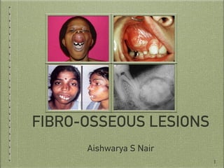
- 1. FIBRO-OSSEOUS LESIONS Aishwarya S Nair 1
- 2. FIBRO-OSSEOUS LESIONS -Contents Introduction Classification Importance of Radiology in the Diagnosis of FOLs Fibrous Dysplasia Cemento-osseous Dysplasia 2
- 4. INTRODUCTION… Fibro-osseous lesions of the jaw comprise a diverse group of conditions in which the normal architecture of bone is replaced by fibrous tissue and may contain varying amounts of mineralised substance which may be bone, cementum or both in appearance. The term fibro-osseous lesion was originally a histo-pathological term 4
- 6. CLASSIFICATION SYSTEMS… The various classifications systems proposed by authors are enumerated as below. Charles Waldron Classification Of Fibro-Osseous Lesions Of The Jaws (1985) Working Classification Of Fibro-Osseous Lesions By Mico M. Malek (1987) Peiter J. Slootweg & Hellmuth Muller (1990) WHO Classification (1992) Waldron Modified Classification Of Fibro-Osseous Lesions Of Jaws (1993) 6
- 7. CLASSIFICATION SYSTEMS… The various classifications systems proposed by authors are enumerated as below. Brannon & Fowler Classification (2001) WHO Classification Of Fibro-Osseous Lesions Of Jaws (2005) Paul M. Speight & Roman Carlos Classification (2006) Eversole Classification (2008) 7
- 8. CLASSIFICATION… Fibro-osseous Lesions of Jaws Classification 1. Fibrous Dysplasia a. Monostotic b. Polyostotic 8 2. Cemento-osseous Dysplasia a. Periapical Cemental Dysplasia b. Focal Cemento-osseous Dysplasia c. Florid Cemento-osseous Dysplasia (Gigantiform Cementoma)
- 9. CLASSIFICATION… 9 3. Familial Gigantiform Cementoma 6. Miscellaneous Osteoblastoma, Osteiod osteoma, Cementoblastoma 5. Juvenile Ossifying Fibroma 4. Ossifying Fibroma
- 10. CLASSIFICATION… Waldron’s Classification (1993) 1. Fibrous Dysplasia a. Monostotic b. Polyostotic 10 2. Fibro-Osseous (Cemental) Lesions Presumably Arising In The Periodontal Ligament a. Periapical Cemental Dysplasia b. Localized Fibro-Osseous-Cemental Lesions (Probably Reactive In Nature) c. Florid Cemento-osseous Dysplasia (Gigantiform Cementoma) d. Ossifying & Cementifying Fibroma
- 11. CLASSIFICATION… Waldron’s Classification (1993) 11 3. Fibro-Osseous Neoplasms Of Uncertain Or Detectable Relationship To Those Arising In The Periodontal Ligament (Category II) a. Cemetoblastoma, Osteoblastoma & Osteoid Osteoma b. Juvenile Active Ossifying Fibroma & Other So Called Aggressive, Active Ossifying/Cementifying Fibromas
- 12. CLASSIFICATION… Brannon & Fowler’s Classification 1. Fibrous Dysplasia a. Monostotic b. Polyostotic c. Craniofacial d. McCune-Albright syndrome 2. Osseous Dysplasia a. Periapical b. Focal c. Florid d. Familial gigantiform cementoma 3. Ossifying Fibroma & Juvenile Ossifying Fibroma 12
- 13. IMPORTANCE OF RADIOLOGY IN DIAGNOSIS OF FIBRO-OSSEOUS LESIONS… The radiologist plays a central role in diagnosis of fibro-osseous lesions. Charles Waldron had stated “In the absence of good clinical & radiologic information, a pathologist can only state a biopsy to be consistent with a fibro-osseous lesion” Therefore, identification of majority of FOLs is based on clinical and radiological features. 13
- 14. IMPORTANCE OF RADIOLOGY IN DIAGNOSIS OF FIBRO-OSSEOUS LESIONS… Many fibro-osseous lesions are symptomless and require no surgery. Therefore, diagnosis of the lesions on clinical & radiological features may obviate the need for surgery This my benefit the patient, as fibrous dysplasia may show exaggerated growth after surgery 14
- 15. IMPORTANCE OF RADIOLOGY IN DIAGNOSIS OF FIBRO-OSSEOUS LESIONS… Radiological evaluation- Plain radiography IOPAR Occlusal radiograph Bitewing radiograph 15
- 16. IMPORTANCE OF RADIOLOGY IN DIAGNOSIS OF FIBRO-OSSEOUS LESIONS… Radiological evaluation- CT- expansile, destructive lesions, cortical breakthrough & extra-osseous extensions MRI- solid from non-solid masses 16
- 18. FIBROUS DYSPLASIA -Introduction WHO (1992) defined fibrous dysplasia as “A benign lesion, presumably developmental in nature, characterised by a presence of fibrous connective tissue with a characteristic whorled pattern and containing trabeculae of immature bone” Asymptomatic regional alteration of bone in which normal architecture of bone is replaced by trabeculae-like osseous structures; lesions may be monostotic or polyostotic, with or without associated endocrinal disturbances - Eversole 18
- 19. FIBROUS DYSPLASIA -Introduction Congenital, skeletal disorder wherein bone is formed and the maturation is arrested in the “woven bone stage”. NON-HEREDITARY DISORDER 19
- 20. FIBROUS DYSPLASIA -History First citation by von Recklinghausen in 1891 Albright pointed out that 2 cases mentioned by von Recklinghausen- fibrous dysplasia Lichtenstein- term FIBROUS DYSPLASIA (1938) Initially considered- Polyostotic Lichtenstein & Jaffe expanded the concept- monostotic 20
- 21. FIBROUS DYSPLASIA -History McCune & Albright (1936 &1937) described an association of abnormal skin pigmentation & precocious puberty (McCune-Albright Syndrome) Jaffe-Lichtenstein described the association of polyostotic fibrous dysplasia with abnormal pigmentation (Jaffe-Lichtenstein Syndrome) 21
- 22. FIBROUS DYSPLASIA -Aetiology & Pathogenesis GNAS1 gene Activation of GS𝞪 subunit of G protein complex Production of cAMP 22 MUTATION CONTINUOUS
- 23. FIBROUS DYSPLASIA -Pathogenesis 1. Hyperfunction of endocrine glands 2. Increased production of melanocytes 3. Affects differentiation of osteoblasts— impairs ability to form mature osteoblasts Fibrous tissue is produced + trabeculae contain “fluid-filled cysts” — GROUND-GLASS APPEARANCE 23
- 24. FIBROUS DYSPLASIA -Pathogenesis Undifferentiated stem cells— Melanocyte progenitor cells Skeletal progenitor cells Endocrinal progenitor cells 24
- 25. FIBROUS DYSPLASIA -Pathogenesis Skeletal progenitor cells— Migrate to form different bones Multiple bones Post natal mutation— Single bone is affected 25
- 26. FIBROUS DYSPLASIA -Classification Monostotic— Juvenile Juvenile, aggressive Adult 26 Polyostotic— Craniofacial M c C u n e - A l b r i g h t Syndrome Ja f f e - L i ch t e n s t e i n Syndrome
- 27. JUVENILE FIBROUS DYSPLASIA -Clinical Features AGE- Early to late childhood SITE- Maxilla > Mandible 27
- 28. JUVENILE FIBROUS DYSPLASIA -Clinical Presentation Asymptomatic Swelling is not prominent & eventually becomes prominent In teenage— swelling may become prominent. Slowly growing ; in aggressive form, growth is more rapid than rest of the bone 28
- 29. JUVENILE FIBROUS DYSPLASIA -Clinical Presentation May cause- Displacement, rotation or malalignment of teeth Malocclusion Facial deformity 29
- 30. JUVENILE FIBROUS DYSPLASIA -Clinical Presentation Aggressive form: Pain, trauma & ulceration 2o to impingement by teeth In maxilla- may extend to involve Floor of the orbit Nasal passages 30
- 31. ADULT MONOSTOTIC FIBROUS DYSPLASIA -Introduction Rare form Spontaneously occurring May appear similar to ossifying fibroma but must be differentiated from it 31
- 32. ADULT MONOSTOTIC FIBROUS DYSPLASIA -Clinical Presentation Asymptomatic Diffuse expansion of cortices Movement of teeth within the area may occur 32
- 33. ADULT MONOSTOTIC FIBROUS DYSPLASIA -Radiographic Features Differs from juvenile form. Less homogenous & p r e s e n t s a s m i x e d r a d i o l u c e n t - r a d i o - o p a q u e l e s i o n — “ COTTON-BALL” pattern 33
- 34. POLYOSTOTIC FIBROUS DYSPLASIA -Introduction Three types under this sub-heading- Craniofacial form McCune-Albright Syndrome Jaffe- Lichtenstein Syndrome 34
- 35. POLYOSTOTIC FIBROUS DYSPLASIA -Clinical Features 15-20% of remaining cases- POLYOSTOTIC in nature SITE- Skull & facial bones, pelvis, femur, tibia, spine & shoulder girdle; Single limb or all the limbs with/ without axial skeleton involvement UNILATERAL, but if disease is generalised— may be BILATERAL 35
- 36. POLYOSTOTIC FIBROUS DYSPLASIA -Clinical Presentation Symptomatic before 10years INITIAL PHASE- Pain in limb with associated limp or spontaneous fracture Leg length discrepancy- 70% cases Weight-bearing bones— BOWED Curvature of femoral shaft & neck- SHEPHERD’S CROOK DEFORMITY (characteristic feature) 36
- 37. POLYOSTOTIC FIBROUS DYSPLASIA -Cafe au lait Macules Pigmentation present in polyostotic fibrous dyspalsia -Café au lait pigmentation Café au lait pigmentation(50%) – irregular margins resembling coast line of Maine ( Neurofibromatosis – coastline of California) Café au lait spots is ipsilateral to side of bony lesion (difference from neurofibromatosis) Pigmentation may occur at birth and may precede other symptoms 37
- 38. JAFFE-LICHTENSTEIN SYNDROME -Clinical Features Variable number of bones are involved Accompanied by abnormal pigmentation in skin (CAFE-AU- LAIT SPOTS) Pigmentation may occur at birth & may precede other symptoms Occurs in same side as that of the bony lesion 38
- 39. McCUNE-ALBRIGHT’S SYNDROME -Clinical Features Severe form Nearly all bones are involved Endocrine disturbances Abnormal pigmentation in skin (CAFE-AU-LAIT SPOTS) 39
- 40. McCUNE-ALBRIGHT’S SYNDROME -Clinical Features McCune Albright syndrome affects females >males The most common endocrinal abnormality: sexual precocity Other endocrinal manifestations are: Accelerated skeletal growth Acromegaly Gigantism Hyperprolactenimia Cushing’s syndrome Hyperthyroidism Diabetes mellitus Hypothalamic hypogonadism Hypophospahtemic rickets Gynecomastia Spermatogenesis 40
- 41. CRANIOFACIAL FORM -Clinical Features Occurs in 50% of polyostotic cases; 10-25% of monostotic cases SITE- Frontal, sphenoid, maxillary, ethmoid; less commonly occipital, temporal
- 42. CRANIOFACIAL FORM -Clinical Presentation SPHENOID WING & TEMPORAL BONE- Vestibular dysfunction Tinnitus Hearing loss 42
- 43. CRANIOFACIAL FORM -Clinical Presentation ORBITAL & PERIORBITAL REGION- Hypertelorism Visual impairment Exophthalmos Cranial asymmetry Facial asymmetry CRIBRIFORM PLATE- Hyposmia or anosmia 43
- 44. FIBROUS DYSPLASIA -Mazabraud’s Syndrome 44 Rare disease Association of fibrous dysplasia and intramuscular myxoma Greater risk of malignant transformation in fibrous dysplasia
- 45. FIBROUS DYSPLASIA -Radiographic Features Course of the disease— Early lesion- Radiolucent Ill-defined borders Surrounding areas- increased density (granular appearance) Unilocular; may appear multilocular 45
- 46. FIBROUS DYSPLASIA -Radiographic Features Course of the disease— Mature lesion- Mixed radiolucent- radio-opaque lesion NEW BONE: small radio- opacities 46
- 47. FIBROUS DYSPLASIA -Radiographic Features Location- Maxilla: mandible = 2:1 Posterior regions of jaw Unilateral (very rarely extensive lesions may be bilateral) 47
- 48. FIBROUS DYSPLASIA -Radiographic Features Periphery- Ill-defined Gradual blending of irregular trabeculae & normal trabeculae OCCASIONALLY, may appear sharp & well- corticated in younger individuals 48
- 49. FIBROUS DYSPLASIA -Radiographic Features Internal Structure- Density & trabecular pattern vary Variation is more in mandible May be Radiolucent, Radio-opaque or a Mixed radiolucent-radio- opaque lesion 49
- 50. FIBROUS DYSPLASIA -Radiographic Features Internal Structure- OBISESAN ET AL’S CLASSIFICATION of Radiographic Features of Fibrous Dysplasia:- PEAU DE ORANGE- resembling surface of an orange GROUND-GLASS- resembling shattered wind-screen PAGETOID- wispy arrangement with alternating areas of radiolucency & radio-opacity 50
- 51. FIBROUS DYSPLASIA -Radiographic Features Internal Structure- OBISESAN ET AL’S CLASSIFICATION of Radiographic Features of Fibrous Dysplasia:- FINGERPRINT— swirling pattern CYST-LIKE RADIOLUCENCY— Radiolucent lesions resembling cysts may occur in mature lesions CHALKY-TYPE— Well-circumscribed lesion with amorphous dense radio-opaque material 51
- 52. FIBROUS DYSPLASIA -Radiographic Features Effects on Surrounding Structures- Small lesions- No effect (SUB-CLINICAL VARIETY) BONE- Expansion Maintenance of thinned outer cortex 52
- 53. FIBROUS DYSPLASIA -Radiographic Features Effects on Surrounding Structures- MAXILLARY SINUS- May expand into it Displaces cortical outline Occupies part or most of the sinus cavity Extension into sinus is through LATERAL WALL 53
- 54. FIBROUS DYSPLASIA -Radiographic Features Effects on Surrounding Structures- MAXILLARY SINUS- Last section to be involved: POSTERO-SUPERIOR PORTION Parallel thickening of cortical border-results in residual air space— approximately the normal anatomic shape of the antrum 54
- 55. FIBROUS DYSPLASIA -Radiographic Features Effects on Surrounding Structures- ALVEOLAR BONE- Bone is altered without affecting the dentition. TEETH- LAMINA DURA IS ABSENT PDL SPACE may appear very narrow— if fibrous dysplasia increases bone density. 55
- 56. FIBROUS DYSPLASIA -Radiographic Features Effects on Surrounding Structures- TEETH- Displaces teeth Interfere with normal eruption RARELY, root resorption may occur Involved teeth may have- HYPERCEMENTOSIS 56
- 57. FIBROUS DYSPLASIA -Radiographic Features Effects on Surrounding Structures- MANDIBULAR CANAL- Unique in its ability to displace the canal SUPERIORLY 57
- 58. FIBROUS DYSPLASIA -Other Imaging Modalities 58 ROLE OF ULTRASOUND: Helpful for assessing extraskeletal manifestations Usually USG of thyroid and gonads done : evaluate activity and structure of glands and gonads ROLE OF MRI: Not useful as CT and plain films. On T1-weighted MRIs, the lesion has low-to-intermediate signal intensity equal to that of muscle. T2-weighted images also show low signal intensity owing to the high content of collagen and bone
- 59. FIBROUS DYSPLASIA -Other Imaging Modalities 59 BONE SCINTIGRAPHY: Accumulation of isotope increases because of the lesion's hypervascularity. Pathologic or stress fractures also can increase isotopic activity in the lesions. The features on the bone scan are nonspecific for a conclusive diagnosis based solely on the distribution of the isotope.
- 60. FIBROUS DYSPLASIA -Other Imaging Modalities BONE SCINTIGRAPHY: Hot spots or increased uptake of the radioisotope tracer technetium-99m methylene diphosphonate (99m Tc MDP) occurs in the spine, pelvis, ribs, and appendicular skeleton.
- 61. FIBROUS DYSPLASIA -Differential Diagnosis Metabolic Bone disease FD Number of bones involved Polyostotic Monostotic Bilateral Unilateral Expansion NO ✔ 61 Paget’s Disease FD Expansion ✔ ✔ Age >40 years Younger individuals Site If mandible- whole of mandible Unilateral
- 62. FIBROUS DYSPLASIA -Differential Diagnosis Periapical Cemental Dysplasia FD Age Older individuals Younger Bilateral Unilateral 62 Osteomyelitis FD Expansion Enlargement- on the surface of outer cortex Evidence of original cortex Expands internal structure Sequestrum ✔ NO
- 63. FIBROUS DYSPLASIA -Differential Diagnosis Cemento-ossifying Fibroma FD Displacement ✔ NO One specific centre NO ✔ Bone alteration around teeth ✔ ✔ 63 Neoplasm FD Expansion Convex extension is noted Extension into antrum causes expansion- but, original contour is maintained
- 64. FIBROUS DYSPLASIA -Histopathological Features Woven bone is present in the form of irregular shaped trabeculae (Chinese script writing) Trabeculae are delicate and are not connected to one another; not sharply defined Bone formed is metaplastic in nature. This form of metaplasia is called a fibro-osseous metaplasia 64
- 65. FIBROUS DYSPLASIA -Histopathological Features Fibrous stroma comprises immature appearing small, slender spindle cells in loose and whorled arrangement. Giant cells are usually not seen in lesions of fibrous dysplasia b u t i f s e e n a r e u s u a l l y associated with the pre-existing mineralized tissue. 65
- 66. FIBROUS DYSPLASIA -Laboratory Investigations Serum Ca2+ & P : normal Serum Alk Phosphatase: ed Urinary hydroxyproline, specific index of bone collagen resorption: ed McCune-Albright’s Syndrome- circulating hormones: ed
- 67. FIBROUS DYSPLASIA -Management 67 Mostly clinical & radiological features are sufficient to make a diagnosis of Fibrous dysplasia Reports- exaggeration of growth of lesion— due to surgical intervention Monitor the lesion Ask patient to report if any changes
- 68. FIBROUS DYSPLASIA -Medical Management 68 Limited use in polyostotic cases 1. Bisphosphonates: Palmidronate 180mg i.v. every 6months/ i.v. 1-1.5mg/kg/day for 3 consecutive days, given every 4months ➡es- bone pain ➡es- bone resorption es- bone mineral density
- 69. FIBROUS DYSPLASIA -Medical Management 69 2. Calcitonin: Calcitonin injections in doses ranging from 50 to100 IU three times weekly for a period of 3-months were given subcutaneously ➡es- bleeding es- bone formation 3. Supportive therapy: Vitamin D and calcium
- 70. FIBROUS DYSPLASIA -Pain in fibrous dysplasia 70 Sprouting of neuroma like structures As a result of GNAS mutation—increased IL-6 secretion
- 71. FIBROUS DYSPLASIA -Pain in fibrous dysplasia 71
- 73. PERIAPICAL CEMENTO-OSSEOUS DYSPLASIA -Introduction Localised change in normal bone metabolism that results in replacement of the components of normal cancellous bone with fibrous tissue and cementum-like material, abnormal bone or a mixture of the two. Lesion located near the apex of the tooth 73
- 74. PERIAPICAL CEMENTO-OSSEOUS DYSPLASIA -Clinical Features AGE- Middle age; >30years and mean age is 39years GENDER- ♀ : ♂ = 9 : 1 RACE- Blacks : Whites= 3 : 1 ; frequently seen in Asians 74
- 75. PERIAPICAL CEMENTO-OSSEOUS DYSPLASIA -Clinical Presentation VITAL teeth No h/o pain, sensitivity Incidental finding May become very large— expansion of bone— slow growth
- 76. PERIAPICAL CEMENTO-OSSEOUS DYSPLASIA -Radiographic Features Location- Epicentre- at the apex, apical-third of root Mandibular anterior teeth Rarely maxillary teeth Multiple or bilateral; occasionally solitary lesion may arise 76
- 77. PERIAPICAL CEMENTO-OSSEOUS DYSPLASIA -Radiographic Features Periphery- Well-defined Radiolucent border may surround the lesion Followed by reactive sclerotic border Shape- May be irregular or have round or oval shape 77
- 78. PERIAPICAL CEMENTO-OSSEOUS DYSPLASIA -Radiographic Features Internal Structure- Varies depending on maturity of lesion EARLY STAGE: Bone is resorbed & continuous with PDL space 78
- 79. PERIAPICAL CEMENTO-OSSEOUS DYSPLASIA -Radiographic Features Internal Structure- MIXED STAGE: Radio- opaque tissue appears in the lesion. Round/ o v a l / i r r e g u l a r . Sometimes cementum- like material may form s w i r l i n g p a t t e r n (CEMENTICLES) 79
- 80. PERIAPICAL CEMENTO-OSSEOUS DYSPLASIA -Radiographic Features Internal Structure- MATURE STAGE: Totally radio-opaque. Thin radiolucent rim 80
- 81. PERIAPICAL CEMENTO-OSSEOUS DYSPLASIA -Radiographic Features Effects on surrounding structures- LAMINA DURA: Loss PDL SPACE: Widened TOOTH: Not affected; rarely r e s o r p t i o n o r hypercementosis JAW BONE: Expansion may be seen with thin intact cortex. Undulating in shape 81
- 82. PERIAPICAL CEMENTO-OSSEOUS DYSPLASIA -Differential Diagnosis EARLY STAGE PCOD Apical periodontitis/PA abscess Pulp vitality test Responsive Not/delayed response 82 MIXED STAGE PCOD Rarefying & condensing osteitis Apical region Lamina dura intact Radiolucent zone next to root apex
- 83. PERIAPICAL CEMENTO-OSSEOUS DYSPLASIA -Differential Diagnosis 83 MIXED STAGE PCOD Rarefying & condensing osteitis Chronic Osteomyelitis Apical region Lamina dura intact Radiolucent zone next to root apex Radiolucent zone next to root apex Well-defined lesion Moth-eaten appearance PCOD Calcifying crowns Age > 30years < 20years
- 84. PERIAPICAL CEMENTO-OSSEOUS DYSPLASIA -Differential Diagnosis 84 MIXED STAGE PCOD Odontoma Radio-opacity Less radio-opaque More radio-opaque Position irt adj. teeth At the apical region Seldom below; usually above uneruted teeth PCOD Post-surgical defect No such history H/o recent enucleation
- 85. PERIAPICAL CEMENTO-OSSEOUS DYSPLASIA -Differential Diagnosis 85 MIXED STAGE PCOD Osteogenic sarcoma/ Chondrosarcoma/ Metastatic osteoblastic carcinoma Rate of growth Slowly growing Rapidly growing Margins Well-defined Irregular Root resorption Rarely May be noted
- 86. PERIAPICAL CEMENTO-OSSEOUS DYSPLASIA -Differential Diagnosis 86 MIXED STAGE PCOD Fibrous Dysplasia Age >30years 1st to 2nd decade Gender ♀: ♂ = 3:1 No gender predilection Site Mandible (90%) Maxilla Jaw expansion Nodular/dome shaped Fusiform shaped Frequency of occurrence Less common More common Margins Well-defined lesion Poorly defined Imperceptibly merges with adj bone
- 87. PERIAPICAL CEMENTO-OSSEOUS DYSPLASIA -Differential Diagnosis 87 MIXED STAGE PCOD Cemento-ossifying fibroma Age >30years <30years Gender Marked ♀ predilection Lesser ♀ predilection Site Mandibular anterior PM-M region Jaw expansion Minimal Tendency for expansion Frequency of occurrence Maximum size- < 1cm More common
- 88. PERIAPICAL CEMENTO-OSSEOUS DYSPLASIA -Management 88 Surgical intervention is not indicated
- 90. FOCAL CEMENTO-OSSEOUS DYSPLASIA -Introduction The term Focal Cemento-osseous Dysplasia was first used by Tomich and Summerlin in 1989 90
- 91. FOCAL CEMENTO-OSSEOUS DYSPLASIA -Clinical Features AGE- 4th to 5th decade GENDER- Female predilection SITE- Edentulous posterior areas of the mandible 91
- 92. FOCAL CEMENTO-OSSEOUS DYSPLASIA -Clinical Presentation Asymptomatic; no swelling unless it is an old lesion- has caused expansion
- 93. FOCAL CEMENTO-OSSEOUS DYSPLASIA -Radiographic Features Well-demarcated, mixed radiolucent-radiopaque lesion <2cm in size 93
- 94. FOCAL CEMENTO-OSSEOUS DYSPLASIA -Histopathological Features Cellular fibrous tissue— c o n t a i n s i r r e g u l a r trabeculae of woven bone o r c e m e n t u m - l i k e calcifications S c a t t e r e d f o c i o f multinucleated giant cells may be seen 94
- 95. FOCAL CEMENTO-OSSEOUS DYSPLASIA -Management 95 Lesion shows NO tendency to recur Partial removal- large lesions (doesn’t recur)
- 96. FIBRO-OSSEOUS LESIONS Aishwarya S Nair 96
- 97. FIBRO-OSSEOUS LESIONS -Contents Florid Osseous Dysplasia Gigantiform Cementoma Cementifying, Cemento-osseous, Ossifying fibroma Juvenile Ossifying Fibroma Diagnostic Clues Conclusion 97
- 99. FLORID CEMENTO-OSSEOUS DYSPLASIA -Introduction Also called Familial Multiple Cementoma The term was introduced by Melrose et al in 1976 It is a widespread form of Periapical Cemento-osseous Dysplasia Diagnosis- PCOD in 3-4 quadrants (or) Extensively formed in one jaw 99
- 100. FLORID CEMENTO-OSSEOUS DYSPLASIA -Aetiology Aetiological factor unknown Waldron et al have proposed that reactive or dysplastic changes in PDL may trigger the disease. 100
- 101. FLORID CEMENTO-OSSEOUS DYSPLASIA -Clinical Features AGE- middle age (mean age= 42years) GENDER- Female predilection; uncommon in males SITE- Bilaterally occurring and symmetrical lesions in jaws Limited to alveolar bone of the jaws 101
- 102. FLORID CEMENTO-OSSEOUS DYSPLASIA -Clinical Presentation Signs & symptoms are generally absent Partially or completely edentulous DULL PAIN- if exposed to oral cavity— 2o infected— OSTEOMYELITIS S u p e r fi c i a l - a s b o n e resorption is faster than the sclerotic masses 102
- 103. FLORID CEMENTO-OSSEOUS DYSPLASIA -Radiographic Features Location- Bilaterally present Both jaws; if only one- mand>> max Epicentre- Apical to teeth within alveolar process Posterior to canines Mandible- above the mandibular canal 103
- 104. FLORID CEMENTO-OSSEOUS DYSPLASIA -Radiographic Features Periphery- Sclerotic border In mature lesions, radiolucent, soft-tissue capsule may NOT be appreciable 104
- 105. FLORID CEMENTO-OSSEOUS DYSPLASIA -Radiographic Features Internal Structure- Varies from mixed radiolucent-radio-opaque to completely radio-opaque Some prominent radiolucent areas— development of simple bone cyst Small, oval/round regions to large amorphous calcified areas 105
- 106. FLORID CEMENTO-OSSEOUS DYSPLASIA -Radiographic Features Effect on Surrounding Structures- Mandibular canal: can be displaced inferiorly Maxillary sinus: can be displaced superiorly Alveolar bone: expansion of cortical plates Roots: May show hypercementosis; may fuse with cementum of abnormal tissue of lesion 106
- 107. FLORID CEMENTO-OSSEOUS DYSPLASIA -Differential Diagnosis 107 Florid Cemento—osseous Dysplasia Paget’s Disease Mandibular canal Above mandibular canal Entire mandible Site Only jaws Polyostotic including jaws Florid Cemento— osseous Dysplasia Chronic Sclerosing Osteomyelitis Only alveolar bone Alveolar & basal bone Cementum-like masses may resemble sequestrum
- 108. FLORID CEMENTO-OSSEOUS DYSPLASIA -Management 108 No T/t required No age limit for cessation of growth Propensity for 2o infection— oral hygiene must be maintained
- 110. GIGANTIFORM CEMENTOMA -Introduction Rare, benign fibro-osseous disease of the jaws Characterised by formation of massive sclerotic masses of disorganised mineralised tissue Norberg in 1931, first described Gigantiform Cementoma Familial cases > Sporadic cases; autosomal dominant WHO in 1971, later re-classified it under cemental lesions 110
- 111. GIGANTIFORM CEMENTOMA -Clinical Features AGE- Younger age group; occasionally older individuals GENDER- No gender predilection SITE- Both the jaws; multifocal & multiquadrant Lesions tend to plateau after cessation of growth 111
- 112. GIGANTIFORM CEMENTOMA -Clinical Presentation Painless swelling Expansile lesions Tooth impaction, malpositioning of teeth, malocclusion Enlargement stops in 5th decade 112
- 113. GIGANTIFORM CEMENTOMA -Radiographic Features Number- Multiple Location- Maxilla or mandible Periphery- Well-defined, well- circumscribed Internal Structure- Lobulated, radio-opaque-radiolucent lesions 113
- 114. GIGANTIFORM CEMENTOMA -Radiographic Features Other features- Expansile C r o s s e s m i d l i n e (sometimes lesion may develop in posterior r e g i o n & e n l a r g e towards the anterior part of the jaws & become confluent 114
- 115. GIGANTIFORM CEMENTOMA -Management 115 Surgical removal Incomplete- recurrence Complete + conservative approach— ideal approach
- 117. OSSIFYING FIBROMA -Introduction Before refining the concept of focal cemento- osseous dysplasia, Ossifying fibroma, was thought to be common Presently— relatively rare Radiographically- focal cemento-osseous dysplasia Histopathologically- neoplasm with significant growth potential 117
- 118. OSSIFYING FIBROMA -Introduction In 1972, WHO classification separated cementifying and ossifying fibroma Cementifying fibroma- spherical calcifications Ossifying fibroma- predominant osseous component 118
- 119. OSSIFYING FIBROMA -Introduction Origin of these cementum-like calcifications— uncertain— extra-gnathic sites Bone & cementum— cannot be distinguished histologically 119
- 120. OSSIFYING FIBROMA -Aetiology & Pathogenesis Occurs mostly in jaws— originate from pluripotent cells- periodontal membrane Recently mutations in HRPT2 gene— rare syndrome called Hyperparathyroidism- jaw tumour syndrome Characterised by Parathyroid adenoma/carcinoma Ossifying fibromas of the jaws Renal cysts Wilm’s tumour 120
- 121. OSSIFYING FIBROMA -Aetiology & Pathogenesis Identification of HRPT2 gene mutation— 2 sporadic cases Function of parafibromin & mechanism of tumour formation— NOT KNOWN 121
- 122. OSSIFYING FIBROMA -Clinical Features AGE- 3rd- 4th decade of life GENDER- Female predilection SITE- Mandible>> Maxilla Premolar-molar region Tooth-bearing areas— may extend into ramus 122
- 123. OSSIFYING FIBROMA -Clinical Presentation Hard, localised, slow-growing painless mass May displace adjacent structures Exfoliation of teeth Expansion of inferior border followed by buccal cortical plate expansion 123
- 124. OSSIFYING FIBROMA -Gross Appearance Hypovascular Well-demarcated— easy separation from surrounding bone 124
- 125. OSSIFYING FIBROMA -Radiographic Features Number- Single Location- Exclusively in facial bones; mandible- PM-M regions; superior to mandibular canal. In maxilla- canine fossa Periphery- Well- defined, radiolucent rim; sclerotic border 125
- 126. OSSIFYING FIBROMA -Radiographic Features Internal Structure- Radio-opaque-radiolucent lesions Sometimes— radiolucent In the type that contains abnormal trabeculae— pattern may be similar to fibrous dysplasia May be flocculent— cotton-ball appearance Lesions with amorphous bone- solid, homogenous radiopaque regions Eversole- unilocular & multilocular appearance 126
- 127. OSSIFYING FIBROMA -Radiographic Features Effect on Surrounding Structures- Concentric growth- within medulla Outward expansion in all directions- cortical plate is thinned out but INTACT Teeth:- Displacement- ✔ Root resorption- ✔ Lamina dura- Loss 127
- 128. OSSIFYING FIBROMA -Radiographic Features Effect on Surrounding Structures- Mandibular canal:- Displacement- ✔ Maxillary sinus:- Can grow into & occupy most of the sinus Expands its wall outwards Bony partition remains between them 128
- 129. OSSIFYING FIBROMA -Differential Diagnosis 129 Ossifying fibroma Fibrous Dysplasia Margins Better defined Blends into surrounding bone Internal structure More variation Less variation; more homogenous in maxilla Displacement ✔- displaces from an epicentre ✔ Root resorption ✔ ✖ Expansion of bone ✔- More concentric about an epicentre ✔- Enlarges bone & distorts shape— normal morphology
- 130. OSSIFYING FIBROMA -Differential Diagnosis 130 Ossifying fibroma PCOD Single lesion Multifocal Displacement ✔ ✖ Expansion ✔ Rarely Pattern of Expansion Concentric Undulating Ossifying fibroma Osteogenic Sarcoma Thinning & INTACT Destroyed Displacement ✖ ✔
- 131. OSSIFYING FIBROMA -Histopathological Features Fibrous stroma- highly cellular Hard-tissue portion— in the form of osteoid or cellular spherules Variation in type of mineralised material- not seen in Fibrous dysplasia 131
- 132. OSSIFYING FIBROMA -Management 132 Surgical enucleation/resection Large lesions- several fragments Recurrence- seldom seen except in younger patients Prognosis- excellent No evidence of malignant transformation
- 134. JUVENILE OSSIFYING FIBROMA -Introduction Controversial lesion Distinguished from the larger group of ossifying fibromas based on Age Common site of involvement Clinical behaviour 2 patterns have been noted- Trabecular & Psammomatoid 134
- 135. JUVENILE OSSIFYING FIBROMA -Introduction According to WHO, “A fibro-osseous lesion that is characterised by cellular rich fibrous tissue, bands of cellular osteoid trabeculae and giant cells.” 135
- 136. JUVENILE OSSIFYING FIBROMA -Clinical Features AGE- Mean age are 11years and 22 years; 2-15 years GENDER- No gender predilection (slight male predilection) SITE- Maxillary predominance Psammomatoid- extra-gnathic sites ( 70%- orbital, frontal bones & paranasal sinuses) 136
- 137. JUVENILE OSSIFYING FIBROMA -Clinical Presentation May be an incidental finding Sometimes- clinically detectable facial deformity Pain & paraesthesia- rarely 137
- 138. JUVENILE OSSIFYING FIBROMA -Clinical Presentation COMPLICATIONS- due to impingement on neighbouring structures Nasal obstruction Exophthalmos Proptosis Temporary/permanent blindness 138
- 139. JUVENILE OSSIFYING FIBROMA -Radiographic Features Unilocular/multilocular Location- In jaws > in maxilla. Psammomatoid- in extra-gnathic sites Periphery- Well-defined, radiolucent rim; sclerotic border Internal structure- Central opacification Effect on Surrounding Structures- Cortical plate thinning & perforation 139
- 140. JUVENILE OSSIFYING FIBROMA -Radiographic Features CT findings- Well-defined borders, thin sclerotic shell. C o r t i c a l d i s r u p t i o n & i n vo l v e m e n t o f a d j a c e n t structures. More aggressive than Fibrous dysplasia or ossifying fibroma MRI findings- Intermediate to low signal intensity on MRI. G r e a t e r s p e c i fi c i t y wh e n n e u r o va s c u l a r o r o c u l a r involvement is there. 140
- 141. JUVENILE OSSIFYING FIBROMA -Histopathological Features Not encapsulated Well-demarcated from surrounding bone Cellular fibrous tissue- some areas are highly cellular whereas some may not be Mitotic figures- are found Mieralised component- Trabecular & psammomatoid 141
- 142. JUVENILE OSSIFYING FIBROMA -Management 142 Non-aggressive forms- conservative approach Aggressive form- enbloc resection Troulis & colleagues- 4 stages of treatment Kaban & colleagues- 2 stages of treatment ( Aggressive JOF in maxilla & orbit) Recurrence- 30-58% No evidence of malignant transformation
- 143. A NOTE ON FEW OTHER SIMILAR APPEARING LESIONS 143
- 144. NOTE ON FEW OTHER LESIONS -Diagnostic Clues 144 CHERUBISM- Appears between 2-7years of age Bilateral & symmetrical swelling of mandible Radiographically- multilocular lesion in the mandible, bilateral Histopathologically- giant cells are preponderant PAGET’S DISEASE- 3 PHASES; >40years of age; Max>> mandible Bilateral presentation; radiolucent— cotton-wool appearance Cortex-intact but thinned; linear horizontal trabecular pattern Lumen of maxillary sinus is spared HYpercementosis is seen loss of lamina dura; Resorption is rare
- 145. NOTE ON FEW OTHER LESIONS -Diagnostic Clues 145 CEMENTOBLASTOMA- 12-65years; relatively young; slight male predilection Mandible>> maxilla— PM-M region Well-defined with a radiolucent rim and a sclerotic border in the surrounding bone Mixed radilucent-radio-opaque- majority are radioopaque. Amorphous/ wheel-spoke pattern If root outline is apparent— root resorption; mostly obscures the root outline May cause expansion with intact outer cortex OSTEOBLASTOMA- Rare in the jaws, 5-22years Most cases- condylar process; If in tooth-bearing areas— root resorption Appears radiolucent solitary lesion
- 146. DIAGNOSING BASED ON VARIOUS PARAMETERS 146
- 147. DIAGNOSING FEATURES -Differential Diagnosis 147 BASED ON AGE Before 30years After 30years Cherubism Cemento ossifying fibroma Fibrous dysplasia Cemento- osseous dysplasia Juvenile ossifying fibroma Paget’s disease BASED ON GENDER FEMALES MALES Cemento- osseous dysplasia Juvenile ossifying fibroma Fibrous dysplasia
- 148. DIAGNOSING FEATURES -Differential Diagnosis 148 BASED ON SITE OF OCCURRENCE ANT. MANDIBLE ANT. MAXILLA POST. MANDIBLE POST. MAXILLA PCOD Juvenile OF Ossifying fibroma Ossifying Fibroma Fibrous Dysplasia Juvenile OF FCOD Florid COD Gigantiform Cementoma
- 149. DIAGNOSING FEATURES -Differential Diagnosis 149 BASED ON CLINICAL PRESENTATION CLINICAL PRESENTATION Lesions associated Swelling and facial disfigurement Fibrous dysplasia Gigantiform cementoma Incidental finding Cemento osseous dysplasia Pain Juvenile Ossifying Fibroma Self limiting Fibrous dysplasia Continuous growth Neoplasms
- 150. FIBRO-OSSEOUS LESIONS -Clinical Evaluation 150 Facial deformity Vestibular obliteration Overlying mucosa- same as the adjacent tissues Bony-hard in consistency Non-tender on palpation May show ulceration due to trauma Displacement, malpositioning of teeth with malocclusion Mobility of teeth H/o pain or paraesthesia— rarely
- 151. RADIOGRAPHIC DIAGNOSING FEATURES -Differential Diagnosis 151 RADIOLUCENT STAGE- UNILOCULAR TEETH NOT ASSOCIATED Periapical granuloma Stafne bone cavity Periapical cyst Osteoblastoma Periapical abscess Cemento-ossifying fibroma PCOD Cemento-ossifying fibroma
- 152. RADIOGRAPHIC DIAGNOSING FEATURES -Differential Diagnosis 152 RADIOLUCENT STAGE- MULTILOCULAR TEETH NOT ASSOCIATED Odontogenic myxoma Central Giant Cell Granuloma Glandular Odontogenic Cyst Juvenile Ossifying Fibroma Ameloblastoma Hyperparathyroidism Juvenile Ossifying Fibroma Metastatic tumours of the jaws Ossifying Fibroma Ossifying Fibroma Cherubism
- 153. RADIOGRAPHIC DIAGNOSING FEATURES -Differential Diagnosis 153 MIXED RADIOLUCENT-RADIOPAQUE STAGE TEETH NOT ASSOCIATED PCOD FCOD Ossifying Fibroma Florid Cemento-osseous Dysplasia Fibrous Dysplasia Paget’s Disease Ossifying Fibroma Osteogenic Sarcoma Desmoplastic Ameloblastoma
- 154. RADIOGRAPHIC DIAGNOSING FEATURES -Differential Diagnosis 154 RADIO-OPAQUE STAGE TEETH GENERALISED Condensing osteitis Florid Cemento-osseous Dysplasia PCOD Paget’s Disease FCOD Familial Gigantiform Cementoma Cemento-ossifying fibroma Fibrous Dysplasia Cementoblastoma Osteopetrosis Complex odontoma Hypercementosis
- 155. RADIOGRAPHIC DIAGNOSING FEATURES -Differential Diagnosis 155 CHARACTERISTIC RADIOGRAPHIC APPEARANCES Radiographic appearance Lesions associated Cotton-Wool 1. Florid Cemento-osseous Dysplasia 2. Paget’s Disease 3. Ossifying Fibroma 4. Fibrous Dysplasia (adult monostotic) Ground-glass Fibrous Dysplasia Paget’s disease Orange-peel Fibrous Dysplasia Chalky type Osteopetrosis Fibrous dysplasia Wheel-spoke pattern Cementoblastoma
- 156. FIBRO-OSSEOUS LESIONS -Conclusions 156 Thus, a working knowledge of the clinical features, presentation and radiographic features is of utmost importance in identifying and diagnosing a fibro-osseous lesion to help in providing guidance in chosing the right treatment modality for the patient.
- 157. Thank you…