Presentation(ch24)lindh
•Download as PPTX, PDF•
0 likes•49 views

Summary of Book Lindh chapter 24

Recommended
Recommended
More Related Content
What's hot
What's hot (20)
Similar to Presentation(ch24)lindh
Similar to Presentation(ch24)lindh (20)
More from DrNadiah ALENAIZAN
Recently uploaded
Recently uploaded (20)
Presentation(ch24)lindh
- 1. Chapter 24: Peri-implant Pathology Dr Nadiah Alenaizan
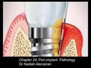
- 3. Healthy peri- implant mucosa
- 4. Peri- Implant Mucosa *Tissues are lined with a keratinized oral epithelium; this is continuous with a thin non- keratinized sulcular epithelium that faces the implant. *In the underlying connective tissue there are small infiltrates of inflammatory cells(neutrophils, macrophages, T cells, B cells) . *Junctional epithelium connects to the dental implant surface via hemidesmosomoes and basal lamina.
- 6. Peri-Implant Complications: A Clinical Guide to Diagnosis and Treatment By Anastasia Kelekis-Cholakis, Reem Atout, Nader Hamdan, Ioannis Tsourounakis
- 7. Teeth Dental Implants periodontal fibers Insert in cementum on the root surfaces of natural teeth Extend parallel to the surface of the implant or abutment Connection Periodontal ligaments Osseointegration Connective tissue Lower percentage of collagen fibers Higher percentage of cells More vascular Higher percentage of collagen fibers Lower percentage of fibroblast Less vascular Blood supply to surrounding gingivae Three different sources (PDL, interdental bone ,supraperiosteal region ) Two different sources (supraperiosteal region,few vesseles from bone ) Biological width JE:0.97-1.14mm CT: 0.77-1.07mm BW:2.04-2.91mm JE:1.88mm CT: 1.05mm BW:3.08mm
- 8. Teeth Dental implants proprioception Periodontal mechanoreceptors Osseoperception Tactile sensitivity High low Axial 25-100 μm 3-5μm Fulcrum when lateral forces applied Apical third of the root crestal bone
- 10. Prevalence of implant diseases Has been reported in several studies ranges from 5-63% * *Introduction to Understanding the Basics of Teeth vs. Dental Implants: Similarities and Differences , Reem Atout1, Nader Hamdan2 and Ioannis Tsourounakis3
- 11. bacterial species associated with peri- implant diseases mainly (Gram –negative anerobic) Staphylococcus aureus are important pathogens in peri- implantitis *Introduction to Understanding the Basics of Teeth vs. Dental Implants: Similarities and Differences , Reem Atout1, Nader Hamdan2 and Ioannis Tsourounakis3
- 12. A disease in which the presence of inflammation is confined to the mucosa surrounding dental implant with no signs of loss of supporting bone.
- 13. Reversible inflammatory process in the soft tissues surrounding a functioning implant.
- 14. 1-signs of redness and swelling. 2-Bleeding on probing . Peri-implant mucositis Clinical features
- 15. Berglundhbet al. (1992) Abrahamsson et al. (1998) Ericsson et al. 1992) Zitzmann et al. (2001) 2- Peri-implant mucositis may or may not progress to peri-implantitis 1-Plaque accumulation on the titanium surface and the formation of a biofilm seem to be essential for the initiation and progression of peri-implant mucositis. Aetiology
- 16. • peri-implant mucositis when effectively treated, can be reversed back to health • So removal of the biofilm from the dental implant surface is the primary objective when treating peri-implant mucositis
- 18. An inflammatory process around dental implant which includes both soft tissue inflammation and loss of supporting bone
- 19. Infl ammatory process additionally characterized by loss of peri-implant bone.
- 20. Clinical features • An inflamatory lesion in the peri-implant mucosa and loss of peri-implant bone. • Peri-implantitis initially affects the marginal part of the peri-implant tissues and the implant may remain stable and in function for varying periods of time. Implant mobility is therefore not an essential symptom for peri-implantitis but may occur in a final stage of disease progression and indicates complete loss of integration.
- 21. Most of the researches related to peri- implantitis suggested that a certain amount of bone loss (≥1.8 mm compared with the 1-year data) together with the finding of BoP was required for the diagnosis peri-implantitis
- 22. Q-Why The rate of progression to the supporting bone is often faster in peri- implantitis compared to periodontitis? A- the periodontal ligament is missing, so inflammation progresses directly to the bone.
- 23. Pathogensis Studies have documented that a biofilm on the implant surface initiates inflammation in the peri-implant mucosa (peri-implant mucositis). This lesion is initially located in the connective tissue immediately lateral to the barrier epithelium. In the continued presence of a submarginal biofilm, the lesion in the marginal mucosa around implants may occasionally spread in an “apical” direction to involve the hard tissue, compromise osseointegration, cause varying degrees of marginal bone loss (peri-implantitis), and eventually the loss of the implant.
- 24. An Individualized Approach to Peri-Mucositis Prevention Oral health professionals well versed in effective diagnostics and prevention strategies can help patients avoid this precursor to peri-implantitis. By Christine DeBaz, BA, Lisa Lang, BSDH, DDS, MS, MBA and Leena Palomo, DDS, MSD
- 25. The End