Aggressive Nodular Melanoma
•
1 like•571 views

Nodular melanoma (NM) is an aggressive form of melanoma that makes up 14-30% of melanoma cases. It is characterized by early vertical growth phase without typical ABCDE signs. A case is presented of a 72-year-old woman with a large NM on her cheek that had grown rapidly over 9 weeks. Her cancer was stage IV with lymph node involvement. She declined further treatment and passed away 3 weeks later. NMs often present at later stages due to their atypical appearance and explosive growth pattern, contributing to higher mortality. Screening high-risk populations and educating about changes over time are important for early detection and management of this deadly skin cancer.

Recommended
More Related Content
What's hot
What's hot (20)
Similar to Aggressive Nodular Melanoma
Similar to Aggressive Nodular Melanoma (20)
More from PLASTIC, COSMETIC, BURNS AND HAND SURGEON
More from PLASTIC, COSMETIC, BURNS AND HAND SURGEON (20)
Recently uploaded
Recently uploaded (20)
Aggressive Nodular Melanoma
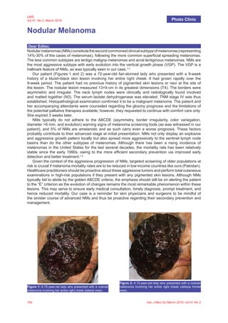
- 1. 164 Iran J Med Sci March 2016; Vol 41 No 2 IJMS Vol 41, No 2, March 2016 Nodular Melanoma Photo Clinic Dear Editor, Nodularmelanomas(NMs)constitutethesecondcommonestclinicalsubtypeofmelanomas(representing 14%-30% of the cases of melanomas), following the more common superficial spreading melanomas. The less common subtypes are lentigo maligna melanomas and acral lentiginous melanomas. NMs are the most aggressive subtype with early evolution into the vertical growth phase (VGP). The VGP is a hallmark feature of NMs, as was typically seen in our case.1-3 Our patient (Figures 1 and 2) was a 72-year-old fair-skinned lady who presented with a 9-week history of a bluish-black skin lesion involving her entire right cheek. It had grown rapidly over the 9-week period. The patient had no previous history of pigmented skin lesions or nevi at the site of the lesion. The nodular lesion measured 13×9 cm in its greatest dimensions (T4). The borders were asymmetric and irregular. The neck lymph nodes were clinically and radiologically found involved and matted together (N3). The serum lactate dehydrogenase was elevated. TNM stage IV was thus established. Histopathological examination confirmed it to be a malignant melanoma. The patient and her accompanying attendants were counseled regarding the gloomy prognosis and the limitations of the potential palliative therapies available; however, they requested to continue with comfort care only. She expired 3 weeks later. NMs typically do not adhere to the ABCDE (asymmetry, border irregularity, color variegation, diameter >6 mm, and evolution) warning signs of melanoma screening tools (as was witnessed in our patient), and 5% of NMs are amelanotic and as such carry even a worse prognosis. These factors probably contribute to their advanced stage at initial presentation. NMs not only display an explosive and aggressive growth pattern locally but also spread more aggressively to the sentinel lymph node basins than do the other subtypes of melanomas. Although there has been a rising incidence of melanomas in the United States for the last several decades, the mortality rate has been relatively stable since the early 1990s, owing to the more efficient secondary prevention via improved early detection and better treatment.1,2 Given the context of the aggressive progression of NMs, targeted screening of older populations at risk is crucial if melanoma mortality rates are to be reduced in low-income countries like ours (Pakistan). Healthcare practitioners should be proactive about these aggressive tumors and perform total cutaneous examinations in high-risk populations if they present with any pigmented skin lesions. Although NMs typically fail to abide by the golden ABCDE criteria, the emphasis should still be on alerting the patient to the “E” criterion as the evolution of changes remains the most remarkable phenomenon within these lesions. This may serve to ensure early medical consultation, timely diagnosis, prompt treatment, and hence reduced mortality. Our case is a reminder for skin physicians and surgeons to be mindful of the sinister course of advanced NMs and thus be proactive regarding their secondary prevention and management. Figure 1: A 72-year-old lady who presented with a nodular melanoma involving her entire right cheek (lateral view). Figure 2: A 72-year-old lady who presented with a nodular melanoma involving her entire right cheek (oblique frontal view).
- 2. Nodular melanoma Iran J Med Sci March 2016; Vol 41 No 2 165 Acknowledgment The authors would like to thank the patient and her attendants for their cooperation and consenting for taking the patient’s photographs and their use for academic and research purposes. Conflict of Interest: None declared. Please cite this article as: Saaiq M, Ashraf B, Siddiqui S. Nodular Melanoma. Iran J Med Sci. 2016;41(2):164-165. Muhammad Saaiq, MBBS, FCPS; Bushra Ashraf, MBBS; Saad Siddiqui, MBBS Pakistan Institute of Medical Sciences (PIMS), Shaheed Zulfiqar Ali Bhutto Medical University (SZABMU), Islamabad, Pakistan Corresponding Author: Muhammad Saaiq, MBBS, FCPS; Department of Plastic Surgery and Burn Care Centre, Pakistan Institute of Medical Sciences (PIMS), Shaheed Zulfiqar Ali Bhutto Medical University (SZABMU), Islamabad-44000, Pakistan Tel: +92 341 5105173 Email: muhammadsaaiq5@gmail.com Received: 17 October 2015 Revised: 10 November 2015 Accepted: 22 November 2015 References 1. Warycha MA, Christos PJ, Mazumdar M, Darvishian F, Shapiro RL, Berman RS, et al. Changes in the presentation of nodular and superficial spreading melanomas over 35 years. Cancer. 2008;113:3341-8. doi: 10.1002/cncr.23955. PubMed PMID: 18988292; PubMed Central PMCID: PMC3624077. 2. Shaikh WR, Xiong M, Weinstock MA.The contribution of nodular subtype to melanoma mortality in the United States, 1978 to 2007. Arch Dermatol. 2012;148:30-6. doi: 10.1001/archdermatol.2011.264. PubMed PMID: 21931016. 3. Moreira J, Moreira E, Azevedo F, Mota A. Cutaneou s malignant melanoma: a retrospective study of seven years (2006-2012). Acta Med Port. 2014;27:480-8. PubMed PMID: 25203957.