MR hazard, protection
•Download as PPTX, PDF•
3 likes•495 views

1. The document discusses safety issues related to magnetic resonance (MR) environments, focusing on the three main electromagnetic fields: static magnetic fields, gradient fields, and radiofrequency fields. 2. It defines potential biological effects and mechanical risks of static magnetic fields, including effects on ECG readings and risks of attractive forces on metallic objects. 3. It also outlines safety considerations and risks related to gradient fields inducing currents, radiofrequency fields causing tissue heating and potential burns, and ensuring proper patient preparation and safety protocols are followed.

Recommended
More Related Content
What's hot
What's hot (20)
Similar to MR hazard, protection
Similar to MR hazard, protection (20)
More from Dr. Mohit Goel
More from Dr. Mohit Goel (20)
Recently uploaded
Recently uploaded (20)
MR hazard, protection
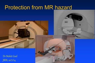
- 1. Protection from MR hazard Dr Mohit Goel JRIII, 10/7/14
- 2. American Society for Testing and Materials (ASTM) published a standard in 2005 for the marking of devices brought into the MR ENVIRONMENT, which includes new safety definitions. Definitions from ASTM International standard
- 4. Static magnetic field biological effects mechanical effects Gradient Field induced currents (PNS) auditory damage RF Field RF power deposition Burns Safety issues centre around the 3 electromagnetic fields and issues indirectly related
- 5. Static magnetic field 1.5 T is 30,000 x the strength of earth’s magnetic field Measured in Gauss or Tesla (10,000G equivalent to 1T)
- 6. Static Magnetic Fields 1. Biological effects (potential risk) - exposure to static magnetic fields of up to 4T are not thought to be harmful Biological effects relevant to clinical imaging - distorted ECG (magnetohydrodynamic effect) - consider prudency with pregnancy 2. Mechanical effects (very real risk) - translational or attractive forces on metallic objects when brought into the field A superconducting magnet is always switched on!
- 7. Static Magnetic Field Biological effects Magnetohydrodynamic effect – augmented T wave Outside field 0.5 Tesla 1.5T
- 8. Magnetohydrodynamic effect seen as augmentation of T-wave • Caused by the effect of the static magnetic field on moving blood (systole) as a conducting fluid. • The gradient and RF fields also affect the configuration of the ECG • Morphological ECG changes are therefore difficult to detect and diagnose, but rhythm is usually recognised • Any concern regarding rhythm, remove patient from scanner and perform 12 lead ECG
- 9. Static Magnetic Field Pregnancy Patients • 1st trimester –avoid MR where possible • 2nd and 3rd trimester – decision made on a risk versus benefit determination. For example if it avoids the patient being subjected to x-rays. Health Care Workers • May enter MR scanning room regardless of trimester • Should not remain in the room when scanner is operational, avoiding exposure to gradient and radiofrequency fields
- 10. Static Magnetic Field Mechanical effects • Projectile or missile effect - the attractive forces exerted by the static magnetic field present the greatest potential for patient injury - objects will be pulled out of hands, pockets etc, and fly into magnet which has caused injury and death. • Effect on ferromagnetic implants - electro-mechanical eg pacemakers - biomedical eg valves, stents
- 11. What is typically ferromagnetic? EQUIPMENT PERSONAL ITEMS (leave outside) • Oxygen cylinders Keys, pens • Wheelchairs Bleeps • Trolleys Mobile phones • IV stands Coins • Monitoring equipment Stethoscopes • Ventilators Scissors It is easy to forget objects, particularly when responding to an emergency!
- 12. Never be complacent – accidents do happen! Oxygen cylinder Infusion pump
- 13. Floor buffer
- 14. • Cerebral aneurysm clips • Metallic foreign body in the eye • Shrapnel, bullets (in critical area) • Ocular implants (containing metal) • Swan-Ganz “Contraindications” to MR* Implants & metal Electromechanical implants • Pacemakers /ICD’s • Pacing wires • Cochlear implants • Neurostimulators • Hydrocephalus shunts Any device electrically or mechanically activated * In some circumstances MR has been performed despite “contraindications”, especially in specialist centres. Work is in progress to make some devices safe. For example, some cerebral aneurysm clips may now be scanned.
- 15. Implants Cerebral clips • modern clips are considered safe (titanium, elgiloy) • obtain operation notes with serial number of clip • take patient consent Foreign bodies • Maybe situated near vascular or nervous tissue • If in doubt – x-ray
- 16. Safety Checklist – comprehensive but concise • Removal of accessories - watch, jewellery (except wedding rings), body piercing rings, hearing aids, glasses, false teeth, artificial limbs and prostheses • Removal of clothes containing metal eg zips, bras Important to know • Previous heart surgery? • Diabetic or epileptic? • Asthmatic or allergies? (in relation to contrast) • Tattoos or permanent eye liner (iron oxide)? Essential to know • Cardiac pacemaker? • Previous neurosurgery? • Implants or metal in the body? • Pregnant (prudent approach)? • Drug patch with foil backing? Essential preparation
- 17. Preparation of patient • Remove watches / jewellery except gold wedding rings • Remove hearing aids, false teeth, glasses, prostheses • Remove all clothes except socks and underpants • Patient gowns - no pockets - no metallic fastenings - ¾ length sleeves for IV access - wrap round for easy chest access • Brass changing room keys • Screen all accompanying personnel • Check any suspicious item with small bar magnet
- 18. Gradient fields - Induced Currents Gradient fields induce an electric field and thus a current in the patient, potentially this can be of sufficient intensity in modern systems to produce a physiological response - peripheral nerve stimulation (PNS) - cardiac stimulation is not considered possible
- 19. Hearing protection mandatory above 90dB time averaged for: • patients • staff remaining in the scanner room • relatives accompanying children or patients Gradient fields – Auditory damage
- 20. Radiofrequency (RF) fields Thermogenic effects - health & safety concern Physiological tissue heating response • most of the transmitted RF power is transferred into heat within the patient’s tissue • all MR systems have safety thresholds to avoid dangerous levels • Patients with compromised thermoregularory systems are at greatest risk
- 21. Specific Absorption Rate (SAR) SAR is the RF power absorbed per unit mass of tissue (expressed in W/kg) • complex function of numerous variables • calculated by software from the average forward power passing into the RF transmitter coil and the body mass situated in the RF transmitting field Therefore an accurate patient weight is vital • SAR increases 9 fold from 0.5T to 1.5T
- 22. Radiofrequency (RF) fields Potential for burns • 1°, 2°, 3° burns have occurred in the past in patients undergoing MRI • This is a result of excessive heat developing in the devices or objects • ECG system is often the culprit • Interventional MRI poses greater risk
- 23. Prevention of burns • Electrodes - carbon fibre studs - placed close together • ECG leads - carbon fibre - fibre optic - high impedance - short as possible (plaited if necessary) • All conductive leads should be placed in a linear fashion coming out of bore of scanner • Avoid crossed limbs where possible
- 24. RF Burn from non-Carbon Electrode
- 25. Quench Cryogens maintain the magnetic field - helium Quenching refers to the events that occur when the liquid cryogens that cool the magnet coils boil off rapidly, which results in helium escaping very rapidly from the cryogen bath. This means that the coils cease to be superconducting and become resistive. A quench will in general be accompanied by a loud bang or thundering or hissing or rushing sound with the cold gas expulsion. Causes - physical - human error (accidental) - intervention (elective) Effects - if no ventilation is present, pressure build up - asphyxiation/frostbite
- 26. Elective Quenching The magnet should only be quenched in two situations:- • If someone is trapped to the scanner by a ferromagnetic object and is injured and/or distressed (eg O2 cylinder, piece of equipment) • If there is a fire in the immediate vicinity on order to reduce risk to the Fire Brigade
- 27. Action to be taken in the event of a Quench • Evacuate the room as quickly as possible • Ensure the door is kept open during evacuation • Close door after evacuation • If trapped in room stay close to floor level • Seek the advice of a senior physicist immediately • Call scanner engineer
- 28. This could be you! Thank you