SlideShare a Scribd company logo
1 of 12
Download to read offline
The new engl and jour nal of medicine
n engl j med 378;8 nejm.org  February 22, 2018740
Review Article
T
he pleural space is defined by the visceral pleura, which covers
the lung, and the parietal pleura, which covers the chest wall, diaphragm,
and mediastinum. It is estimated that pleural effusion develops in more
than 1.5 million patients each year in the United States, with the majority of
cases resulting from congestive heart failure, pneumonia, and cancer.1
Spontane­
ous pneumothorax affects approximately 20,000 patients annually in the United
States, and the incidence of iatrogenic pneumothorax is similar.1
Over the past
several years, substantial advances have been made in our understanding of pleural
biology and related pathophysiology, as well as in the treatment of parapneumonic
effusions, empyema, and malignant pleural effusions and in our understanding of
the high mortality associated with nonmalignant and transudative effusions. In
addition, the definitions and management of pneumothorax have also recently
evolved. For these conditions, the goals of patient care are expeditious and efficient
diagnosis with minimally invasive interventions that avoid the need for multiple
procedures, that minimize hospital days, and that maximize quality of life. This
review considers these various aspects of pleural disease.
Pleur al Anatomy and Pathophysiology
When normal lungs are removed from the chest cavity, their gas volume decreases
as a result of elastic recoil. The chest wall, in contrast, when opened to atmos­
pheric pressure at the end of a normal breath (i.e., at functional residual capacity),
tends to expand. This balance of physical forces keeps the pressure in the pleural
space slightly negative, at approximately −3 to −5 cm of water.2,3
The physiological
function of the pleural space in humans is unclear. One theory maintains that the
pleura serves as an elastic serous membrane to allow changes in lung shape with
respiration, whereas others suggest that the slightly negative pleural pressure at
functional residual capacity prevents atelectasis by maintaining positive transpul­
monary pressure.2,4
Elephants, however, do not have a pleural space; they instead
have layers of loose and dense connective tissue between the lung and chest wall,
and they seem to do just fine. It is postulated that if elephants did have a pleural
space, the pressure gradient between the atmosphere and their submerged thorax
(approximately 150 mm Hg) when they are “snorkeling” across a river would both
rupture the small pleural capillaries and create large transudative pleural effusions.5,6
In fact, humans fare quite well after obliteration of the pleural space (pleurodesis),
with substantial alleviation of dyspnea if a pleural effusion or pneumothorax had
been present. In humans, the parietal and visceral pleura merge at the hilum of
the lungs, separating the thorax into two noncontiguous spaces (the hemithoraxes).
The North American bison, in contrast, has in some cases been found to have an
incomplete mediastinum; this makes it possible to kill these large animals with a
single arrow or gunshot to the chest, which creates bilateral pneumothoraxes.7
From the Division of Pulmonary, Critical
Care, and Sleep Medicine, Johns Hop-
kins University, Baltimore (D.F.-K.); and
the Division of Allergy, Pulmonary, and
Critical Care Medicine, Vanderbilt Uni-
versity, Nashville (R.L.). Address reprint
requests to Dr. Feller-Kopman at the Sec-
tion of Interventional Pulmonology, Johns
Hopkins Hospital, 1800 Orleans St.,
Suite 7-125, Baltimore, MD 21287, or at
­dfk@​­jhmi​.­edu.
N Engl J Med 2018;378:740-51.
DOI: 10.1056/NEJMra1403503
Copyright © 2018 Massachusetts Medical Society.
Julie R. Ingelfinger, M.D., Editor
Pleural Disease
David Feller‑Kopman, M.D., and Richard Light, M.D.​​
The New England Journal of Medicine
Downloaded from nejm.org on February 21, 2018. For personal use only. No other uses without permission.
Copyright © 2018 Massachusetts Medical Society. All rights reserved.
n engl j med 378;8 nejm.org  February 22, 2018 741
Pleural Disease
When considering the pleura, it is important
not to think only of the pleural space, since both
the visceral and parietal pleurae play important
roles in maintaining normal homeostasis. The
pleurae are covered by mesothelial cells, which
are metabolically active and produce many sub­
stances, including hyaluronic acid–rich glycopro­
teins, nitric oxide, and transforming growth
factor β.1
Research over the past several years
has greatly enhanced our understanding of pleu­
ral liquid formation and resorption.8,9
In typical humans, it is estimated that ap­
proximately 0.26 ml of fluid per kilogram of body
weight is contained within each pleural cavity.4,10,11
This fluid is both produced and absorbed pri­
marily on the parietal surface2,12
and is depen­
dent on the balance of hydrostatic and oncotic
pressure differences between the systemic and
pulmonary circulations and the pleural space
(Fig. 1). Lymphatic vessels lying in the parietal
pleura are responsible for pleural fluid resorp­
tion, and the flow rate of these vessels can in­
crease by a factor of approximately 20 in response
to increases in pleural liquid formation.12
Thus,
a clinically significant effusion will be seen only
when fluid production substantially overwhelms
the ability of the lymphatic vessels to resorb fluid,
because of high production, diminished resorp­
tion, or a combination of these two factors.
Evaluation of Pleur al Effusions
The differential diagnosis for pleural effusions is
extensive; a list of potential causes is shown in
Table 1. A systematic and expeditious evaluation
is essential, since a delay in making some diag­
noses (e.g., empyema) is associated with increased
morbidity and mortality.13
The use of point-of-
care ultrasonography in the evaluation of pleural
effusions has been associated with a higher rate
of successful aspiration of fluid from the pleural
space than when no imaging is used, more ac­
curate quantitation of the volume of effusion
than can be obtained with chest radiography,
more accurate detection of septations than can
be obtained with computed tomography (CT) of
the chest, an improvement over radiography in
the ability to identify exudative effusions and
malignant effusions, and perhaps most impor­
tant, fewer complications than when ultrasonog­
raphy is not used to guide pleural procedures.14-20
Thus, ultrasonography is strongly recommended
by the British Thoracic Society to guide pleural
intervention.21,22
Unless the cause of the effusion
is relatively straightforward (e.g., in a patient who
presents with shortness of breath, paroxysmal
nocturnal dyspnea, orthopnea, and lower-extrem­
ity edema with elevated jugular venous disten­
tion and an effusion that is more pronounced on
the right side than on the left, all of which are
suggestive of congestive heart failure), a chest
physician should be involved to help ensure the
timely evaluation of pleural effusion,21
to decrease
the likelihood of associated complications, and to
ensure appropriate follow-up based on the results
of pleural fluid analysis.20,23
Tr ansudates versus Exudates
One of the first steps in the evaluation of patients
with pleural fluid is to distinguish those who
have inflammatory (exudative) effusions from
those who have noninflammatory (transudative)
effusions.24
The use of Light’s criteria for differ­
entiating exudative from transudative effusion,
initially described in 1972, has remained the
standard method over the past 45 years.25
Ac­
cording to Light’s criteria, a patient is consid­
ered to have an exudative effusion when any one
of the following findings is present: a ratio of
pleural fluid protein to serum protein higher
than 0.5, a ratio of pleural fluid lactate dehydro­
genase (LDH) level to serum LDH level higher
than 0.6, or a pleural fluid LDH level higher than
200 IU per liter (or >67% of the upper limit of
the normal range for serum LDH level).25,26
Although these criteria correctly identify near­
ly all exudates, approximately 25% of transu­
dates are misclassified as exudates, especially in
patients who have underlying congestive heart
failure and have received diuretics.27,28
In patients
with suspected congestive heart failure who are
receiving diuretic therapy, a serum protein level that
is more than 3.1 g per deciliter higher than that
in pleural fluid or a serum albumin level that is
more than 1.2 g per deciliter higher than that in
pleural fluid has been suggested to help identify
transudates that were misclassified as exudates
with the use of Light’s criteria. However, the over­
all accuracy of that approach has not been found
to be significantly higher than that with Light’s
criteria.28,29
The New England Journal of Medicine
Downloaded from nejm.org on February 21, 2018. For personal use only. No other uses without permission.
Copyright © 2018 Massachusetts Medical Society. All rights reserved.
n engl j med 378;8 nejm.org February 22, 2018742
The new engl and jour nal of medicine
Similarly, a pleural fluid level of N­terminal
pro–B­type natriuretic peptide (NT­proBNP) high­
er than 1500 pg per milliliter has been shown to
accurately identify effusions due to heart disease
such as congestive heart failure; however, since
serum levels of NT­proBNP are nearly identical
to pleural fluid levels, current recommendations
suggest using the serum NT­proBNP level and
clinical judgment to correctly identify transudates
in patients who have been undergoing active
Figure 1. Balance of Forces Regulating Pleural Fluid Formation.
The amount of fluid in the pleural space is dependent on the balance of hydrostatic and oncotic pressures between the
parietal and visceral pleura and the pleural space. Because hydrostatic pressures are higher on the parietal pleura than
on the visceral pleura and the oncotic pressures are equivalent, pleural fluid is primarily produced from the parietal
pleura. Likewise, the lymphatic vessels on the parietal pleura are responsible for the majority of pleural fluid resorption.
Stoma
35 29
29 29
6
Parietal Space Pleural Space Visceral Space
Pulmonary
capillary
Systemic
capillary
Hydrostatic pressure
+30 cm of water
Hydrostatic pressure
+24 cm of water
Oncotic pressure
+34 cm of water
Net driving force
Hydrostatic pressure
–5 cm of water
Oncotic pressure
+5 cm of water
Oncotic pressure
+34 cm of water
Pulmonary lymphatic
PULMONARY
INTERSTITIUM
Basal lamina
EXTRAPLEURAL PARIETAL
INTERSTITIUM
ALVEOLUS
Visceral
pleura
Parietal
pleura
Net driving force
ALVEOLUS
0
INTERSTITIUM
Parietal lymphatic
The New England Journal of Medicine
Downloaded from nejm.org on February 21, 2018. For personal use only. No other uses without permission.
Copyright © 2018 Massachusetts Medical Society. All rights reserved.
n engl j med 378;8 nejm.org  February 22, 2018 743
Pleural Disease
diuresis for congestive heart failure in the con­
text of a pleural fluid collection.30,31
If serum
levels of protein and albumin are not available
(e.g., in an outpatient who hopes to avoid veni­
puncture), a pleural fluid protein level higher
than 3 g per deciliter or a pleural fluid choles­
terol level higher than 45 mg per deciliter has
been shown to indicate the presence of an exuda­
tive effusion as accurately as Light’s criteria.26,32-34
Common Exudates
Parapneumonic Effusions and Empyema
The most common exudative effusions are
those associated with an underlying pneumonia,
so-called parapneumonic effusions.1
Empyema
refers to frank infection or pus in the pleural
space. The clinical significance of empyema and
the importance of its drainage have been known
for more than 2000 years, and Hippocrates has
been quoted as saying, “Persons who become
affected with empyema after pleurisy, if they get
clear of it in forty days from the breaking of it,
escape the disease; but if not, it passes into
phthisis.”35,36
Despite advances in the treatment
of pneumonia, however, mortality is higher among
patients who have an associated parapneumonic
effusion than among patients with pneumonia
and no effusion,37,38
and delays in drainage are
associated with substantially higher mortality.13
In addition, both the incidence of and mortality
due to parapneumonic effusion and empyema
continue to rise.39,40
Of note, elderly patients often
do not present with the classic symptoms of
cough, fever, sputum, and chest pain, but rather
with anemia, fatigue, and failure to thrive. Prob­
ably in part because of underdiagnosis, elderly
patients also often have more complicated effu­
sions when they are diagnosed, as well as higher
rates of failure of nonsurgical therapy.41
Thus, it is
crucial to consider parapneumonic effusion and
empyema in all elderly patients with pneumonia.
A cornerstone of treating parapneumonic ef­
fusion and empyema is the selection of appropri­
ate antibiotics on the basis of local microbiology
and antibiotic resistance. Patients with commu­
nity-acquired pneumonia tend to be infected with
streptococcus species and anaerobes (e.g., bacte­
roides and peptostreptococcus), whereas patients
with hospital-acquired infection are more like­
ly to have methicillin-resistant staphylococcus
and gram-negative bacteria (e.g., enterobacter).42
Transudative effusions
Congestive heart failure
Cirrhosis
Nephrotic syndrome
Glomerulonephritis
Peritoneal dialysis
Hypoalbuminemia (typical serum albumin, <1.5 mg/dl)
Atelectasis
Superior vena cava obstruction
Trapped lung
Sarcoidosis
Peritoneal dialysis
Myxedema
Cerebrospinal fluid leak or ventriculopleural shunt
Urinothorax
Pulmonary arterial hypertension
Pulmonary embolism
Pericardial disease
Extravascular migration of central venous catheter
Exudative effusions
Infectious: bacterial, viral, tuberculosis-related, fungal, parasitic
Neoplastic: metastatic disease (e.g., lung cancer, breast cancer, lymphoma,
myeloma, ovarian cancer, pancreatic cancer, cholangiocarcinoma), meso-
thelioma, primary body-cavity lymphoma
Paramalignant effusions: reactive pleuritis due to underlying lung cancer, airway
obstruction or atelectasis, radiation-induced pleuritis
Reactive: reactive pleuritis due to underlying pneumonia (i.e., parapneumonic)
Embolic disease: pulmonary embolism
Abdominal disease: pancreatitis, cholecystitis, hepatic or splenic abscess,
esophageal perforation after esophageal varix sclerotherapy
Cardiac or pericardial injury, including myocardial infarction (after coronary-
artery bypass, cardiac surgery, or cardiac ablation procedures), pulmonary-
vein stenosis
Gynecologic: ovarian hyperstimulation, Meigs’ syndrome, endometriosis,
postpartum complications
Collagen vascular disease: rheumatoid arthritis, systemic lupus erythematosus,
Sjögren’s syndrome, familial Mediterranean fever, eosinophilic granulo-
matosis, granulomatosis with polyangiitis
Medications: nitrofurantoin, dantrolene, methysergide, dasatinib, amiodarone,
interleukin-2, procarbazine, methotrexate, clozapine, phenytoin, β-blockers,
ergot drugs
Hemothorax
Chylothorax (most commonly seen after trauma or in patients with lymphoma)
Sarcoidosis
Lymphoplasmacytic lymphoma
Cholesterol effusions (commonly seen in tuberculosis, rheumatoid effusions,
and any other chronic pleural effusion)
Miscellaneous: benign asbestos pleural effusion, yellow nail syndrome, ure-
mia, drowning, amyloidosis, electrical burns, iatrogenic effusion, capillary
leak syndromes, extramedullary hematopoiesis
Table 1. Causes of Pleural Effusions.
The New England Journal of Medicine
Downloaded from nejm.org on February 21, 2018. For personal use only. No other uses without permission.
Copyright © 2018 Massachusetts Medical Society. All rights reserved.
n engl j med 378;8 nejm.org  February 22, 2018744
The new engl and jour nal of medicine
Mortality is significantly higher among patients
with hospital-acquired infection than among
those with community-acquired infection (47%
vs. 17%).42
Rahman and colleagues have devel­
oped a scoring system called RAPID (renal func­
tion, age, purulence, infection source, and dietary
factors) to help identify patients who are at risk
for a poor outcome at the time of their presenta­
tion.43
Scores range from 0 to 7, with values be­
tween 0 and 2 assigned for renal function and
age (with higher scores given for worse renal
function or older age) and scores of 0 or 1 as­
signed for purulence of the effusion (a nonpuru­
lent effusion receives a score of 1), whether the
infection was hospital acquired (score of 1) or
not (score of 0), and dietary factors (a score of
0 is assigned for an albumin level ≥2.7 g per
deciliter and a score of 1 is assigned for a value
below that threshold). In one study, patients in
the high-risk category (those with a RAPID score
of 5 to 7) were found to have at least a 30%
chance of dying in the subsequent 12 weeks, and
thus similar patients may warrant more invasive
initial therapy.
Patients with parapneumonic effusions or em­
pyema have the potential for a deterioration in
their condition, and, given their underlying in­
flammatory state, all such patients should under­
go peripheral-blood culture and should receive
adequate nutrition and prophylaxis for deep-vein
thrombosis.44
As with any other infection in a
closed space, empyema needs to be drained.
Although data from randomized trials are lack­
ing, studies of large retrospective series have
shown that small-bore tubes (≤14-French) per­
form on par with larger-bore tubes in terms of
subsequent mortality and the need for surgery
and are associated with less pain during inser­
tion and while in place.45
However, since tubes
smaller than 12-French have a higher failure rate46
in empyema, our practice is to use a 14-French
pigtail catheter placed with the modified Seld­
inger technique. If the pleural space is not
drained with a small-bore tube, the instillation
of tissue plasminogen activator (t-PA) and DNase
has been successful and in one trial was found
to be associated with significantly better fluid
drainage, a lower likelihood of being referred for
surgery, and a shorter mean hospital stay.47
It
should be noted, however, that t-PA and DNase
have not been shown to decrease mortality, and
the cost of six doses of t-PA–DNase is approxi­
mately $7,000 (Rowden A, Johns Hopkins Hospi­
tal: personal communication). The mean hospital
stay among patients in the t-PA–DNase group in
that trial was 12 days.47
In addition, the avoidance of surgery may not
be the most important outcome measure, since
video-assisted thoracoscopic surgery (VATS) is far
less invasive than thoracotomy. Older, although
smaller, randomized trials showed that VATS
can be the definitive treatment for empyema in
up to 91% of cases,48
and more recent data on
VATS suggest that hospital stays of approximate­
ly 5 to 7 days are usual.49-51
Furthermore, when
performed later in the course of the disease,
surgery is associated with a higher conversion
rate to thoracotomy and more complications than
when it is performed earlier in the disease pro­
cess.52
Our general approach to patients with
parapneumonic effusion or empyema is based on
the recommendations from the British Thoracic
Society44
and is shown in Figure 2. There is cur­
rently a planned randomized trial evaluating
t-PA–DNase versus early VATS for the treatment
of parapneumonic effusion or empyema and
studies examining whether reduced dosing regi­
mens of t-PA–DNase and even irrigation with
normal saline can achieve similar results.54
Malignant Pleur al Effusions
Malignant pleural effusions are the second lead­
ing cause of exudative effusions and the leading
cause of exudates among patients who under­
go thoracentesis; they account for more than
125,000 hospital admissions per year in the
United States, with estimated inpatient mortality
of 11.6% and associated hospital charges of more
than $5 billion per year.55
The majority of malig­
nant pleural effusions arise from lung cancer,
breast cancer, and lymphoma, and it is estimated
that 15% of patients with lung cancer will have
a malignant pleural effusion at presentation and
up to 50% will have a malignant pleural effusion
during the course of their illness. Malignant
pleural effusion is associated with a poor prog­
nosis, with a median survival of 4 to 7 months
from the time of diagnosis.56,57
Even among pa­
tients whose effusions are considered “too small
to tap,” survival is significantly shorter than
among patients without any effusion.57
Survival
The New England Journal of Medicine
Downloaded from nejm.org on February 21, 2018. For personal use only. No other uses without permission.
Copyright © 2018 Massachusetts Medical Society. All rights reserved.
n engl j med 378;8 nejm.org  February 22, 2018 745
Pleural Disease
depends primarily on tumor subtype, with lung
cancer and gastrointestinal cancers having the
worst outcomes (median survival, 2 to 3 months)
and mesothelioma and hematologic cancers hav­
ing the best prognosis, with survival approaching
1 year.58,59
To appropriately treat patients with malignant
pleural effusions, it is crucial to understand the
mechanisms by which pleural effusions cause
dyspnea. Although pleural effusions mildly in­
crease shunt fraction, it is rare to find patients
with substantial hypoxemia.60
The related dys­
pnea is generally not a lung problem due to lung
collapse or to a reduction in pulmonary-function
measures.61
Rather, the dyspnea is a chest-wall
issue caused by the diaphragm being displaced
caudally, which is mechanically disadvantageous
for its length–tension relationship.61,62
The gene­
sis of the dyspnea is important, since the most
clinically relevant questions after large-volume
Figure 2. Management of Parapneumonic Effusions.
Poor prognostic factors after incomplete removal of fluid by means of therapeutic thoracentesis include pus in the pleural space, positive
Gram’s stain or culture, pleural fluid glucose level less than 40 mg per deciliter, pleural fluid pH lower than 7.15, and pleural fluid lactate
dehydrogenase level more than 3 times the upper limit of the normal range for serum.1,53
A decision regarding surgery depends on the
patient’s clinical status and ability to undergo surgery, as well as on local resources and the availability of a skilled surgeon. The figure is
modified from Davies et al.44
The abbreviation t-PA denotes tissue plasminogen activator, and VATS video-assisted thoracoscopic surgery.
Are septations or loculation seen?
Pleural effusion with underlying pneumonia
All patients: antibiotics, venous thromboembolism
prophylaxis, nutrition
Therapeutic thoracentesis
Consider pulmonary and thoracic
surgical consultation for multi-
disciplinary discussion
Chest ultrasound
All fluid removed?
No Yes
Fluid recurs?
Return to chest ultra-
sound box above
Place 14-French chest
tube and consider
t-PA–DNase
Consider early VATS
Continue antibiotics
Yes No
No Yes No Yes
Poor prognostic
factors?
Continue antibiotics,
consider repeat ultra-
sound with or without
thoracentesis in 48 hr
Poor or borderline
surgical candidate
Good surgical
candidate
The New England Journal of Medicine
Downloaded from nejm.org on February 21, 2018. For personal use only. No other uses without permission.
Copyright © 2018 Massachusetts Medical Society. All rights reserved.
n engl j med 378;8 nejm.org  February 22, 2018746
The new engl and jour nal of medicine
thoracentesis are “Is the patient’s breathing bet­
ter?” and “Did the lung fully reexpand?” If the
patient does not feel better after a therapeutic
thoracentesis, something else is causing the dys­
pnea (e.g., pulmonary embolism or lymphangitic
carcinomatosis). In such instances, further diag­
nostic testing should be performed; however,
procedures that address the pleural space should
not be performed. If the patient’s dyspnea has
been alleviated with thoracentesis, the effusion
was at least a major contributor to the dyspnea,
and the dyspnea can be diminished regardless of
whether the lung has reexpanded. If the lung
has reexpanded, the patient can be considered
for pleurodesis, placement of a tunneled pleural
catheter, or combination approaches, whereas if
the lung is nonexpandable, a tunneled pleural
catheter is the treatment of choice (Fig. 3).1,63
Tunneled pleural catheters are small-bore
tubes that are tunneled subcutaneously into the
pleural space, can be placed in the outpatient
setting, and allow patients or caregivers to drain
pleural fluid without subjecting the patient to
additional invasive procedures. Because at least
30% of patients with malignant pleural effusion
do not have reexpansion of the lung, which may
not be evident even at the time of thoracoscopy,64,65
it may be important to perform a large-volume
thoracentesis before deciding on definitive ther­
apy for such patients. The goals of treating pa­
tients with malignant pleural effusion are to
improve quality of life, primarily by minimizing
dyspnea, and to minimize pleural procedures and
the need for repeated hospital or doctor visits.
Given the poor prognosis of these patients, early
and definitive pleural palliation — as opposed to
multiple thoracenteses, which expose the patient
to both risk and inconvenience — is recom­
mended.66
The LENT score (LDH in pleural fluid, Eastern
Cooperative Oncology Group [ECOG] performance
status, neutrophil–lymphocyte ratio in the serum,
and tumor type) has been shown to accurately
stratify patients into high-, moderate-, and low-
risk groups and may be helpful in guiding
therapy.59
A score of 0 is assigned for a pleural
fluid LDH level lower than 1500 IU per liter and
a score of 1 is assigned for a level above that
threshold, scores of 0 to 3 for worsening ECOG
status, a score of 0 for a serum neutrophil–lym­
phocyte ratio of less than 9 and a score of 1 for
a ratio above that threshold, and scores of 0 to
3 based on tumor type. Total scores of 0 or 1 are
considered to indicate low risk and are associ­
ated with a median survival of 319 days, as com­
pared with a median survival of 130 days in the
medium-risk category (scores of 2 through 4)
and 44 days in the high-risk category (scores of
5 through 7).59
For patients in the high-risk cate­
gory, less invasive approaches, such as placement
of a tunneled pleural catheter or even thoracen­
tesis, may be most useful, whereas patients who
are considered at low risk can be treated with
tunneled pleural catheters, pleurodesis, or com­
bination approaches. When discussing options
for patients who have expandable lungs, the risks
and benefits of each procedure should be re­
viewed. The benefits of tunneled pleural cathe­
ters include clinically significant improvement
in dyspnea, placement in the outpatient setting,
and the ability of many patients and families to
care for the catheter themselves at home. How­
ever, patients need to drain such a catheter re­
peatedly until the effusion resolves or until death.
Spontaneous pleurodesis is estimated to occur
in approximately 50% of patients; among pa­
tients in whom it does occur, it occurs at a mean
of approximately 60 days after insertion of the
catheter.67,68
The benefits of pleurodesis include substan­
tial alleviation of dyspnea and not having to man­
age a catheter. However, many centers will keep
patients in the hospital for 3 to 5 days after the
instillation of talc to effect pleural surface fu­
sion, and there is a small risk of transient hypox­
emia associated with the use of nongraded talc.64,69
An unblinded randomized trial examined tun­
neled pleural catheters versus talc-slurry pleurode­
sis for the treatment of persistent effusions and
showed no significant differences in dyspnea
or in quality of life.70
Patients in the talc group
underwent more additional procedures, whereas
the patients in the tunneled pleural catheter
group had a higher incidence of nonserious ad­
verse events. Although it is often a fear of refer­
ring health care providers, infection related to
the tunneled pleural catheter occurs approxi­
mately 5% of the time and can usually be treated
without removing the catheter.71
Trials have sug­
gested that the combination of tunneled pleural
catheters with sclerosing agents (talc or silver
nitrate) as well as daily drainage (as opposed to
The New England Journal of Medicine
Downloaded from nejm.org on February 21, 2018. For personal use only. No other uses without permission.
Copyright © 2018 Massachusetts Medical Society. All rights reserved.
n engl j med 378;8 nejm.org  February 22, 2018 747
Pleural Disease
drainage every other day) can result in substan­
tially fewer days with a catheter.72-74
As with all
procedures, we recommend that the risks, ben­
efits, and alternatives always be discussed with
the patient in detail and that therapy be indi­
vidualized.
Complicated Tr ansudates
Congestive heart failure, cirrhosis, and the ne­
phrotic syndrome underlie most transudative
effusions.1
Although often considered benign
conditions, effusions associated with congestive
heart failure, hepatic failure, and renal failure
have recently been shown to be associated with
1-year mortality rates of 50%, 25%, and 46%,
respectively.75
Patients with congestive heart fail­
ure and pleural effusion have a 1-year risk of
death similar to that of patients who are admit­
ted to the intensive care unit with acute decom­
pensated heart failure, patients with hepatic hy­
drothorax have a risk of death similar to that of
patients with a Model for End-Stage Liver Dis­
ease (MELD) score of 20 to 29 (a typical indica­
tion for transplantation; MELD scores range
from 6 to 40, with higher scores indicating more
advanced liver disease), and patients with renal
failure and effusion have a 1-year risk of death
that is triple that among patients undergoing
hemodialysis who do not have effusions.75
Al­
though in the majority of patients, transudative
effusions can be managed by treatment of the
underlying condition, refractory effusions deserve
prompt and aggressive pleural palliation that
will minimize repeat procedures and breathless­
ness and maximize quality of life. As with malig­
Figure 3. Management of Malignant Pleural Effusions.
The figure is modified from Roberts et al.63
Improvement in dyspnea?
Pleural effusion with underlying cancer
Investigate for other
causes of dyspnea
Ultrasound-guided therapeutic
thoracentesis
Lung reexpansion?
No Yes
Suspected survival
>1 wk?
Consider placement of tunneled
pleural catheter
Palliate dyspnea with repeat
thoracentesis if needed, oxygen,
or morphine
No Yes
No Yes
Discuss relative risks and
benefits of tunneled pleural
catheter vs. pleurodesis
vs. combination approaches
The New England Journal of Medicine
Downloaded from nejm.org on February 21, 2018. For personal use only. No other uses without permission.
Copyright © 2018 Massachusetts Medical Society. All rights reserved.
n engl j med 378;8 nejm.org  February 22, 2018748
The new engl and jour nal of medicine
nant pleural effusions, tunneled pleural cathe­
ters, pleurodesis, or both may be indicated for
these patients,76-79
and we recommend careful
discussion with relevant teams (e.g., hepatology
and liver transplantation) to develop a multidis­
ciplinary plan.
Current Concepts in the
Treatment of Pneumothor ax
Pneumothorax has traditionally been categorized
as primary (no underlying lung disease), second­
ary (underlying lung disease present), traumatic,
and iatrogenic. Because of advances in chest
imaging (CT and thoracoscopy), patients with a
pneumothorax who had previously been con­
sidered free of parenchymal disease have been
found to have emphysema-like pulmonary
changes and increased pleural porosity, or de­
fects in the visceral pleura that are independent
of blebs or bullae.80
These findings suggest that
the distinction between primary and secondary
effusion is perhaps an artificial construct and
that therapy should be guided by size of the
pneumothorax and by the patient’s symptoms.81
Furthermore, there is no standard definition of
pneumothorax size; the American College of Chest
Physicians defines “large” as a distance of 3 cm
or more from the apex of the lung to the cupula
of the chest wall,82
whereas the British Thoracic
Society defines it as an intrapleural distance of
at least 2 cm at the level of the hilum.83
In fact,
agreement on size based on these definitions
occurs less than 50% of the time in clinical set­
tings, which leads to substantial variation in
treatment recommendations.84
Up to 70% of patients with a clinically stable
pneumothorax can be treated with simple needle
aspiration, which avoids hospitalization. As with
parapneumonic effusion and empyema, guide­
lines currently recommend the use of small-bore
(14-French) chest tubes rather than large-bore
chest tubes for patients with pneumothorax who
have treatment failure or are not candidates for
simple needle aspiration.83
The latter group in­
cludes patients who live far from the treating
center, who have minimal social support, or who
have more substantial underlying lung disease.
Over the past several years, more conservative
therapy has been a trend associated with reserv­
ing surgical therapy for patients with the highest
risk of recurrent effusions. Digital air-leak mon­
itoring devices have been shown to reduce the
number of days a chest tube is in place and to
shorten the length of stay in the hospital after
lobectomy or segmentectomy.85
When patients with a pneumothorax are treat­
ed with chest tubes, the lung usually expands
and the air leak ceases within 3 days. If the lung
does not expand fully within 3 to 5 days, consid­
eration should be given to thoracoscopy. At thora­
coscopy, blebs are stapled and an effort is made
to create a pleurodesis, usually with pleural
abrasion. Another method of treating prolonged
air leak is to instill 1 ml of the patient’s own
blood per kilogram of body weight through the
chest tube.86
Alternatively, pleurodesis can be
attempted through instillation of a sclerosing
agent or the placement of endobronchial one-way
valves, with the goal of reducing air flow across
the visceral pleura. The valves are then removed
after the pleural defect has healed, typically in
6 weeks.87
After a patient has had a spontaneous pneu­
mothorax, the likelihood of a recurrence exceeds
50%.88
Prevention of recurrence is key, especially
in patients with markedly decreased lung func­
tion, for whom a recurrence can be fatal. If a
patient has a first recurrence, then the likeli­
hood of a second recurrence is very high. Recur­
rence rates can be reduced to approximately 25%
if an agent such as talc or doxycycline is instilled
through a chest tube and can be reduced to less
than 5% with thoracoscopy and the insufflation
of talc, stapling of blebs, or pleural abrasion to
create a pleurodesis.89
However, a bullectomy
alone, without attempts at pleurodesis, is associ­
ated with a higher recurrence rate, and therefore
pleurodesis should always be considered an inte­
gral part of the procedure.81
Areas for Future Research
Over the past few years, large, multicenter, ran­
domized trials have been published from centers
that have large clinical and research pleural ser­
vices. These trials may lead to improvements in
diagnostic tests to establish the underlying cause
of a pleural effusion, more compounds to de­
crease the rate of pleural fluid production or
increase the rate of pleural fluid reabsorption,
an improved compound for pleurodesis, and fur­
The New England Journal of Medicine
Downloaded from nejm.org on February 21, 2018. For personal use only. No other uses without permission.
Copyright © 2018 Massachusetts Medical Society. All rights reserved.
n engl j med 378;8 nejm.org  February 22, 2018 749
Pleural Disease
ther development of interventional pulmonology
services and dedicated multidisciplinary pleural
disease services. Furthermore, trials investigating
the clinical effect of pleural manometry, studies
to better understand the pharmacodynamics of
drugs in the pleural space, and investigation of
how pleural disease is related to the genetic
makeup of affected patients may be forthcom­
ing. Patient-centered (and caregiver-centered) out­
comes, such as the effect on daily quality of life,
are also being investigated. Pleural disease re­
mains a common clinical problem, and expedi­
tious, multidisciplinary evaluation and treatment
will maximize the care of our patients.
Dr. Feller-Kopman reports receiving grant support and consult­
ing fees from CareFusion/BD; and Dr. Light, serving on an advi­
sory committee for CareFusion and holding a patent (US 6103695
A) on transforming growth factor β for pleurodesis. No other
potential conflict of interest relevant to this article was reported.
Disclosure forms provided by the authors are available with
the full text of this article at NEJM.org.
References
1.	 Light R. Pleural disease. 6th ed. Phila­
delphia:​Lippincott Williams & Wilkins,
2013.
2.	 Agostoni E. Mechanics of the pleural
space. Physiol Rev 1972;​52:​57-128.
3.	 Lai-Fook SJ. Pleural mechanics and
fluid exchange. Physiol Rev 2004;​84:​385-
410.
4.	 Wang NS. Anatomy of the pleura. Clin
Chest Med 1998;​19:​229-40.
5.	 West JB. Why doesn’t the elephant have
a pleural space? News Physiol Sci 2002;​17:​
47-50.
6.	 West JB. Snorkel breathing in the ele­
phant explains the unique anatomy of its
pleura. Respir Physiol 2001;​126:​1-8.
7.	 Grathwohl KW, Derdak S. Buffalo
chest. N Engl J Med 2003;​349:​1829.
8.	 Stathopoulos GT, Moschos C, Loutrari
H, et al. Zoledronic acid is effective
against experimental malignant pleural
effusion. Am J Respir Crit Care Med 2008;​
178:​50-9.
9.	 Psallidas I, Kalomenidis I, Porcel JM,
Robinson BW, Stathopoulos GT. Malig­
nant pleural effusion: from bench to bed­
side. Eur Respir Rev 2016;​25:​189-98.
10.	Agostoni E, D’Angelo E. Thickness
and pressure of the pleural liquid at vari­
ous heights and with various hydrotho­
races. Respir Physiol 1969;​6:​330-42.
11.	Noppen M, De Waele M, Li R, et al.
Volume and cellular content of normal
pleural fluid in humans examined by
pleural lavage. Am J Respir Crit Care Med
2000;​162:​1023-6.
12.	Miserocchi G. Physiology and patho­
physiology of pleural fluid turnover. Eur
Respir J 1997;​10:​219-25.
13.	 Ashbaugh DG. Empyema thoracis: fac­
tors influencing morbidity and mortality.
Chest 1991;​99:​1162-5.
14.	Kohan JM, Poe RH, Israel RH, et al.
Value of chest ultrasonography versus de­
cubitus roentgenography for thoracente­
sis. Am Rev Respir Dis 1986;​133:​1124-6.
15.	Diacon AH, Brutsche MH, Solèr M.
Accuracy of pleural puncture sites: a pro­
spective comparison of clinical examina­
tion with ultrasound. Chest 2003;​123:​436-
41.
16.	Eibenberger KL, Dock WI, Ammann
ME, Dorffner R, Hörmann MF, Graben­
wöger F. Quantification of pleural effu­
sions: sonography versus radiography.
Radiology 1994;​191:​681-4.
17.	Grogan DR, Irwin RS, Channick R,
et al. Complications associated with thora­
centesis: a prospective, randomized study
comparing three different methods. Arch
Intern Med 1990;​150:​873-7.
18.	Yang PC, Luh KT, Chang DB, Wu HD,
Yu CJ, Kuo SH. Value of sonography in de­
termining the nature of pleural effusion:
analysis of 320 cases. AJR Am J Roent­
genol 1992;​159:​29-33.
19.	 Qureshi NR, Rahman NM, Gleeson FV.
Thoracic ultrasound in the diagnosis of
malignant pleural effusion. Thorax 2009;​
64:​139-43.
20.	Gordon CE, Feller-Kopman D, Balk
EM, Smetana GW. Pneumothorax follow­
ing thoracentesis: a systematic review and
meta-analysis. Arch Intern Med 2010;​170:​
332-9.
21.	 Hooper C, Lee YCG, Maskell N. Inves­
tigation of a unilateral pleural effusion in
adults: British Thoracic Society Pleural
Disease Guideline 2010. Thorax 2010;​65:​
Suppl 2:​ii4-ii17.
22.	Havelock T, Teoh R, Laws D, Gleeson
F. Pleural procedures and thoracic ultra­
sound: British Thoracic Society Pleural
Disease Guideline 2010. Thorax 2010;​65:​
Suppl 2:​ii61-ii76.
23.	Hooper CE, Lee YC, Maskell NA. Set­
ting up a specialist pleural disease service.
Respirology 2010;​15:​1028-36.
24.	Paddock FK. The diagnostic signifi­
cance of serous fluids in disease. N Engl J
Med 1940;​223:​1010-5.
25.	Light RW, Macgregor MI, Luchsinger
PC, Ball WC Jr. Pleural effusions: the diag­
nostic separation of transudates and exu­
dates. Ann Intern Med 1972;​77:​507-13.
26.	Heffner JE, Brown LK, Barbieri CA.
Diagnostic value of tests that discrimi­
nate between exudative and transudative
pleural effusions. Chest 1997;​111:​970-80.
27.	Romero-Candeira S, Fernández C,
Martín C, Sánchez-Paya J, Hernández L.
Influence of diuretics on the concentra­
tion of proteins and other components of
pleural transudates in patients with heart
failure. Am J Med 2001;​110:​681-6.
28.	Romero-Candeira S, Hernández L,
Romero-Brufao S, Orts D, Fernández C,
Martín C. Is it meaningful to use bio­
chemical parameters to discriminate be­
tween transudative and exudative pleural
effusions? Chest 2002;​122:​1524-9.
29.	Bielsa S, Porcel JM, Castellote J, Mas
E, Esquerda A, Light RW. Solving the
Light’s criteria misclassification rate of
cardiac and hepatic transudates. Respirol­
ogy 2012;​17:​721-6.
30.	Porcel JM, Vives M, Cao G, Esquerda
A, Rubio M, Rivas MC. Measurement of
pro-brain natriuretic peptide in pleural
fluid for the diagnosis of pleural effu­
sions due to heart failure. Am J Med 2004;​
116:​417-20.
31.	 Kolditz M, Halank M, Schiemanck CS,
Schmeisser A, Höffken G. High diagnos­
tic accuracy of NT-proBNP for cardiac
origin of pleural effusions. Eur Respir J
2006;​28:​144-50.
32.	Carr DT, Power MH. Clinical value of
measurements of concentration of pro­
tein in pleural fluid. N Engl J Med 1958;​
259:​926-7.
33.	 Costa M, Quiroga T, Cruz E. Measure­
ment of pleural fluid cholesterol and lac­
tate dehydrogenase: a simple and accurate
set of indicators for separating exudates
from transudates. Chest 1995;​108:​1260-3.
34.	Porcel JM, Vives M, Vicente de Vera
MC, Cao G, Rubio M, Rivas MC. Useful
tests on pleural fluid that distinguish
transudates from exudates. Ann Clin Bio­
chem 2001;​38:​671-5.
35.	 Internet Classics Archive. Aphorisms
by Hippocrates (Adams F, translator) (http://
classics​.mit​.edu/​Hippocrates/​aphorisms​
.mb​.txt).
36.	Adams F. The genuine works of Hip­
pocrates. London:​C. and J. Adlard Print­
ers, 1848.
37.	 Hasley PB, Albaum MN, Li YH, et al.
Do pulmonary radiographic findings at
presentation predict mortality in patients
with community-acquired pneumonia?
Arch Intern Med 1996;​156:​2206-12.
The New England Journal of Medicine
Downloaded from nejm.org on February 21, 2018. For personal use only. No other uses without permission.
Copyright © 2018 Massachusetts Medical Society. All rights reserved.
n engl j med 378;8 nejm.org  February 22, 2018750
The new engl and jour nal of medicine
38.	Menéndez R, Torres A, Zalacaín R,
et al. Risk factors of treatment failure in
community acquired pneumonia: implica­
tions for disease outcome. Thorax 2004;​
59:​960-5.
39.	 Grijalva CG, Zhu Y, Nuorti JP, Griffin
MR. Emergence of parapneumonic empy­
ema in the USA. Thorax 2011;​66:​663-8.
40.	Bender JM, Ampofo K, Sheng X, Pavia
AT, Cannon-Albright L, Byington CL. Para­
pneumonic empyema deaths during past
century, Utah. Emerg Infect Dis 2009;​15:​
44-8.
41.	El Solh AA, Alhajjhasan A, Ramadan
FH, Pineda LA. A comparative study of
community- and nursing home-acquired
empyema thoracis. J Am Geriatr Soc 2007;​
55:​1847-52.
42.	 Maskell NA, Batt S, Hedley EL, Davies
CW, Gillespie SH, Davies RJ. The bacteri­
ology of pleural infection by genetic and
standard methods and its mortality sig­
nificance. Am J Respir Crit Care Med
2006;​174:​817-23.
43.	Rahman NM, Kahan BC, Miller RF,
Gleeson FV, Nunn AJ, Maskell NA. A clin­
ical score (RAPID) to identify those at risk
for poor outcome at presentation in pa­
tients with pleural infection. Chest 2014;​
145:​848-55.
44.	Davies HE, Davies RJO, Davies CWH.
Management of pleural infection in adults:
British Thoracic Society Pleural Disease
Guideline 2010. Thorax 2010;​65:​Suppl 2:​
ii41-ii53.
45.	Rahman NM, Maskell NA, Davies
CWH, et al. The relationship between
chest tube size and clinical outcome in
pleural infection. Chest 2010;​137:​536-43.
46.	Keeling AN, Leong S, Logan PM, Lee
MJ. Empyema and effusion: outcome of
image-guided small-bore catheter drain­
age. Cardiovasc Intervent Radiol 2008;​31:​
135-41.
47.	 Rahman NM, Maskell NA, West A, et al.
Intrapleural use of tissue plasminogen
activator and DNase in pleural infection.
N Engl J Med 2011;​365:​518-26.
48.	Wait MA, Sharma S, Hohn J, Dal
Nogare A. A randomized trial of empy­
ema therapy. Chest 1997;​111:​1548-51.
49.	 Kim BY, Oh BS, Jang WC, Min YI, Park
YK, Park JC. Video-assisted thoracoscopic
decortication for management of post­
pneumonic pleural empyema. Am J Surg
2004;​188:​321-4.
50.	Marks DJ, Fisk MD, Koo CY, et al.
Thoracic empyema: a 12-year study from a
UK tertiary cardiothoracic referral centre.
PLoS One 2012;​7(1):​e30074.
51.	Shahin Y, Duffy J, Beggs D, Black E,
Majewski A. Surgical management of pri­
mary empyema of the pleural cavity: out­
come of 81 patients. Interact Cardiovasc
Thorac Surg 2010;​10:​565-7.
52.	 Scarci M, Abah U, Solli P, et al. EACTS
expert consensus statement for surgical
management of pleural empyema. Eur J
Cardiothorac Surg 2015;​48:​642-53.
53.	Porcel JM, Valencia H, Bielsa S. Fac­
tors influencing pleural drainage in para­
pneumonic effusions. Rev Clin Esp 2016;​
216:​361-6.
54.	Hooper CE, Edey AJ, Wallis A, et al.
Pleural Irrigation Trial (PIT): a random­
ised controlled trial of pleural irrigation
with normal saline versus standard care
in patients with pleural infection. Eur
Respir J 2015;​46:​456-63.
55.	 Taghizadeh N, Fortin M, Tremblay A.
US hospitalizations for malignant pleural
effusions: data from the 2012 National
Inpatient Sample. Chest 2017;​151:​845-54.
56.	Heffner JE, Nietert PJ, Barbieri C.
Pleural fluid pH as a predictor of pleurode­
sis failure: analysis of primary data. Chest
2000;​117:​87-95.
57.	 Porcel JM, Gasol A, Bielsa S, Civit C,
Light RW, Salud A. Clinical features and
survival of lung cancer patients with pleu­
ral effusions. Respirology 2015;​20:​654-9.
58.	Postmus PE, Brambilla E, Chansky K,
et al. The IASLC Lung Cancer Staging
Project: proposals for revision of the M
descriptors in the forthcoming (seventh)
edition of the TNM classification of lung
cancer. J Thorac Oncol 2007;​2:​686-93.
59.	 Clive AO, Kahan BC, Hooper CE, et al.
Predicting survival in malignant pleural
effusion: development and validation of
the LENT prognostic score. Thorax 2014;​
69:​1098-104.
60.	Agustí AGN, Cardús J, Roca J, Grau
JM, Xaubet A, Rodriguez-Roisin R. Venti­
lation-perfusion mismatch in patients with
pleural effusion: effects of thoracentesis.
Am J Respir Crit Care Med 1997;​156:​1205-9.
61.	Estenne M, Yernault JC, De Troyer A.
Mechanism of relief of dyspnea after thora­
cocentesis in patients with large pleural
effusions. Am J Med 1983;​74:​813-9.
62.	Krell WS, Rodarte JR. Effects of acute
pleural effusion on respiratory system
mechanics in dogs. J Appl Physiol (1985)
1985;​59:​1458-63.
63.	 Roberts ME, Neville E, Berrisford RG,
Antunes G, Ali NJ. Management of a ma­
lignant pleural effusion: British Thoracic
Society Pleural Disease Guideline 2010.
Thorax 2010;​65:​Suppl 2:​ii32-ii40.
64.	Dresler CM, Olak J, Herndon JE II, et al.
Phase III intergroup study of talc poudrage
vs talc slurry sclerosis for malignant pleu­
ral effusion. Chest 2005;​127:​909-15.
65.	Hallifax RJ, Corcoran JP, Psallidas I,
Rahman NM. Medical thoracoscopy: sur­
vey of current practice — how successful
are medical thoracoscopists at predicting
malignancy? Respirology 2016;​21:​958-60.
66.	Ost DE, Niu J, Zhao H, Grosu HB,
Giordano SH. Quality gaps and compara­
tive effectiveness of management strate­
gies for recurrent malignant pleural effu­
sions. Chest 2017 August 31 (Epub ahead
of print).
67.	 Tremblay A, Michaud G. Single-center
experience with 250 tunnelled pleural
catheter insertions for malignant pleural
effusion. Chest 2006;​129:​362-8.
68.	Kheir F, Shawwa K, Alokla K, Om­
balli M, Alraiyes AH. Tunneled pleural
catheter for the treatment of malignant
pleural effusion: a systematic review and
meta-analysis. Am J Ther 2016;​23(6):​e1300-
e1306.
69.	Gonzalez AV, Bezwada V, Beamis JF
Jr, Villanueva AG. Lung injury following
thoracoscopic talc insufflation: experience
of a single North American center. Chest
2010;​137:​1375-81.
70.	 Davies HE, Mishra EK, Kahan BC, et al.
Effect of an indwelling pleural catheter vs
chest tube and talc pleurodesis for reliev­
ing dyspnea in patients with malignant
pleural effusion: the TIME2 randomized
controlled trial. JAMA 2012;​307:​2383-9.
71.	 Fysh ETH, Tremblay A, Feller-Kopman
D, et al. Clinical outcomes of indwelling
pleural catheter-related pleural infections:
an international multicenter study. Chest
2013;​144:​1597-602.
72.	 Reddy C, Ernst A, Lamb C, Feller-Kop­
man D. Rapid pleurodesis for malignant
pleural effusions: a pilot study. Chest 2011;​
139:​1419-23.
73.	 Bhatnagar R, Zahan-Evans N, Kearney
C, et al. A novel drug-eluting indwelling
pleural catheter for the management of
malignant effusions. Am J Respir Crit Care
Med 2018;​197:​136-8.
74.	 Wahidi MM, Reddy C, Yarmus L, et al.
Randomized trial of pleural fluid drain­
age frequency in patients with malignant
pleural effusions: the ASAP Trial. Am J
Respir Crit Care Med 2017;​195:​1050-7.
75.	Walker SP, Morley AJ, Stadon L, et al.
Nonmalignant pleural effusions: a pro­
spective study of 356 consecutive unselect­
ed patients. Chest 2017;​151:​1099-105.
76.	 Patil M, Dhillon SS, Attwood K, Saoud
M, Alraiyes AH, Harris K. Management of
benign pleural effusions using indwelling
pleural catheters: a systematic review and
meta-analysis. Chest 2017;​151:​626-35.
77.	 Chen A, Massoni J, Jung D, Crippin J.
Indwelling tunneled pleural catheters for
the management of hepatic hydrothorax:
a pilot study. Ann Am Thorac Soc 2016;​13:​
862-6.
78.	Majid A, Kheir F, Fashjian M, et al.
Tunneled pleural catheter placement with
and without talc poudrage for treatment
of pleural effusions due to congestive heart
failure. Ann Am Thorac Soc 2016;​13:​212-6.
79.	Bintcliffe OJ, Lee GY, Rahman NM,
Maskell NA. The management of benign
non-infective pleural effusions. Eur Respir
Rev 2016;​25:​303-16.
80.	Noppen M, Stratakos G, Verbanck S,
D’Haese J, Meysman M, Vincken W. Fluo­
rescein-enhanced autofluorescence thora­
coscopy in primary spontaneous pneumo­
thorax. Am J Respir Crit Care Med 2004;​
170:​680-2.
81.	 Bintcliffe OJ, Hallifax RJ, Edey A, et al.
Spontaneous pneumothorax: time to re­
think management? Lancet Respir Med
2015;​3:​578-88.
82.	Baumann MH, Strange C, Heffner JE,
et al. Management of spontaneous pneu­
The New England Journal of Medicine
Downloaded from nejm.org on February 21, 2018. For personal use only. No other uses without permission.
Copyright © 2018 Massachusetts Medical Society. All rights reserved.
n engl j med 378;8 nejm.org  February 22, 2018 751
Pleural Disease	
mothorax: an American College of Chest
Physicians Delphi consensus statement.
Chest 2001;​119:​590-602.
83.	 MacDuff A, Arnold A, Harvey J. Man­
agement of spontaneous pneumothorax:
British Thoracic Society Pleural Disease
Guideline 2010. Thorax 2010;​65:​Suppl 2:​
ii18-ii31.
84.	 Kelly AM, Druda D. Comparison of
size classification of primary spontaneous
pneumothorax by three international
guidelines: a case for international con­
sensus? Respir Med 2008;​102:​1830-2.
85.	 Pompili C, Detterbeck F, Papagian­
nopoulos K, et al. Multicenter interna­
tional randomized comparison of objec­
tive and subjective outcomes between
electronic and traditional chest drainage
systems. Ann Thorac Surg 2014;​98:​490-6.
86.	 Cao G, Kang J, Wang F, Wang H. Intra­
pleural instillation of autologous blood for
persistent air leak in spontaneous pneumo­
thorax in patients with advanced chronic
obstructive pulmonary disease. Ann Thorac
Surg 2012;​93:​1652-7.
87.	 Reed MF, Gilbert CR, Taylor MD, Toth
JW. Endobronchial valves for challenging
air leaks. Ann Thorac Surg 2015;​100:​1181-6.
88.	 Sadikot RT, Greene T, Meadows K,
Arnold AG. Recurrence of primary spon­
taneous pneumothorax. Thorax 1997;​52:​
805-9.
89.	 Shaikhrezai K, Thompson AI, Parkin C,
Stamenkovic S, Walker WS. Video-assisted
thoracoscopic surgery management of
spontaneous pneumothorax — long-term
results. Eur J Cardiothorac Surg 2011;​40:​
120-3.
Copyright © 2018 Massachusetts Medical Society.
The New England Journal of Medicine
Downloaded from nejm.org on February 21, 2018. For personal use only. No other uses without permission.
Copyright © 2018 Massachusetts Medical Society. All rights reserved.

More Related Content

What's hot

Principales patrones radiológicos en la placa simple de tórax
Principales patrones radiológicos en la placa simple de tóraxPrincipales patrones radiológicos en la placa simple de tórax
Principales patrones radiológicos en la placa simple de tóraxMercedes Calleja
 
NODULO PULMONAR SOLITARIO (NPS) BUAP MEDICINA
NODULO PULMONAR SOLITARIO (NPS) BUAP MEDICINA NODULO PULMONAR SOLITARIO (NPS) BUAP MEDICINA
NODULO PULMONAR SOLITARIO (NPS) BUAP MEDICINA Marisol Mora López
 
Derrame Paraneumónico y Empiema
Derrame Paraneumónico y EmpiemaDerrame Paraneumónico y Empiema
Derrame Paraneumónico y EmpiemaAlexaCantoral
 
HOMELPAVI - EGRESO - CorPulmonar Crónico
HOMELPAVI - EGRESO - CorPulmonar CrónicoHOMELPAVI - EGRESO - CorPulmonar Crónico
HOMELPAVI - EGRESO - CorPulmonar CrónicoCarmelo Gallardo
 
Clasificación braunwald de angina inestable
Clasificación braunwald de angina inestableClasificación braunwald de angina inestable
Clasificación braunwald de angina inestableDrMandingo WEB
 
Derrame pleural, atualización y tratamiento
Derrame pleural, atualización y tratamientoDerrame pleural, atualización y tratamiento
Derrame pleural, atualización y tratamientoformaciossibe
 
Síndrome de Distrés Respiratorio Agudo Pediátrico
Síndrome de Distrés Respiratorio Agudo PediátricoSíndrome de Distrés Respiratorio Agudo Pediátrico
Síndrome de Distrés Respiratorio Agudo PediátricoFelipe Cadena Suàrez
 
Cardiopatias acianoticas md6 1
Cardiopatias acianoticas md6 1Cardiopatias acianoticas md6 1
Cardiopatias acianoticas md6 1lewis1886
 
4 bloqueos de rama hh
4 bloqueos de rama hh4 bloqueos de rama hh
4 bloqueos de rama hhMocte Salaiza
 
12. CARDIOPATÍAS CONGÉNITAS ACI Y CIANÓTICAS.pdf
12. CARDIOPATÍAS CONGÉNITAS ACI Y CIANÓTICAS.pdf12. CARDIOPATÍAS CONGÉNITAS ACI Y CIANÓTICAS.pdf
12. CARDIOPATÍAS CONGÉNITAS ACI Y CIANÓTICAS.pdfPoolPrez
 

What's hot (20)

Principales patrones radiológicos en la placa simple de tórax
Principales patrones radiológicos en la placa simple de tóraxPrincipales patrones radiológicos en la placa simple de tórax
Principales patrones radiológicos en la placa simple de tórax
 
Neumonias intersticiales idiopáticas dr casanova
Neumonias intersticiales idiopáticas dr casanova Neumonias intersticiales idiopáticas dr casanova
Neumonias intersticiales idiopáticas dr casanova
 
Hipertension pulmonar
Hipertension pulmonarHipertension pulmonar
Hipertension pulmonar
 
NODULO PULMONAR SOLITARIO (NPS) BUAP MEDICINA
NODULO PULMONAR SOLITARIO (NPS) BUAP MEDICINA NODULO PULMONAR SOLITARIO (NPS) BUAP MEDICINA
NODULO PULMONAR SOLITARIO (NPS) BUAP MEDICINA
 
Derrame Paraneumónico y Empiema
Derrame Paraneumónico y EmpiemaDerrame Paraneumónico y Empiema
Derrame Paraneumónico y Empiema
 
HOMELPAVI - EGRESO - CorPulmonar Crónico
HOMELPAVI - EGRESO - CorPulmonar CrónicoHOMELPAVI - EGRESO - CorPulmonar Crónico
HOMELPAVI - EGRESO - CorPulmonar Crónico
 
Atresia tricuspidea
Atresia tricuspideaAtresia tricuspidea
Atresia tricuspidea
 
Clasificación braunwald de angina inestable
Clasificación braunwald de angina inestableClasificación braunwald de angina inestable
Clasificación braunwald de angina inestable
 
Derrame pleural maligno
Derrame pleural malignoDerrame pleural maligno
Derrame pleural maligno
 
Taquipnea Transitoria del Recién Nacido
Taquipnea Transitoria del Recién NacidoTaquipnea Transitoria del Recién Nacido
Taquipnea Transitoria del Recién Nacido
 
Cardiaco Cia Civ Dap
Cardiaco Cia Civ DapCardiaco Cia Civ Dap
Cardiaco Cia Civ Dap
 
Derrame pleural, atualización y tratamiento
Derrame pleural, atualización y tratamientoDerrame pleural, atualización y tratamiento
Derrame pleural, atualización y tratamiento
 
Atresia Tricuspidea Expo
Atresia Tricuspidea ExpoAtresia Tricuspidea Expo
Atresia Tricuspidea Expo
 
Metastasis pulmonares
Metastasis pulmonaresMetastasis pulmonares
Metastasis pulmonares
 
Síndrome de Distrés Respiratorio Agudo Pediátrico
Síndrome de Distrés Respiratorio Agudo PediátricoSíndrome de Distrés Respiratorio Agudo Pediátrico
Síndrome de Distrés Respiratorio Agudo Pediátrico
 
Tbc
TbcTbc
Tbc
 
Cardiopatias acianoticas md6 1
Cardiopatias acianoticas md6 1Cardiopatias acianoticas md6 1
Cardiopatias acianoticas md6 1
 
Soplos Cardiacos
Soplos CardiacosSoplos Cardiacos
Soplos Cardiacos
 
4 bloqueos de rama hh
4 bloqueos de rama hh4 bloqueos de rama hh
4 bloqueos de rama hh
 
12. CARDIOPATÍAS CONGÉNITAS ACI Y CIANÓTICAS.pdf
12. CARDIOPATÍAS CONGÉNITAS ACI Y CIANÓTICAS.pdf12. CARDIOPATÍAS CONGÉNITAS ACI Y CIANÓTICAS.pdf
12. CARDIOPATÍAS CONGÉNITAS ACI Y CIANÓTICAS.pdf
 

Similar to Revisión derrame pleural, NEJM 2018

Anaesthesia for septic patient
Anaesthesia for septic patientAnaesthesia for septic patient
Anaesthesia for septic patientArun Gupta
 
Pulmonary Edema: Fluids or Diuretics?
Pulmonary Edema: Fluids or Diuretics?Pulmonary Edema: Fluids or Diuretics?
Pulmonary Edema: Fluids or Diuretics?Dr.Mahmoud Abbas
 
Resuscitaion global vs_regional
Resuscitaion global vs_regionalResuscitaion global vs_regional
Resuscitaion global vs_regionalOSCHOCA
 
Fluid Management in Patients with Chronic Heart Failure (2).pdf
Fluid Management in Patients with Chronic Heart Failure (2).pdfFluid Management in Patients with Chronic Heart Failure (2).pdf
Fluid Management in Patients with Chronic Heart Failure (2).pdfJohn Nguyen
 
Fluid Management in Patients with Chronic Heart Failure.pdf
Fluid Management in Patients with Chronic Heart Failure.pdfFluid Management in Patients with Chronic Heart Failure.pdf
Fluid Management in Patients with Chronic Heart Failure.pdfJohn Nguyen
 
Clinical and haemodynamic evaluation of chronic thromboembolic pulmonary hype...
Clinical and haemodynamic evaluation of chronic thromboembolic pulmonary hype...Clinical and haemodynamic evaluation of chronic thromboembolic pulmonary hype...
Clinical and haemodynamic evaluation of chronic thromboembolic pulmonary hype...Dra. Mônica Lapa
 
Treatment of pulmonary arterial hypertension
Treatment of pulmonary arterial hypertensionTreatment of pulmonary arterial hypertension
Treatment of pulmonary arterial hypertensionbsphamphong
 
6.Pleural Effusions
6.Pleural Effusions6.Pleural Effusions
6.Pleural Effusionsghalan
 
Pleural disease 2010 investigation
Pleural disease 2010 investigationPleural disease 2010 investigation
Pleural disease 2010 investigationSoM
 
Management of-pulmonary-embolism--a 2016-journal-of-the-american-college-of-
Management of-pulmonary-embolism--a 2016-journal-of-the-american-college-of-Management of-pulmonary-embolism--a 2016-journal-of-the-american-college-of-
Management of-pulmonary-embolism--a 2016-journal-of-the-american-college-of-Pitchya Wangmeesri
 
Outpatient talc administration by indwelling pleural catheter for malignant e...
Outpatient talc administration by indwelling pleural catheter for malignant e...Outpatient talc administration by indwelling pleural catheter for malignant e...
Outpatient talc administration by indwelling pleural catheter for malignant e...marcela maria morinigo kober
 
Pulmonary embolism update on management and controversies.pdf
Pulmonary embolism update on management and controversies.pdfPulmonary embolism update on management and controversies.pdf
Pulmonary embolism update on management and controversies.pdfjosepabloqueroreyes
 
approachtopleuraleffusion-140205151929-phpapp01.pdf
approachtopleuraleffusion-140205151929-phpapp01.pdfapproachtopleuraleffusion-140205151929-phpapp01.pdf
approachtopleuraleffusion-140205151929-phpapp01.pdfNellyPhiri5
 
Letter 2 Dr. Paul W. Corey
Letter 2 Dr. Paul W. CoreyLetter 2 Dr. Paul W. Corey
Letter 2 Dr. Paul W. Coreyfast.track
 
accurate monitoring of intravascular fluid volume
accurate monitoring of intravascular fluid volumeaccurate monitoring of intravascular fluid volume
accurate monitoring of intravascular fluid volumePhilip Binkley MD, MPH
 
Behcet s Disease, Case presentation
Behcet s Disease, Case presentationBehcet s Disease, Case presentation
Behcet s Disease, Case presentationGamal Agmy
 
Linking HFpEF and Chronic kidney disease
Linking HFpEF and Chronic kidney disease    Linking HFpEF and Chronic kidney disease
Linking HFpEF and Chronic kidney disease magdy elmasry
 

Similar to Revisión derrame pleural, NEJM 2018 (20)

Pleural effusion
Pleural effusionPleural effusion
Pleural effusion
 
Anaesthesia for septic patient
Anaesthesia for septic patientAnaesthesia for septic patient
Anaesthesia for septic patient
 
Pulmonary Edema: Fluids or Diuretics?
Pulmonary Edema: Fluids or Diuretics?Pulmonary Edema: Fluids or Diuretics?
Pulmonary Edema: Fluids or Diuretics?
 
Resuscitaion global vs_regional
Resuscitaion global vs_regionalResuscitaion global vs_regional
Resuscitaion global vs_regional
 
Fluid Management in Patients with Chronic Heart Failure (2).pdf
Fluid Management in Patients with Chronic Heart Failure (2).pdfFluid Management in Patients with Chronic Heart Failure (2).pdf
Fluid Management in Patients with Chronic Heart Failure (2).pdf
 
Fluid Management in Patients with Chronic Heart Failure.pdf
Fluid Management in Patients with Chronic Heart Failure.pdfFluid Management in Patients with Chronic Heart Failure.pdf
Fluid Management in Patients with Chronic Heart Failure.pdf
 
Clinical and haemodynamic evaluation of chronic thromboembolic pulmonary hype...
Clinical and haemodynamic evaluation of chronic thromboembolic pulmonary hype...Clinical and haemodynamic evaluation of chronic thromboembolic pulmonary hype...
Clinical and haemodynamic evaluation of chronic thromboembolic pulmonary hype...
 
Treatment of pulmonary arterial hypertension
Treatment of pulmonary arterial hypertensionTreatment of pulmonary arterial hypertension
Treatment of pulmonary arterial hypertension
 
6.Pleural Effusions
6.Pleural Effusions6.Pleural Effusions
6.Pleural Effusions
 
Pediatric pulmonary hypertension
Pediatric pulmonary hypertensionPediatric pulmonary hypertension
Pediatric pulmonary hypertension
 
Pleural disease 2010 investigation
Pleural disease 2010 investigationPleural disease 2010 investigation
Pleural disease 2010 investigation
 
Management of-pulmonary-embolism--a 2016-journal-of-the-american-college-of-
Management of-pulmonary-embolism--a 2016-journal-of-the-american-college-of-Management of-pulmonary-embolism--a 2016-journal-of-the-american-college-of-
Management of-pulmonary-embolism--a 2016-journal-of-the-american-college-of-
 
Outpatient talc administration by indwelling pleural catheter for malignant e...
Outpatient talc administration by indwelling pleural catheter for malignant e...Outpatient talc administration by indwelling pleural catheter for malignant e...
Outpatient talc administration by indwelling pleural catheter for malignant e...
 
Pulmonary embolism update on management and controversies.pdf
Pulmonary embolism update on management and controversies.pdfPulmonary embolism update on management and controversies.pdf
Pulmonary embolism update on management and controversies.pdf
 
approachtopleuraleffusion-140205151929-phpapp01.pdf
approachtopleuraleffusion-140205151929-phpapp01.pdfapproachtopleuraleffusion-140205151929-phpapp01.pdf
approachtopleuraleffusion-140205151929-phpapp01.pdf
 
Letter 2 Dr. Paul W. Corey
Letter 2 Dr. Paul W. CoreyLetter 2 Dr. Paul W. Corey
Letter 2 Dr. Paul W. Corey
 
accurate monitoring of intravascular fluid volume
accurate monitoring of intravascular fluid volumeaccurate monitoring of intravascular fluid volume
accurate monitoring of intravascular fluid volume
 
Behcet s Disease, Case presentation
Behcet s Disease, Case presentationBehcet s Disease, Case presentation
Behcet s Disease, Case presentation
 
Linking HFpEF and Chronic kidney disease
Linking HFpEF and Chronic kidney disease    Linking HFpEF and Chronic kidney disease
Linking HFpEF and Chronic kidney disease
 
seminar PDA
seminar PDAseminar PDA
seminar PDA
 

More from marcela maria morinigo kober

Outpatient management of fever and neutropenia in adults treated for malignan...
Outpatient management of fever and neutropenia in adults treated for malignan...Outpatient management of fever and neutropenia in adults treated for malignan...
Outpatient management of fever and neutropenia in adults treated for malignan...marcela maria morinigo kober
 
HIV associated cancers and related diseases, NEJM 2018
HIV associated cancers and related diseases, NEJM 2018HIV associated cancers and related diseases, NEJM 2018
HIV associated cancers and related diseases, NEJM 2018marcela maria morinigo kober
 
Educating undergraduate medical students about oncology. a literature review,...
Educating undergraduate medical students about oncology. a literature review,...Educating undergraduate medical students about oncology. a literature review,...
Educating undergraduate medical students about oncology. a literature review,...marcela maria morinigo kober
 
Intervenciones para problemas sexuales en pacientes con cáncer, jco 2018
Intervenciones para problemas sexuales en pacientes con cáncer, jco 2018Intervenciones para problemas sexuales en pacientes con cáncer, jco 2018
Intervenciones para problemas sexuales en pacientes con cáncer, jco 2018marcela maria morinigo kober
 
Alcohol factor de riesgo para Cáncer, Journal of Clinical Oncology, 2017
Alcohol factor de riesgo para Cáncer, Journal of Clinical Oncology, 2017Alcohol factor de riesgo para Cáncer, Journal of Clinical Oncology, 2017
Alcohol factor de riesgo para Cáncer, Journal of Clinical Oncology, 2017marcela maria morinigo kober
 
Biopsia de ganglio centinela (técnica combinada) en cáncer de mama
Biopsia de ganglio centinela (técnica combinada) en cáncer de mamaBiopsia de ganglio centinela (técnica combinada) en cáncer de mama
Biopsia de ganglio centinela (técnica combinada) en cáncer de mamamarcela maria morinigo kober
 
Tumor Germinal asociado a HIV y Patologías Oportunistas
Tumor Germinal asociado a HIV y Patologías OportunistasTumor Germinal asociado a HIV y Patologías Oportunistas
Tumor Germinal asociado a HIV y Patologías Oportunistasmarcela maria morinigo kober
 
Informe Descriptivo Registro Institucional de Tumores
Informe Descriptivo Registro Institucional de TumoresInforme Descriptivo Registro Institucional de Tumores
Informe Descriptivo Registro Institucional de Tumoresmarcela maria morinigo kober
 
Analisis monoinstitucional de pacientes con cancer cuello uterino tratadas co...
Analisis monoinstitucional de pacientes con cancer cuello uterino tratadas co...Analisis monoinstitucional de pacientes con cancer cuello uterino tratadas co...
Analisis monoinstitucional de pacientes con cancer cuello uterino tratadas co...marcela maria morinigo kober
 

More from marcela maria morinigo kober (20)

Metastatic prostate cancer, nejm 2018
Metastatic prostate cancer, nejm 2018Metastatic prostate cancer, nejm 2018
Metastatic prostate cancer, nejm 2018
 
2018 integrative-tx-slides
2018 integrative-tx-slides2018 integrative-tx-slides
2018 integrative-tx-slides
 
To fight burnout, organize
To fight burnout, organizeTo fight burnout, organize
To fight burnout, organize
 
Outpatient management of fever and neutropenia in adults treated for malignan...
Outpatient management of fever and neutropenia in adults treated for malignan...Outpatient management of fever and neutropenia in adults treated for malignan...
Outpatient management of fever and neutropenia in adults treated for malignan...
 
Leadership development in medicine, nejm 2018
Leadership development in medicine, nejm 2018Leadership development in medicine, nejm 2018
Leadership development in medicine, nejm 2018
 
HIV associated cancers and related diseases, NEJM 2018
HIV associated cancers and related diseases, NEJM 2018HIV associated cancers and related diseases, NEJM 2018
HIV associated cancers and related diseases, NEJM 2018
 
Larotrectinib, fase 1 2, nejm febrero 2018
Larotrectinib, fase 1 2, nejm febrero 2018Larotrectinib, fase 1 2, nejm febrero 2018
Larotrectinib, fase 1 2, nejm febrero 2018
 
Educating undergraduate medical students about oncology. a literature review,...
Educating undergraduate medical students about oncology. a literature review,...Educating undergraduate medical students about oncology. a literature review,...
Educating undergraduate medical students about oncology. a literature review,...
 
Intervenciones para problemas sexuales en pacientes con cáncer, jco 2018
Intervenciones para problemas sexuales en pacientes con cáncer, jco 2018Intervenciones para problemas sexuales en pacientes con cáncer, jco 2018
Intervenciones para problemas sexuales en pacientes con cáncer, jco 2018
 
Alcohol factor de riesgo para Cáncer, Journal of Clinical Oncology, 2017
Alcohol factor de riesgo para Cáncer, Journal of Clinical Oncology, 2017Alcohol factor de riesgo para Cáncer, Journal of Clinical Oncology, 2017
Alcohol factor de riesgo para Cáncer, Journal of Clinical Oncology, 2017
 
Cáncer de cérvix y embarazo
Cáncer de cérvix y embarazoCáncer de cérvix y embarazo
Cáncer de cérvix y embarazo
 
Biopsia de ganglio centinela (técnica combinada) en cáncer de mama
Biopsia de ganglio centinela (técnica combinada) en cáncer de mamaBiopsia de ganglio centinela (técnica combinada) en cáncer de mama
Biopsia de ganglio centinela (técnica combinada) en cáncer de mama
 
Guia para entender el cáncer de mama
Guia para entender el cáncer de mamaGuia para entender el cáncer de mama
Guia para entender el cáncer de mama
 
Tumor Germinal asociado a HIV y Patologías Oportunistas
Tumor Germinal asociado a HIV y Patologías OportunistasTumor Germinal asociado a HIV y Patologías Oportunistas
Tumor Germinal asociado a HIV y Patologías Oportunistas
 
Informe Descriptivo Registro Institucional de Tumores
Informe Descriptivo Registro Institucional de TumoresInforme Descriptivo Registro Institucional de Tumores
Informe Descriptivo Registro Institucional de Tumores
 
Cirugía Robótica Cáncer de Cérvix
Cirugía Robótica Cáncer de CérvixCirugía Robótica Cáncer de Cérvix
Cirugía Robótica Cáncer de Cérvix
 
Analisis monoinstitucional de pacientes con cancer cuello uterino tratadas co...
Analisis monoinstitucional de pacientes con cancer cuello uterino tratadas co...Analisis monoinstitucional de pacientes con cancer cuello uterino tratadas co...
Analisis monoinstitucional de pacientes con cancer cuello uterino tratadas co...
 
Screening cáncer próstata, nejm, marzo 2017
Screening cáncer próstata, nejm, marzo 2017Screening cáncer próstata, nejm, marzo 2017
Screening cáncer próstata, nejm, marzo 2017
 
Vacuna hpv y embarazo, nejm marzo 2017
Vacuna hpv y embarazo, nejm marzo 2017Vacuna hpv y embarazo, nejm marzo 2017
Vacuna hpv y embarazo, nejm marzo 2017
 
Información para paciente, porta cath
Información para paciente, porta cathInformación para paciente, porta cath
Información para paciente, porta cath
 

Recently uploaded

Call Girl Coimbatore Prisha☎️ 8250192130 Independent Escort Service Coimbatore
Call Girl Coimbatore Prisha☎️  8250192130 Independent Escort Service CoimbatoreCall Girl Coimbatore Prisha☎️  8250192130 Independent Escort Service Coimbatore
Call Girl Coimbatore Prisha☎️ 8250192130 Independent Escort Service Coimbatorenarwatsonia7
 
Call Girls Service Chennai Jiya 7001305949 Independent Escort Service Chennai
Call Girls Service Chennai Jiya 7001305949 Independent Escort Service ChennaiCall Girls Service Chennai Jiya 7001305949 Independent Escort Service Chennai
Call Girls Service Chennai Jiya 7001305949 Independent Escort Service ChennaiNehru place Escorts
 
VIP Call Girls Tirunelveli Aaradhya 8250192130 Independent Escort Service Tir...
VIP Call Girls Tirunelveli Aaradhya 8250192130 Independent Escort Service Tir...VIP Call Girls Tirunelveli Aaradhya 8250192130 Independent Escort Service Tir...
VIP Call Girls Tirunelveli Aaradhya 8250192130 Independent Escort Service Tir...narwatsonia7
 
Bangalore Call Girls Marathahalli 📞 9907093804 High Profile Service 100% Safe
Bangalore Call Girls Marathahalli 📞 9907093804 High Profile Service 100% SafeBangalore Call Girls Marathahalli 📞 9907093804 High Profile Service 100% Safe
Bangalore Call Girls Marathahalli 📞 9907093804 High Profile Service 100% Safenarwatsonia7
 
Best Rate (Hyderabad) Call Girls Jahanuma ⟟ 8250192130 ⟟ High Class Call Girl...
Best Rate (Hyderabad) Call Girls Jahanuma ⟟ 8250192130 ⟟ High Class Call Girl...Best Rate (Hyderabad) Call Girls Jahanuma ⟟ 8250192130 ⟟ High Class Call Girl...
Best Rate (Hyderabad) Call Girls Jahanuma ⟟ 8250192130 ⟟ High Class Call Girl...astropune
 
VIP Call Girls Indore Kirti 💚😋 9256729539 🚀 Indore Escorts
VIP Call Girls Indore Kirti 💚😋  9256729539 🚀 Indore EscortsVIP Call Girls Indore Kirti 💚😋  9256729539 🚀 Indore Escorts
VIP Call Girls Indore Kirti 💚😋 9256729539 🚀 Indore Escortsaditipandeya
 
VIP Call Girls Pune Sanjana 9907093804 Short 1500 Night 6000 Best call girls ...
VIP Call Girls Pune Sanjana 9907093804 Short 1500 Night 6000 Best call girls ...VIP Call Girls Pune Sanjana 9907093804 Short 1500 Night 6000 Best call girls ...
VIP Call Girls Pune Sanjana 9907093804 Short 1500 Night 6000 Best call girls ...Miss joya
 
College Call Girls Pune Mira 9907093804 Short 1500 Night 6000 Best call girls...
College Call Girls Pune Mira 9907093804 Short 1500 Night 6000 Best call girls...College Call Girls Pune Mira 9907093804 Short 1500 Night 6000 Best call girls...
College Call Girls Pune Mira 9907093804 Short 1500 Night 6000 Best call girls...Miss joya
 
Call Girls Colaba Mumbai ❤️ 9920874524 👈 Cash on Delivery
Call Girls Colaba Mumbai ❤️ 9920874524 👈 Cash on DeliveryCall Girls Colaba Mumbai ❤️ 9920874524 👈 Cash on Delivery
Call Girls Colaba Mumbai ❤️ 9920874524 👈 Cash on Deliverynehamumbai
 
Kesar Bagh Call Girl Price 9548273370 , Lucknow Call Girls Service
Kesar Bagh Call Girl Price 9548273370 , Lucknow Call Girls ServiceKesar Bagh Call Girl Price 9548273370 , Lucknow Call Girls Service
Kesar Bagh Call Girl Price 9548273370 , Lucknow Call Girls Servicemakika9823
 
Call Girl Number in Panvel Mumbai📲 9833363713 💞 Full Night Enjoy
Call Girl Number in Panvel Mumbai📲 9833363713 💞 Full Night EnjoyCall Girl Number in Panvel Mumbai📲 9833363713 💞 Full Night Enjoy
Call Girl Number in Panvel Mumbai📲 9833363713 💞 Full Night Enjoybabeytanya
 
Low Rate Call Girls Pune Esha 9907093804 Short 1500 Night 6000 Best call girl...
Low Rate Call Girls Pune Esha 9907093804 Short 1500 Night 6000 Best call girl...Low Rate Call Girls Pune Esha 9907093804 Short 1500 Night 6000 Best call girl...
Low Rate Call Girls Pune Esha 9907093804 Short 1500 Night 6000 Best call girl...Miss joya
 
Russian Call Girls in Pune Riya 9907093804 Short 1500 Night 6000 Best call gi...
Russian Call Girls in Pune Riya 9907093804 Short 1500 Night 6000 Best call gi...Russian Call Girls in Pune Riya 9907093804 Short 1500 Night 6000 Best call gi...
Russian Call Girls in Pune Riya 9907093804 Short 1500 Night 6000 Best call gi...Miss joya
 
Call Girls Service Jaipur Grishma WhatsApp ❤8445551418 VIP Call Girls Jaipur
Call Girls Service Jaipur Grishma WhatsApp ❤8445551418 VIP Call Girls JaipurCall Girls Service Jaipur Grishma WhatsApp ❤8445551418 VIP Call Girls Jaipur
Call Girls Service Jaipur Grishma WhatsApp ❤8445551418 VIP Call Girls Jaipurparulsinha
 
Russian Call Girls in Bangalore Manisha 7001305949 Independent Escort Service...
Russian Call Girls in Bangalore Manisha 7001305949 Independent Escort Service...Russian Call Girls in Bangalore Manisha 7001305949 Independent Escort Service...
Russian Call Girls in Bangalore Manisha 7001305949 Independent Escort Service...narwatsonia7
 
Call Girls Service Bellary Road Just Call 7001305949 Enjoy College Girls Service
Call Girls Service Bellary Road Just Call 7001305949 Enjoy College Girls ServiceCall Girls Service Bellary Road Just Call 7001305949 Enjoy College Girls Service
Call Girls Service Bellary Road Just Call 7001305949 Enjoy College Girls Servicenarwatsonia7
 
Russian Call Girls in Pune Tanvi 9907093804 Short 1500 Night 6000 Best call g...
Russian Call Girls in Pune Tanvi 9907093804 Short 1500 Night 6000 Best call g...Russian Call Girls in Pune Tanvi 9907093804 Short 1500 Night 6000 Best call g...
Russian Call Girls in Pune Tanvi 9907093804 Short 1500 Night 6000 Best call g...Miss joya
 
Aspirin presentation slides by Dr. Rewas Ali
Aspirin presentation slides by Dr. Rewas AliAspirin presentation slides by Dr. Rewas Ali
Aspirin presentation slides by Dr. Rewas AliRewAs ALI
 
VIP Call Girls Pune Vani 9907093804 Short 1500 Night 6000 Best call girls Ser...
VIP Call Girls Pune Vani 9907093804 Short 1500 Night 6000 Best call girls Ser...VIP Call Girls Pune Vani 9907093804 Short 1500 Night 6000 Best call girls Ser...
VIP Call Girls Pune Vani 9907093804 Short 1500 Night 6000 Best call girls Ser...Miss joya
 

Recently uploaded (20)

Call Girl Coimbatore Prisha☎️ 8250192130 Independent Escort Service Coimbatore
Call Girl Coimbatore Prisha☎️  8250192130 Independent Escort Service CoimbatoreCall Girl Coimbatore Prisha☎️  8250192130 Independent Escort Service Coimbatore
Call Girl Coimbatore Prisha☎️ 8250192130 Independent Escort Service Coimbatore
 
Call Girls Service Chennai Jiya 7001305949 Independent Escort Service Chennai
Call Girls Service Chennai Jiya 7001305949 Independent Escort Service ChennaiCall Girls Service Chennai Jiya 7001305949 Independent Escort Service Chennai
Call Girls Service Chennai Jiya 7001305949 Independent Escort Service Chennai
 
VIP Call Girls Tirunelveli Aaradhya 8250192130 Independent Escort Service Tir...
VIP Call Girls Tirunelveli Aaradhya 8250192130 Independent Escort Service Tir...VIP Call Girls Tirunelveli Aaradhya 8250192130 Independent Escort Service Tir...
VIP Call Girls Tirunelveli Aaradhya 8250192130 Independent Escort Service Tir...
 
Bangalore Call Girls Marathahalli 📞 9907093804 High Profile Service 100% Safe
Bangalore Call Girls Marathahalli 📞 9907093804 High Profile Service 100% SafeBangalore Call Girls Marathahalli 📞 9907093804 High Profile Service 100% Safe
Bangalore Call Girls Marathahalli 📞 9907093804 High Profile Service 100% Safe
 
Best Rate (Hyderabad) Call Girls Jahanuma ⟟ 8250192130 ⟟ High Class Call Girl...
Best Rate (Hyderabad) Call Girls Jahanuma ⟟ 8250192130 ⟟ High Class Call Girl...Best Rate (Hyderabad) Call Girls Jahanuma ⟟ 8250192130 ⟟ High Class Call Girl...
Best Rate (Hyderabad) Call Girls Jahanuma ⟟ 8250192130 ⟟ High Class Call Girl...
 
VIP Call Girls Indore Kirti 💚😋 9256729539 🚀 Indore Escorts
VIP Call Girls Indore Kirti 💚😋  9256729539 🚀 Indore EscortsVIP Call Girls Indore Kirti 💚😋  9256729539 🚀 Indore Escorts
VIP Call Girls Indore Kirti 💚😋 9256729539 🚀 Indore Escorts
 
VIP Call Girls Pune Sanjana 9907093804 Short 1500 Night 6000 Best call girls ...
VIP Call Girls Pune Sanjana 9907093804 Short 1500 Night 6000 Best call girls ...VIP Call Girls Pune Sanjana 9907093804 Short 1500 Night 6000 Best call girls ...
VIP Call Girls Pune Sanjana 9907093804 Short 1500 Night 6000 Best call girls ...
 
College Call Girls Pune Mira 9907093804 Short 1500 Night 6000 Best call girls...
College Call Girls Pune Mira 9907093804 Short 1500 Night 6000 Best call girls...College Call Girls Pune Mira 9907093804 Short 1500 Night 6000 Best call girls...
College Call Girls Pune Mira 9907093804 Short 1500 Night 6000 Best call girls...
 
Call Girls Colaba Mumbai ❤️ 9920874524 👈 Cash on Delivery
Call Girls Colaba Mumbai ❤️ 9920874524 👈 Cash on DeliveryCall Girls Colaba Mumbai ❤️ 9920874524 👈 Cash on Delivery
Call Girls Colaba Mumbai ❤️ 9920874524 👈 Cash on Delivery
 
Kesar Bagh Call Girl Price 9548273370 , Lucknow Call Girls Service
Kesar Bagh Call Girl Price 9548273370 , Lucknow Call Girls ServiceKesar Bagh Call Girl Price 9548273370 , Lucknow Call Girls Service
Kesar Bagh Call Girl Price 9548273370 , Lucknow Call Girls Service
 
Call Girl Number in Panvel Mumbai📲 9833363713 💞 Full Night Enjoy
Call Girl Number in Panvel Mumbai📲 9833363713 💞 Full Night EnjoyCall Girl Number in Panvel Mumbai📲 9833363713 💞 Full Night Enjoy
Call Girl Number in Panvel Mumbai📲 9833363713 💞 Full Night Enjoy
 
Low Rate Call Girls Pune Esha 9907093804 Short 1500 Night 6000 Best call girl...
Low Rate Call Girls Pune Esha 9907093804 Short 1500 Night 6000 Best call girl...Low Rate Call Girls Pune Esha 9907093804 Short 1500 Night 6000 Best call girl...
Low Rate Call Girls Pune Esha 9907093804 Short 1500 Night 6000 Best call girl...
 
Russian Call Girls in Pune Riya 9907093804 Short 1500 Night 6000 Best call gi...
Russian Call Girls in Pune Riya 9907093804 Short 1500 Night 6000 Best call gi...Russian Call Girls in Pune Riya 9907093804 Short 1500 Night 6000 Best call gi...
Russian Call Girls in Pune Riya 9907093804 Short 1500 Night 6000 Best call gi...
 
Call Girls Service Jaipur Grishma WhatsApp ❤8445551418 VIP Call Girls Jaipur
Call Girls Service Jaipur Grishma WhatsApp ❤8445551418 VIP Call Girls JaipurCall Girls Service Jaipur Grishma WhatsApp ❤8445551418 VIP Call Girls Jaipur
Call Girls Service Jaipur Grishma WhatsApp ❤8445551418 VIP Call Girls Jaipur
 
Russian Call Girls in Bangalore Manisha 7001305949 Independent Escort Service...
Russian Call Girls in Bangalore Manisha 7001305949 Independent Escort Service...Russian Call Girls in Bangalore Manisha 7001305949 Independent Escort Service...
Russian Call Girls in Bangalore Manisha 7001305949 Independent Escort Service...
 
Escort Service Call Girls In Sarita Vihar,, 99530°56974 Delhi NCR
Escort Service Call Girls In Sarita Vihar,, 99530°56974 Delhi NCREscort Service Call Girls In Sarita Vihar,, 99530°56974 Delhi NCR
Escort Service Call Girls In Sarita Vihar,, 99530°56974 Delhi NCR
 
Call Girls Service Bellary Road Just Call 7001305949 Enjoy College Girls Service
Call Girls Service Bellary Road Just Call 7001305949 Enjoy College Girls ServiceCall Girls Service Bellary Road Just Call 7001305949 Enjoy College Girls Service
Call Girls Service Bellary Road Just Call 7001305949 Enjoy College Girls Service
 
Russian Call Girls in Pune Tanvi 9907093804 Short 1500 Night 6000 Best call g...
Russian Call Girls in Pune Tanvi 9907093804 Short 1500 Night 6000 Best call g...Russian Call Girls in Pune Tanvi 9907093804 Short 1500 Night 6000 Best call g...
Russian Call Girls in Pune Tanvi 9907093804 Short 1500 Night 6000 Best call g...
 
Aspirin presentation slides by Dr. Rewas Ali
Aspirin presentation slides by Dr. Rewas AliAspirin presentation slides by Dr. Rewas Ali
Aspirin presentation slides by Dr. Rewas Ali
 
VIP Call Girls Pune Vani 9907093804 Short 1500 Night 6000 Best call girls Ser...
VIP Call Girls Pune Vani 9907093804 Short 1500 Night 6000 Best call girls Ser...VIP Call Girls Pune Vani 9907093804 Short 1500 Night 6000 Best call girls Ser...
VIP Call Girls Pune Vani 9907093804 Short 1500 Night 6000 Best call girls Ser...
 

Revisión derrame pleural, NEJM 2018

  • 1. The new engl and jour nal of medicine n engl j med 378;8 nejm.org  February 22, 2018740 Review Article T he pleural space is defined by the visceral pleura, which covers the lung, and the parietal pleura, which covers the chest wall, diaphragm, and mediastinum. It is estimated that pleural effusion develops in more than 1.5 million patients each year in the United States, with the majority of cases resulting from congestive heart failure, pneumonia, and cancer.1 Spontane­ ous pneumothorax affects approximately 20,000 patients annually in the United States, and the incidence of iatrogenic pneumothorax is similar.1 Over the past several years, substantial advances have been made in our understanding of pleural biology and related pathophysiology, as well as in the treatment of parapneumonic effusions, empyema, and malignant pleural effusions and in our understanding of the high mortality associated with nonmalignant and transudative effusions. In addition, the definitions and management of pneumothorax have also recently evolved. For these conditions, the goals of patient care are expeditious and efficient diagnosis with minimally invasive interventions that avoid the need for multiple procedures, that minimize hospital days, and that maximize quality of life. This review considers these various aspects of pleural disease. Pleur al Anatomy and Pathophysiology When normal lungs are removed from the chest cavity, their gas volume decreases as a result of elastic recoil. The chest wall, in contrast, when opened to atmos­ pheric pressure at the end of a normal breath (i.e., at functional residual capacity), tends to expand. This balance of physical forces keeps the pressure in the pleural space slightly negative, at approximately −3 to −5 cm of water.2,3 The physiological function of the pleural space in humans is unclear. One theory maintains that the pleura serves as an elastic serous membrane to allow changes in lung shape with respiration, whereas others suggest that the slightly negative pleural pressure at functional residual capacity prevents atelectasis by maintaining positive transpul­ monary pressure.2,4 Elephants, however, do not have a pleural space; they instead have layers of loose and dense connective tissue between the lung and chest wall, and they seem to do just fine. It is postulated that if elephants did have a pleural space, the pressure gradient between the atmosphere and their submerged thorax (approximately 150 mm Hg) when they are “snorkeling” across a river would both rupture the small pleural capillaries and create large transudative pleural effusions.5,6 In fact, humans fare quite well after obliteration of the pleural space (pleurodesis), with substantial alleviation of dyspnea if a pleural effusion or pneumothorax had been present. In humans, the parietal and visceral pleura merge at the hilum of the lungs, separating the thorax into two noncontiguous spaces (the hemithoraxes). The North American bison, in contrast, has in some cases been found to have an incomplete mediastinum; this makes it possible to kill these large animals with a single arrow or gunshot to the chest, which creates bilateral pneumothoraxes.7 From the Division of Pulmonary, Critical Care, and Sleep Medicine, Johns Hop- kins University, Baltimore (D.F.-K.); and the Division of Allergy, Pulmonary, and Critical Care Medicine, Vanderbilt Uni- versity, Nashville (R.L.). Address reprint requests to Dr. Feller-Kopman at the Sec- tion of Interventional Pulmonology, Johns Hopkins Hospital, 1800 Orleans St., Suite 7-125, Baltimore, MD 21287, or at ­dfk@​­jhmi​.­edu. N Engl J Med 2018;378:740-51. DOI: 10.1056/NEJMra1403503 Copyright © 2018 Massachusetts Medical Society. Julie R. Ingelfinger, M.D., Editor Pleural Disease David Feller‑Kopman, M.D., and Richard Light, M.D.​​ The New England Journal of Medicine Downloaded from nejm.org on February 21, 2018. For personal use only. No other uses without permission. Copyright © 2018 Massachusetts Medical Society. All rights reserved.
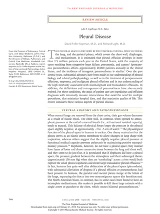
  • 2. n engl j med 378;8 nejm.org  February 22, 2018 741 Pleural Disease When considering the pleura, it is important not to think only of the pleural space, since both the visceral and parietal pleurae play important roles in maintaining normal homeostasis. The pleurae are covered by mesothelial cells, which are metabolically active and produce many sub­ stances, including hyaluronic acid–rich glycopro­ teins, nitric oxide, and transforming growth factor β.1 Research over the past several years has greatly enhanced our understanding of pleu­ ral liquid formation and resorption.8,9 In typical humans, it is estimated that ap­ proximately 0.26 ml of fluid per kilogram of body weight is contained within each pleural cavity.4,10,11 This fluid is both produced and absorbed pri­ marily on the parietal surface2,12 and is depen­ dent on the balance of hydrostatic and oncotic pressure differences between the systemic and pulmonary circulations and the pleural space (Fig. 1). Lymphatic vessels lying in the parietal pleura are responsible for pleural fluid resorp­ tion, and the flow rate of these vessels can in­ crease by a factor of approximately 20 in response to increases in pleural liquid formation.12 Thus, a clinically significant effusion will be seen only when fluid production substantially overwhelms the ability of the lymphatic vessels to resorb fluid, because of high production, diminished resorp­ tion, or a combination of these two factors. Evaluation of Pleur al Effusions The differential diagnosis for pleural effusions is extensive; a list of potential causes is shown in Table 1. A systematic and expeditious evaluation is essential, since a delay in making some diag­ noses (e.g., empyema) is associated with increased morbidity and mortality.13 The use of point-of- care ultrasonography in the evaluation of pleural effusions has been associated with a higher rate of successful aspiration of fluid from the pleural space than when no imaging is used, more ac­ curate quantitation of the volume of effusion than can be obtained with chest radiography, more accurate detection of septations than can be obtained with computed tomography (CT) of the chest, an improvement over radiography in the ability to identify exudative effusions and malignant effusions, and perhaps most impor­ tant, fewer complications than when ultrasonog­ raphy is not used to guide pleural procedures.14-20 Thus, ultrasonography is strongly recommended by the British Thoracic Society to guide pleural intervention.21,22 Unless the cause of the effusion is relatively straightforward (e.g., in a patient who presents with shortness of breath, paroxysmal nocturnal dyspnea, orthopnea, and lower-extrem­ ity edema with elevated jugular venous disten­ tion and an effusion that is more pronounced on the right side than on the left, all of which are suggestive of congestive heart failure), a chest physician should be involved to help ensure the timely evaluation of pleural effusion,21 to decrease the likelihood of associated complications, and to ensure appropriate follow-up based on the results of pleural fluid analysis.20,23 Tr ansudates versus Exudates One of the first steps in the evaluation of patients with pleural fluid is to distinguish those who have inflammatory (exudative) effusions from those who have noninflammatory (transudative) effusions.24 The use of Light’s criteria for differ­ entiating exudative from transudative effusion, initially described in 1972, has remained the standard method over the past 45 years.25 Ac­ cording to Light’s criteria, a patient is consid­ ered to have an exudative effusion when any one of the following findings is present: a ratio of pleural fluid protein to serum protein higher than 0.5, a ratio of pleural fluid lactate dehydro­ genase (LDH) level to serum LDH level higher than 0.6, or a pleural fluid LDH level higher than 200 IU per liter (or >67% of the upper limit of the normal range for serum LDH level).25,26 Although these criteria correctly identify near­ ly all exudates, approximately 25% of transu­ dates are misclassified as exudates, especially in patients who have underlying congestive heart failure and have received diuretics.27,28 In patients with suspected congestive heart failure who are receiving diuretic therapy, a serum protein level that is more than 3.1 g per deciliter higher than that in pleural fluid or a serum albumin level that is more than 1.2 g per deciliter higher than that in pleural fluid has been suggested to help identify transudates that were misclassified as exudates with the use of Light’s criteria. However, the over­ all accuracy of that approach has not been found to be significantly higher than that with Light’s criteria.28,29 The New England Journal of Medicine Downloaded from nejm.org on February 21, 2018. For personal use only. No other uses without permission. Copyright © 2018 Massachusetts Medical Society. All rights reserved.
  • 3. n engl j med 378;8 nejm.org February 22, 2018742 The new engl and jour nal of medicine Similarly, a pleural fluid level of N­terminal pro–B­type natriuretic peptide (NT­proBNP) high­ er than 1500 pg per milliliter has been shown to accurately identify effusions due to heart disease such as congestive heart failure; however, since serum levels of NT­proBNP are nearly identical to pleural fluid levels, current recommendations suggest using the serum NT­proBNP level and clinical judgment to correctly identify transudates in patients who have been undergoing active Figure 1. Balance of Forces Regulating Pleural Fluid Formation. The amount of fluid in the pleural space is dependent on the balance of hydrostatic and oncotic pressures between the parietal and visceral pleura and the pleural space. Because hydrostatic pressures are higher on the parietal pleura than on the visceral pleura and the oncotic pressures are equivalent, pleural fluid is primarily produced from the parietal pleura. Likewise, the lymphatic vessels on the parietal pleura are responsible for the majority of pleural fluid resorption. Stoma 35 29 29 29 6 Parietal Space Pleural Space Visceral Space Pulmonary capillary Systemic capillary Hydrostatic pressure +30 cm of water Hydrostatic pressure +24 cm of water Oncotic pressure +34 cm of water Net driving force Hydrostatic pressure –5 cm of water Oncotic pressure +5 cm of water Oncotic pressure +34 cm of water Pulmonary lymphatic PULMONARY INTERSTITIUM Basal lamina EXTRAPLEURAL PARIETAL INTERSTITIUM ALVEOLUS Visceral pleura Parietal pleura Net driving force ALVEOLUS 0 INTERSTITIUM Parietal lymphatic The New England Journal of Medicine Downloaded from nejm.org on February 21, 2018. For personal use only. No other uses without permission. Copyright © 2018 Massachusetts Medical Society. All rights reserved.
  • 4. n engl j med 378;8 nejm.org  February 22, 2018 743 Pleural Disease diuresis for congestive heart failure in the con­ text of a pleural fluid collection.30,31 If serum levels of protein and albumin are not available (e.g., in an outpatient who hopes to avoid veni­ puncture), a pleural fluid protein level higher than 3 g per deciliter or a pleural fluid choles­ terol level higher than 45 mg per deciliter has been shown to indicate the presence of an exuda­ tive effusion as accurately as Light’s criteria.26,32-34 Common Exudates Parapneumonic Effusions and Empyema The most common exudative effusions are those associated with an underlying pneumonia, so-called parapneumonic effusions.1 Empyema refers to frank infection or pus in the pleural space. The clinical significance of empyema and the importance of its drainage have been known for more than 2000 years, and Hippocrates has been quoted as saying, “Persons who become affected with empyema after pleurisy, if they get clear of it in forty days from the breaking of it, escape the disease; but if not, it passes into phthisis.”35,36 Despite advances in the treatment of pneumonia, however, mortality is higher among patients who have an associated parapneumonic effusion than among patients with pneumonia and no effusion,37,38 and delays in drainage are associated with substantially higher mortality.13 In addition, both the incidence of and mortality due to parapneumonic effusion and empyema continue to rise.39,40 Of note, elderly patients often do not present with the classic symptoms of cough, fever, sputum, and chest pain, but rather with anemia, fatigue, and failure to thrive. Prob­ ably in part because of underdiagnosis, elderly patients also often have more complicated effu­ sions when they are diagnosed, as well as higher rates of failure of nonsurgical therapy.41 Thus, it is crucial to consider parapneumonic effusion and empyema in all elderly patients with pneumonia. A cornerstone of treating parapneumonic ef­ fusion and empyema is the selection of appropri­ ate antibiotics on the basis of local microbiology and antibiotic resistance. Patients with commu­ nity-acquired pneumonia tend to be infected with streptococcus species and anaerobes (e.g., bacte­ roides and peptostreptococcus), whereas patients with hospital-acquired infection are more like­ ly to have methicillin-resistant staphylococcus and gram-negative bacteria (e.g., enterobacter).42 Transudative effusions Congestive heart failure Cirrhosis Nephrotic syndrome Glomerulonephritis Peritoneal dialysis Hypoalbuminemia (typical serum albumin, <1.5 mg/dl) Atelectasis Superior vena cava obstruction Trapped lung Sarcoidosis Peritoneal dialysis Myxedema Cerebrospinal fluid leak or ventriculopleural shunt Urinothorax Pulmonary arterial hypertension Pulmonary embolism Pericardial disease Extravascular migration of central venous catheter Exudative effusions Infectious: bacterial, viral, tuberculosis-related, fungal, parasitic Neoplastic: metastatic disease (e.g., lung cancer, breast cancer, lymphoma, myeloma, ovarian cancer, pancreatic cancer, cholangiocarcinoma), meso- thelioma, primary body-cavity lymphoma Paramalignant effusions: reactive pleuritis due to underlying lung cancer, airway obstruction or atelectasis, radiation-induced pleuritis Reactive: reactive pleuritis due to underlying pneumonia (i.e., parapneumonic) Embolic disease: pulmonary embolism Abdominal disease: pancreatitis, cholecystitis, hepatic or splenic abscess, esophageal perforation after esophageal varix sclerotherapy Cardiac or pericardial injury, including myocardial infarction (after coronary- artery bypass, cardiac surgery, or cardiac ablation procedures), pulmonary- vein stenosis Gynecologic: ovarian hyperstimulation, Meigs’ syndrome, endometriosis, postpartum complications Collagen vascular disease: rheumatoid arthritis, systemic lupus erythematosus, Sjögren’s syndrome, familial Mediterranean fever, eosinophilic granulo- matosis, granulomatosis with polyangiitis Medications: nitrofurantoin, dantrolene, methysergide, dasatinib, amiodarone, interleukin-2, procarbazine, methotrexate, clozapine, phenytoin, β-blockers, ergot drugs Hemothorax Chylothorax (most commonly seen after trauma or in patients with lymphoma) Sarcoidosis Lymphoplasmacytic lymphoma Cholesterol effusions (commonly seen in tuberculosis, rheumatoid effusions, and any other chronic pleural effusion) Miscellaneous: benign asbestos pleural effusion, yellow nail syndrome, ure- mia, drowning, amyloidosis, electrical burns, iatrogenic effusion, capillary leak syndromes, extramedullary hematopoiesis Table 1. Causes of Pleural Effusions. The New England Journal of Medicine Downloaded from nejm.org on February 21, 2018. For personal use only. No other uses without permission. Copyright © 2018 Massachusetts Medical Society. All rights reserved.
  • 5. n engl j med 378;8 nejm.org  February 22, 2018744 The new engl and jour nal of medicine Mortality is significantly higher among patients with hospital-acquired infection than among those with community-acquired infection (47% vs. 17%).42 Rahman and colleagues have devel­ oped a scoring system called RAPID (renal func­ tion, age, purulence, infection source, and dietary factors) to help identify patients who are at risk for a poor outcome at the time of their presenta­ tion.43 Scores range from 0 to 7, with values be­ tween 0 and 2 assigned for renal function and age (with higher scores given for worse renal function or older age) and scores of 0 or 1 as­ signed for purulence of the effusion (a nonpuru­ lent effusion receives a score of 1), whether the infection was hospital acquired (score of 1) or not (score of 0), and dietary factors (a score of 0 is assigned for an albumin level ≥2.7 g per deciliter and a score of 1 is assigned for a value below that threshold). In one study, patients in the high-risk category (those with a RAPID score of 5 to 7) were found to have at least a 30% chance of dying in the subsequent 12 weeks, and thus similar patients may warrant more invasive initial therapy. Patients with parapneumonic effusions or em­ pyema have the potential for a deterioration in their condition, and, given their underlying in­ flammatory state, all such patients should under­ go peripheral-blood culture and should receive adequate nutrition and prophylaxis for deep-vein thrombosis.44 As with any other infection in a closed space, empyema needs to be drained. Although data from randomized trials are lack­ ing, studies of large retrospective series have shown that small-bore tubes (≤14-French) per­ form on par with larger-bore tubes in terms of subsequent mortality and the need for surgery and are associated with less pain during inser­ tion and while in place.45 However, since tubes smaller than 12-French have a higher failure rate46 in empyema, our practice is to use a 14-French pigtail catheter placed with the modified Seld­ inger technique. If the pleural space is not drained with a small-bore tube, the instillation of tissue plasminogen activator (t-PA) and DNase has been successful and in one trial was found to be associated with significantly better fluid drainage, a lower likelihood of being referred for surgery, and a shorter mean hospital stay.47 It should be noted, however, that t-PA and DNase have not been shown to decrease mortality, and the cost of six doses of t-PA–DNase is approxi­ mately $7,000 (Rowden A, Johns Hopkins Hospi­ tal: personal communication). The mean hospital stay among patients in the t-PA–DNase group in that trial was 12 days.47 In addition, the avoidance of surgery may not be the most important outcome measure, since video-assisted thoracoscopic surgery (VATS) is far less invasive than thoracotomy. Older, although smaller, randomized trials showed that VATS can be the definitive treatment for empyema in up to 91% of cases,48 and more recent data on VATS suggest that hospital stays of approximate­ ly 5 to 7 days are usual.49-51 Furthermore, when performed later in the course of the disease, surgery is associated with a higher conversion rate to thoracotomy and more complications than when it is performed earlier in the disease pro­ cess.52 Our general approach to patients with parapneumonic effusion or empyema is based on the recommendations from the British Thoracic Society44 and is shown in Figure 2. There is cur­ rently a planned randomized trial evaluating t-PA–DNase versus early VATS for the treatment of parapneumonic effusion or empyema and studies examining whether reduced dosing regi­ mens of t-PA–DNase and even irrigation with normal saline can achieve similar results.54 Malignant Pleur al Effusions Malignant pleural effusions are the second lead­ ing cause of exudative effusions and the leading cause of exudates among patients who under­ go thoracentesis; they account for more than 125,000 hospital admissions per year in the United States, with estimated inpatient mortality of 11.6% and associated hospital charges of more than $5 billion per year.55 The majority of malig­ nant pleural effusions arise from lung cancer, breast cancer, and lymphoma, and it is estimated that 15% of patients with lung cancer will have a malignant pleural effusion at presentation and up to 50% will have a malignant pleural effusion during the course of their illness. Malignant pleural effusion is associated with a poor prog­ nosis, with a median survival of 4 to 7 months from the time of diagnosis.56,57 Even among pa­ tients whose effusions are considered “too small to tap,” survival is significantly shorter than among patients without any effusion.57 Survival The New England Journal of Medicine Downloaded from nejm.org on February 21, 2018. For personal use only. No other uses without permission. Copyright © 2018 Massachusetts Medical Society. All rights reserved.
  • 6. n engl j med 378;8 nejm.org  February 22, 2018 745 Pleural Disease depends primarily on tumor subtype, with lung cancer and gastrointestinal cancers having the worst outcomes (median survival, 2 to 3 months) and mesothelioma and hematologic cancers hav­ ing the best prognosis, with survival approaching 1 year.58,59 To appropriately treat patients with malignant pleural effusions, it is crucial to understand the mechanisms by which pleural effusions cause dyspnea. Although pleural effusions mildly in­ crease shunt fraction, it is rare to find patients with substantial hypoxemia.60 The related dys­ pnea is generally not a lung problem due to lung collapse or to a reduction in pulmonary-function measures.61 Rather, the dyspnea is a chest-wall issue caused by the diaphragm being displaced caudally, which is mechanically disadvantageous for its length–tension relationship.61,62 The gene­ sis of the dyspnea is important, since the most clinically relevant questions after large-volume Figure 2. Management of Parapneumonic Effusions. Poor prognostic factors after incomplete removal of fluid by means of therapeutic thoracentesis include pus in the pleural space, positive Gram’s stain or culture, pleural fluid glucose level less than 40 mg per deciliter, pleural fluid pH lower than 7.15, and pleural fluid lactate dehydrogenase level more than 3 times the upper limit of the normal range for serum.1,53 A decision regarding surgery depends on the patient’s clinical status and ability to undergo surgery, as well as on local resources and the availability of a skilled surgeon. The figure is modified from Davies et al.44 The abbreviation t-PA denotes tissue plasminogen activator, and VATS video-assisted thoracoscopic surgery. Are septations or loculation seen? Pleural effusion with underlying pneumonia All patients: antibiotics, venous thromboembolism prophylaxis, nutrition Therapeutic thoracentesis Consider pulmonary and thoracic surgical consultation for multi- disciplinary discussion Chest ultrasound All fluid removed? No Yes Fluid recurs? Return to chest ultra- sound box above Place 14-French chest tube and consider t-PA–DNase Consider early VATS Continue antibiotics Yes No No Yes No Yes Poor prognostic factors? Continue antibiotics, consider repeat ultra- sound with or without thoracentesis in 48 hr Poor or borderline surgical candidate Good surgical candidate The New England Journal of Medicine Downloaded from nejm.org on February 21, 2018. For personal use only. No other uses without permission. Copyright © 2018 Massachusetts Medical Society. All rights reserved.
  • 7. n engl j med 378;8 nejm.org  February 22, 2018746 The new engl and jour nal of medicine thoracentesis are “Is the patient’s breathing bet­ ter?” and “Did the lung fully reexpand?” If the patient does not feel better after a therapeutic thoracentesis, something else is causing the dys­ pnea (e.g., pulmonary embolism or lymphangitic carcinomatosis). In such instances, further diag­ nostic testing should be performed; however, procedures that address the pleural space should not be performed. If the patient’s dyspnea has been alleviated with thoracentesis, the effusion was at least a major contributor to the dyspnea, and the dyspnea can be diminished regardless of whether the lung has reexpanded. If the lung has reexpanded, the patient can be considered for pleurodesis, placement of a tunneled pleural catheter, or combination approaches, whereas if the lung is nonexpandable, a tunneled pleural catheter is the treatment of choice (Fig. 3).1,63 Tunneled pleural catheters are small-bore tubes that are tunneled subcutaneously into the pleural space, can be placed in the outpatient setting, and allow patients or caregivers to drain pleural fluid without subjecting the patient to additional invasive procedures. Because at least 30% of patients with malignant pleural effusion do not have reexpansion of the lung, which may not be evident even at the time of thoracoscopy,64,65 it may be important to perform a large-volume thoracentesis before deciding on definitive ther­ apy for such patients. The goals of treating pa­ tients with malignant pleural effusion are to improve quality of life, primarily by minimizing dyspnea, and to minimize pleural procedures and the need for repeated hospital or doctor visits. Given the poor prognosis of these patients, early and definitive pleural palliation — as opposed to multiple thoracenteses, which expose the patient to both risk and inconvenience — is recom­ mended.66 The LENT score (LDH in pleural fluid, Eastern Cooperative Oncology Group [ECOG] performance status, neutrophil–lymphocyte ratio in the serum, and tumor type) has been shown to accurately stratify patients into high-, moderate-, and low- risk groups and may be helpful in guiding therapy.59 A score of 0 is assigned for a pleural fluid LDH level lower than 1500 IU per liter and a score of 1 is assigned for a level above that threshold, scores of 0 to 3 for worsening ECOG status, a score of 0 for a serum neutrophil–lym­ phocyte ratio of less than 9 and a score of 1 for a ratio above that threshold, and scores of 0 to 3 based on tumor type. Total scores of 0 or 1 are considered to indicate low risk and are associ­ ated with a median survival of 319 days, as com­ pared with a median survival of 130 days in the medium-risk category (scores of 2 through 4) and 44 days in the high-risk category (scores of 5 through 7).59 For patients in the high-risk cate­ gory, less invasive approaches, such as placement of a tunneled pleural catheter or even thoracen­ tesis, may be most useful, whereas patients who are considered at low risk can be treated with tunneled pleural catheters, pleurodesis, or com­ bination approaches. When discussing options for patients who have expandable lungs, the risks and benefits of each procedure should be re­ viewed. The benefits of tunneled pleural cathe­ ters include clinically significant improvement in dyspnea, placement in the outpatient setting, and the ability of many patients and families to care for the catheter themselves at home. How­ ever, patients need to drain such a catheter re­ peatedly until the effusion resolves or until death. Spontaneous pleurodesis is estimated to occur in approximately 50% of patients; among pa­ tients in whom it does occur, it occurs at a mean of approximately 60 days after insertion of the catheter.67,68 The benefits of pleurodesis include substan­ tial alleviation of dyspnea and not having to man­ age a catheter. However, many centers will keep patients in the hospital for 3 to 5 days after the instillation of talc to effect pleural surface fu­ sion, and there is a small risk of transient hypox­ emia associated with the use of nongraded talc.64,69 An unblinded randomized trial examined tun­ neled pleural catheters versus talc-slurry pleurode­ sis for the treatment of persistent effusions and showed no significant differences in dyspnea or in quality of life.70 Patients in the talc group underwent more additional procedures, whereas the patients in the tunneled pleural catheter group had a higher incidence of nonserious ad­ verse events. Although it is often a fear of refer­ ring health care providers, infection related to the tunneled pleural catheter occurs approxi­ mately 5% of the time and can usually be treated without removing the catheter.71 Trials have sug­ gested that the combination of tunneled pleural catheters with sclerosing agents (talc or silver nitrate) as well as daily drainage (as opposed to The New England Journal of Medicine Downloaded from nejm.org on February 21, 2018. For personal use only. No other uses without permission. Copyright © 2018 Massachusetts Medical Society. All rights reserved.
  • 8. n engl j med 378;8 nejm.org  February 22, 2018 747 Pleural Disease drainage every other day) can result in substan­ tially fewer days with a catheter.72-74 As with all procedures, we recommend that the risks, ben­ efits, and alternatives always be discussed with the patient in detail and that therapy be indi­ vidualized. Complicated Tr ansudates Congestive heart failure, cirrhosis, and the ne­ phrotic syndrome underlie most transudative effusions.1 Although often considered benign conditions, effusions associated with congestive heart failure, hepatic failure, and renal failure have recently been shown to be associated with 1-year mortality rates of 50%, 25%, and 46%, respectively.75 Patients with congestive heart fail­ ure and pleural effusion have a 1-year risk of death similar to that of patients who are admit­ ted to the intensive care unit with acute decom­ pensated heart failure, patients with hepatic hy­ drothorax have a risk of death similar to that of patients with a Model for End-Stage Liver Dis­ ease (MELD) score of 20 to 29 (a typical indica­ tion for transplantation; MELD scores range from 6 to 40, with higher scores indicating more advanced liver disease), and patients with renal failure and effusion have a 1-year risk of death that is triple that among patients undergoing hemodialysis who do not have effusions.75 Al­ though in the majority of patients, transudative effusions can be managed by treatment of the underlying condition, refractory effusions deserve prompt and aggressive pleural palliation that will minimize repeat procedures and breathless­ ness and maximize quality of life. As with malig­ Figure 3. Management of Malignant Pleural Effusions. The figure is modified from Roberts et al.63 Improvement in dyspnea? Pleural effusion with underlying cancer Investigate for other causes of dyspnea Ultrasound-guided therapeutic thoracentesis Lung reexpansion? No Yes Suspected survival >1 wk? Consider placement of tunneled pleural catheter Palliate dyspnea with repeat thoracentesis if needed, oxygen, or morphine No Yes No Yes Discuss relative risks and benefits of tunneled pleural catheter vs. pleurodesis vs. combination approaches The New England Journal of Medicine Downloaded from nejm.org on February 21, 2018. For personal use only. No other uses without permission. Copyright © 2018 Massachusetts Medical Society. All rights reserved.
  • 9. n engl j med 378;8 nejm.org  February 22, 2018748 The new engl and jour nal of medicine nant pleural effusions, tunneled pleural cathe­ ters, pleurodesis, or both may be indicated for these patients,76-79 and we recommend careful discussion with relevant teams (e.g., hepatology and liver transplantation) to develop a multidis­ ciplinary plan. Current Concepts in the Treatment of Pneumothor ax Pneumothorax has traditionally been categorized as primary (no underlying lung disease), second­ ary (underlying lung disease present), traumatic, and iatrogenic. Because of advances in chest imaging (CT and thoracoscopy), patients with a pneumothorax who had previously been con­ sidered free of parenchymal disease have been found to have emphysema-like pulmonary changes and increased pleural porosity, or de­ fects in the visceral pleura that are independent of blebs or bullae.80 These findings suggest that the distinction between primary and secondary effusion is perhaps an artificial construct and that therapy should be guided by size of the pneumothorax and by the patient’s symptoms.81 Furthermore, there is no standard definition of pneumothorax size; the American College of Chest Physicians defines “large” as a distance of 3 cm or more from the apex of the lung to the cupula of the chest wall,82 whereas the British Thoracic Society defines it as an intrapleural distance of at least 2 cm at the level of the hilum.83 In fact, agreement on size based on these definitions occurs less than 50% of the time in clinical set­ tings, which leads to substantial variation in treatment recommendations.84 Up to 70% of patients with a clinically stable pneumothorax can be treated with simple needle aspiration, which avoids hospitalization. As with parapneumonic effusion and empyema, guide­ lines currently recommend the use of small-bore (14-French) chest tubes rather than large-bore chest tubes for patients with pneumothorax who have treatment failure or are not candidates for simple needle aspiration.83 The latter group in­ cludes patients who live far from the treating center, who have minimal social support, or who have more substantial underlying lung disease. Over the past several years, more conservative therapy has been a trend associated with reserv­ ing surgical therapy for patients with the highest risk of recurrent effusions. Digital air-leak mon­ itoring devices have been shown to reduce the number of days a chest tube is in place and to shorten the length of stay in the hospital after lobectomy or segmentectomy.85 When patients with a pneumothorax are treat­ ed with chest tubes, the lung usually expands and the air leak ceases within 3 days. If the lung does not expand fully within 3 to 5 days, consid­ eration should be given to thoracoscopy. At thora­ coscopy, blebs are stapled and an effort is made to create a pleurodesis, usually with pleural abrasion. Another method of treating prolonged air leak is to instill 1 ml of the patient’s own blood per kilogram of body weight through the chest tube.86 Alternatively, pleurodesis can be attempted through instillation of a sclerosing agent or the placement of endobronchial one-way valves, with the goal of reducing air flow across the visceral pleura. The valves are then removed after the pleural defect has healed, typically in 6 weeks.87 After a patient has had a spontaneous pneu­ mothorax, the likelihood of a recurrence exceeds 50%.88 Prevention of recurrence is key, especially in patients with markedly decreased lung func­ tion, for whom a recurrence can be fatal. If a patient has a first recurrence, then the likeli­ hood of a second recurrence is very high. Recur­ rence rates can be reduced to approximately 25% if an agent such as talc or doxycycline is instilled through a chest tube and can be reduced to less than 5% with thoracoscopy and the insufflation of talc, stapling of blebs, or pleural abrasion to create a pleurodesis.89 However, a bullectomy alone, without attempts at pleurodesis, is associ­ ated with a higher recurrence rate, and therefore pleurodesis should always be considered an inte­ gral part of the procedure.81 Areas for Future Research Over the past few years, large, multicenter, ran­ domized trials have been published from centers that have large clinical and research pleural ser­ vices. These trials may lead to improvements in diagnostic tests to establish the underlying cause of a pleural effusion, more compounds to de­ crease the rate of pleural fluid production or increase the rate of pleural fluid reabsorption, an improved compound for pleurodesis, and fur­ The New England Journal of Medicine Downloaded from nejm.org on February 21, 2018. For personal use only. No other uses without permission. Copyright © 2018 Massachusetts Medical Society. All rights reserved.
  • 10. n engl j med 378;8 nejm.org  February 22, 2018 749 Pleural Disease ther development of interventional pulmonology services and dedicated multidisciplinary pleural disease services. Furthermore, trials investigating the clinical effect of pleural manometry, studies to better understand the pharmacodynamics of drugs in the pleural space, and investigation of how pleural disease is related to the genetic makeup of affected patients may be forthcom­ ing. Patient-centered (and caregiver-centered) out­ comes, such as the effect on daily quality of life, are also being investigated. Pleural disease re­ mains a common clinical problem, and expedi­ tious, multidisciplinary evaluation and treatment will maximize the care of our patients. Dr. Feller-Kopman reports receiving grant support and consult­ ing fees from CareFusion/BD; and Dr. Light, serving on an advi­ sory committee for CareFusion and holding a patent (US 6103695 A) on transforming growth factor β for pleurodesis. No other potential conflict of interest relevant to this article was reported. Disclosure forms provided by the authors are available with the full text of this article at NEJM.org. References 1. Light R. Pleural disease. 6th ed. Phila­ delphia:​Lippincott Williams & Wilkins, 2013. 2. Agostoni E. Mechanics of the pleural space. Physiol Rev 1972;​52:​57-128. 3. Lai-Fook SJ. Pleural mechanics and fluid exchange. Physiol Rev 2004;​84:​385- 410. 4. Wang NS. Anatomy of the pleura. Clin Chest Med 1998;​19:​229-40. 5. West JB. Why doesn’t the elephant have a pleural space? News Physiol Sci 2002;​17:​ 47-50. 6. West JB. Snorkel breathing in the ele­ phant explains the unique anatomy of its pleura. Respir Physiol 2001;​126:​1-8. 7. Grathwohl KW, Derdak S. Buffalo chest. N Engl J Med 2003;​349:​1829. 8. Stathopoulos GT, Moschos C, Loutrari H, et al. Zoledronic acid is effective against experimental malignant pleural effusion. Am J Respir Crit Care Med 2008;​ 178:​50-9. 9. Psallidas I, Kalomenidis I, Porcel JM, Robinson BW, Stathopoulos GT. Malig­ nant pleural effusion: from bench to bed­ side. Eur Respir Rev 2016;​25:​189-98. 10. Agostoni E, D’Angelo E. Thickness and pressure of the pleural liquid at vari­ ous heights and with various hydrotho­ races. Respir Physiol 1969;​6:​330-42. 11. Noppen M, De Waele M, Li R, et al. Volume and cellular content of normal pleural fluid in humans examined by pleural lavage. Am J Respir Crit Care Med 2000;​162:​1023-6. 12. Miserocchi G. Physiology and patho­ physiology of pleural fluid turnover. Eur Respir J 1997;​10:​219-25. 13. Ashbaugh DG. Empyema thoracis: fac­ tors influencing morbidity and mortality. Chest 1991;​99:​1162-5. 14. Kohan JM, Poe RH, Israel RH, et al. Value of chest ultrasonography versus de­ cubitus roentgenography for thoracente­ sis. Am Rev Respir Dis 1986;​133:​1124-6. 15. Diacon AH, Brutsche MH, Solèr M. Accuracy of pleural puncture sites: a pro­ spective comparison of clinical examina­ tion with ultrasound. Chest 2003;​123:​436- 41. 16. Eibenberger KL, Dock WI, Ammann ME, Dorffner R, Hörmann MF, Graben­ wöger F. Quantification of pleural effu­ sions: sonography versus radiography. Radiology 1994;​191:​681-4. 17. Grogan DR, Irwin RS, Channick R, et al. Complications associated with thora­ centesis: a prospective, randomized study comparing three different methods. Arch Intern Med 1990;​150:​873-7. 18. Yang PC, Luh KT, Chang DB, Wu HD, Yu CJ, Kuo SH. Value of sonography in de­ termining the nature of pleural effusion: analysis of 320 cases. AJR Am J Roent­ genol 1992;​159:​29-33. 19. Qureshi NR, Rahman NM, Gleeson FV. Thoracic ultrasound in the diagnosis of malignant pleural effusion. Thorax 2009;​ 64:​139-43. 20. Gordon CE, Feller-Kopman D, Balk EM, Smetana GW. Pneumothorax follow­ ing thoracentesis: a systematic review and meta-analysis. Arch Intern Med 2010;​170:​ 332-9. 21. Hooper C, Lee YCG, Maskell N. Inves­ tigation of a unilateral pleural effusion in adults: British Thoracic Society Pleural Disease Guideline 2010. Thorax 2010;​65:​ Suppl 2:​ii4-ii17. 22. Havelock T, Teoh R, Laws D, Gleeson F. Pleural procedures and thoracic ultra­ sound: British Thoracic Society Pleural Disease Guideline 2010. Thorax 2010;​65:​ Suppl 2:​ii61-ii76. 23. Hooper CE, Lee YC, Maskell NA. Set­ ting up a specialist pleural disease service. Respirology 2010;​15:​1028-36. 24. Paddock FK. The diagnostic signifi­ cance of serous fluids in disease. N Engl J Med 1940;​223:​1010-5. 25. Light RW, Macgregor MI, Luchsinger PC, Ball WC Jr. Pleural effusions: the diag­ nostic separation of transudates and exu­ dates. Ann Intern Med 1972;​77:​507-13. 26. Heffner JE, Brown LK, Barbieri CA. Diagnostic value of tests that discrimi­ nate between exudative and transudative pleural effusions. Chest 1997;​111:​970-80. 27. Romero-Candeira S, Fernández C, Martín C, Sánchez-Paya J, Hernández L. Influence of diuretics on the concentra­ tion of proteins and other components of pleural transudates in patients with heart failure. Am J Med 2001;​110:​681-6. 28. Romero-Candeira S, Hernández L, Romero-Brufao S, Orts D, Fernández C, Martín C. Is it meaningful to use bio­ chemical parameters to discriminate be­ tween transudative and exudative pleural effusions? Chest 2002;​122:​1524-9. 29. Bielsa S, Porcel JM, Castellote J, Mas E, Esquerda A, Light RW. Solving the Light’s criteria misclassification rate of cardiac and hepatic transudates. Respirol­ ogy 2012;​17:​721-6. 30. Porcel JM, Vives M, Cao G, Esquerda A, Rubio M, Rivas MC. Measurement of pro-brain natriuretic peptide in pleural fluid for the diagnosis of pleural effu­ sions due to heart failure. Am J Med 2004;​ 116:​417-20. 31. Kolditz M, Halank M, Schiemanck CS, Schmeisser A, Höffken G. High diagnos­ tic accuracy of NT-proBNP for cardiac origin of pleural effusions. Eur Respir J 2006;​28:​144-50. 32. Carr DT, Power MH. Clinical value of measurements of concentration of pro­ tein in pleural fluid. N Engl J Med 1958;​ 259:​926-7. 33. Costa M, Quiroga T, Cruz E. Measure­ ment of pleural fluid cholesterol and lac­ tate dehydrogenase: a simple and accurate set of indicators for separating exudates from transudates. Chest 1995;​108:​1260-3. 34. Porcel JM, Vives M, Vicente de Vera MC, Cao G, Rubio M, Rivas MC. Useful tests on pleural fluid that distinguish transudates from exudates. Ann Clin Bio­ chem 2001;​38:​671-5. 35. Internet Classics Archive. Aphorisms by Hippocrates (Adams F, translator) (http:// classics​.mit​.edu/​Hippocrates/​aphorisms​ .mb​.txt). 36. Adams F. The genuine works of Hip­ pocrates. London:​C. and J. Adlard Print­ ers, 1848. 37. Hasley PB, Albaum MN, Li YH, et al. Do pulmonary radiographic findings at presentation predict mortality in patients with community-acquired pneumonia? Arch Intern Med 1996;​156:​2206-12. The New England Journal of Medicine Downloaded from nejm.org on February 21, 2018. For personal use only. No other uses without permission. Copyright © 2018 Massachusetts Medical Society. All rights reserved.
  • 11. n engl j med 378;8 nejm.org  February 22, 2018750 The new engl and jour nal of medicine 38. Menéndez R, Torres A, Zalacaín R, et al. Risk factors of treatment failure in community acquired pneumonia: implica­ tions for disease outcome. Thorax 2004;​ 59:​960-5. 39. Grijalva CG, Zhu Y, Nuorti JP, Griffin MR. Emergence of parapneumonic empy­ ema in the USA. Thorax 2011;​66:​663-8. 40. Bender JM, Ampofo K, Sheng X, Pavia AT, Cannon-Albright L, Byington CL. Para­ pneumonic empyema deaths during past century, Utah. Emerg Infect Dis 2009;​15:​ 44-8. 41. El Solh AA, Alhajjhasan A, Ramadan FH, Pineda LA. A comparative study of community- and nursing home-acquired empyema thoracis. J Am Geriatr Soc 2007;​ 55:​1847-52. 42. Maskell NA, Batt S, Hedley EL, Davies CW, Gillespie SH, Davies RJ. The bacteri­ ology of pleural infection by genetic and standard methods and its mortality sig­ nificance. Am J Respir Crit Care Med 2006;​174:​817-23. 43. Rahman NM, Kahan BC, Miller RF, Gleeson FV, Nunn AJ, Maskell NA. A clin­ ical score (RAPID) to identify those at risk for poor outcome at presentation in pa­ tients with pleural infection. Chest 2014;​ 145:​848-55. 44. Davies HE, Davies RJO, Davies CWH. Management of pleural infection in adults: British Thoracic Society Pleural Disease Guideline 2010. Thorax 2010;​65:​Suppl 2:​ ii41-ii53. 45. Rahman NM, Maskell NA, Davies CWH, et al. The relationship between chest tube size and clinical outcome in pleural infection. Chest 2010;​137:​536-43. 46. Keeling AN, Leong S, Logan PM, Lee MJ. Empyema and effusion: outcome of image-guided small-bore catheter drain­ age. Cardiovasc Intervent Radiol 2008;​31:​ 135-41. 47. Rahman NM, Maskell NA, West A, et al. Intrapleural use of tissue plasminogen activator and DNase in pleural infection. N Engl J Med 2011;​365:​518-26. 48. Wait MA, Sharma S, Hohn J, Dal Nogare A. A randomized trial of empy­ ema therapy. Chest 1997;​111:​1548-51. 49. Kim BY, Oh BS, Jang WC, Min YI, Park YK, Park JC. Video-assisted thoracoscopic decortication for management of post­ pneumonic pleural empyema. Am J Surg 2004;​188:​321-4. 50. Marks DJ, Fisk MD, Koo CY, et al. Thoracic empyema: a 12-year study from a UK tertiary cardiothoracic referral centre. PLoS One 2012;​7(1):​e30074. 51. Shahin Y, Duffy J, Beggs D, Black E, Majewski A. Surgical management of pri­ mary empyema of the pleural cavity: out­ come of 81 patients. Interact Cardiovasc Thorac Surg 2010;​10:​565-7. 52. Scarci M, Abah U, Solli P, et al. EACTS expert consensus statement for surgical management of pleural empyema. Eur J Cardiothorac Surg 2015;​48:​642-53. 53. Porcel JM, Valencia H, Bielsa S. Fac­ tors influencing pleural drainage in para­ pneumonic effusions. Rev Clin Esp 2016;​ 216:​361-6. 54. Hooper CE, Edey AJ, Wallis A, et al. Pleural Irrigation Trial (PIT): a random­ ised controlled trial of pleural irrigation with normal saline versus standard care in patients with pleural infection. Eur Respir J 2015;​46:​456-63. 55. Taghizadeh N, Fortin M, Tremblay A. US hospitalizations for malignant pleural effusions: data from the 2012 National Inpatient Sample. Chest 2017;​151:​845-54. 56. Heffner JE, Nietert PJ, Barbieri C. Pleural fluid pH as a predictor of pleurode­ sis failure: analysis of primary data. Chest 2000;​117:​87-95. 57. Porcel JM, Gasol A, Bielsa S, Civit C, Light RW, Salud A. Clinical features and survival of lung cancer patients with pleu­ ral effusions. Respirology 2015;​20:​654-9. 58. Postmus PE, Brambilla E, Chansky K, et al. The IASLC Lung Cancer Staging Project: proposals for revision of the M descriptors in the forthcoming (seventh) edition of the TNM classification of lung cancer. J Thorac Oncol 2007;​2:​686-93. 59. Clive AO, Kahan BC, Hooper CE, et al. Predicting survival in malignant pleural effusion: development and validation of the LENT prognostic score. Thorax 2014;​ 69:​1098-104. 60. Agustí AGN, Cardús J, Roca J, Grau JM, Xaubet A, Rodriguez-Roisin R. Venti­ lation-perfusion mismatch in patients with pleural effusion: effects of thoracentesis. Am J Respir Crit Care Med 1997;​156:​1205-9. 61. Estenne M, Yernault JC, De Troyer A. Mechanism of relief of dyspnea after thora­ cocentesis in patients with large pleural effusions. Am J Med 1983;​74:​813-9. 62. Krell WS, Rodarte JR. Effects of acute pleural effusion on respiratory system mechanics in dogs. J Appl Physiol (1985) 1985;​59:​1458-63. 63. Roberts ME, Neville E, Berrisford RG, Antunes G, Ali NJ. Management of a ma­ lignant pleural effusion: British Thoracic Society Pleural Disease Guideline 2010. Thorax 2010;​65:​Suppl 2:​ii32-ii40. 64. Dresler CM, Olak J, Herndon JE II, et al. Phase III intergroup study of talc poudrage vs talc slurry sclerosis for malignant pleu­ ral effusion. Chest 2005;​127:​909-15. 65. Hallifax RJ, Corcoran JP, Psallidas I, Rahman NM. Medical thoracoscopy: sur­ vey of current practice — how successful are medical thoracoscopists at predicting malignancy? Respirology 2016;​21:​958-60. 66. Ost DE, Niu J, Zhao H, Grosu HB, Giordano SH. Quality gaps and compara­ tive effectiveness of management strate­ gies for recurrent malignant pleural effu­ sions. Chest 2017 August 31 (Epub ahead of print). 67. Tremblay A, Michaud G. Single-center experience with 250 tunnelled pleural catheter insertions for malignant pleural effusion. Chest 2006;​129:​362-8. 68. Kheir F, Shawwa K, Alokla K, Om­ balli M, Alraiyes AH. Tunneled pleural catheter for the treatment of malignant pleural effusion: a systematic review and meta-analysis. Am J Ther 2016;​23(6):​e1300- e1306. 69. Gonzalez AV, Bezwada V, Beamis JF Jr, Villanueva AG. Lung injury following thoracoscopic talc insufflation: experience of a single North American center. Chest 2010;​137:​1375-81. 70. Davies HE, Mishra EK, Kahan BC, et al. Effect of an indwelling pleural catheter vs chest tube and talc pleurodesis for reliev­ ing dyspnea in patients with malignant pleural effusion: the TIME2 randomized controlled trial. JAMA 2012;​307:​2383-9. 71. Fysh ETH, Tremblay A, Feller-Kopman D, et al. Clinical outcomes of indwelling pleural catheter-related pleural infections: an international multicenter study. Chest 2013;​144:​1597-602. 72. Reddy C, Ernst A, Lamb C, Feller-Kop­ man D. Rapid pleurodesis for malignant pleural effusions: a pilot study. Chest 2011;​ 139:​1419-23. 73. Bhatnagar R, Zahan-Evans N, Kearney C, et al. A novel drug-eluting indwelling pleural catheter for the management of malignant effusions. Am J Respir Crit Care Med 2018;​197:​136-8. 74. Wahidi MM, Reddy C, Yarmus L, et al. Randomized trial of pleural fluid drain­ age frequency in patients with malignant pleural effusions: the ASAP Trial. Am J Respir Crit Care Med 2017;​195:​1050-7. 75. Walker SP, Morley AJ, Stadon L, et al. Nonmalignant pleural effusions: a pro­ spective study of 356 consecutive unselect­ ed patients. Chest 2017;​151:​1099-105. 76. Patil M, Dhillon SS, Attwood K, Saoud M, Alraiyes AH, Harris K. Management of benign pleural effusions using indwelling pleural catheters: a systematic review and meta-analysis. Chest 2017;​151:​626-35. 77. Chen A, Massoni J, Jung D, Crippin J. Indwelling tunneled pleural catheters for the management of hepatic hydrothorax: a pilot study. Ann Am Thorac Soc 2016;​13:​ 862-6. 78. Majid A, Kheir F, Fashjian M, et al. Tunneled pleural catheter placement with and without talc poudrage for treatment of pleural effusions due to congestive heart failure. Ann Am Thorac Soc 2016;​13:​212-6. 79. Bintcliffe OJ, Lee GY, Rahman NM, Maskell NA. The management of benign non-infective pleural effusions. Eur Respir Rev 2016;​25:​303-16. 80. Noppen M, Stratakos G, Verbanck S, D’Haese J, Meysman M, Vincken W. Fluo­ rescein-enhanced autofluorescence thora­ coscopy in primary spontaneous pneumo­ thorax. Am J Respir Crit Care Med 2004;​ 170:​680-2. 81. Bintcliffe OJ, Hallifax RJ, Edey A, et al. Spontaneous pneumothorax: time to re­ think management? Lancet Respir Med 2015;​3:​578-88. 82. Baumann MH, Strange C, Heffner JE, et al. Management of spontaneous pneu­ The New England Journal of Medicine Downloaded from nejm.org on February 21, 2018. For personal use only. No other uses without permission. Copyright © 2018 Massachusetts Medical Society. All rights reserved.
  • 12. n engl j med 378;8 nejm.org  February 22, 2018 751 Pleural Disease mothorax: an American College of Chest Physicians Delphi consensus statement. Chest 2001;​119:​590-602. 83. MacDuff A, Arnold A, Harvey J. Man­ agement of spontaneous pneumothorax: British Thoracic Society Pleural Disease Guideline 2010. Thorax 2010;​65:​Suppl 2:​ ii18-ii31. 84. Kelly AM, Druda D. Comparison of size classification of primary spontaneous pneumothorax by three international guidelines: a case for international con­ sensus? Respir Med 2008;​102:​1830-2. 85. Pompili C, Detterbeck F, Papagian­ nopoulos K, et al. Multicenter interna­ tional randomized comparison of objec­ tive and subjective outcomes between electronic and traditional chest drainage systems. Ann Thorac Surg 2014;​98:​490-6. 86. Cao G, Kang J, Wang F, Wang H. Intra­ pleural instillation of autologous blood for persistent air leak in spontaneous pneumo­ thorax in patients with advanced chronic obstructive pulmonary disease. Ann Thorac Surg 2012;​93:​1652-7. 87. Reed MF, Gilbert CR, Taylor MD, Toth JW. Endobronchial valves for challenging air leaks. Ann Thorac Surg 2015;​100:​1181-6. 88. Sadikot RT, Greene T, Meadows K, Arnold AG. Recurrence of primary spon­ taneous pneumothorax. Thorax 1997;​52:​ 805-9. 89. Shaikhrezai K, Thompson AI, Parkin C, Stamenkovic S, Walker WS. Video-assisted thoracoscopic surgery management of spontaneous pneumothorax — long-term results. Eur J Cardiothorac Surg 2011;​40:​ 120-3. Copyright © 2018 Massachusetts Medical Society. The New England Journal of Medicine Downloaded from nejm.org on February 21, 2018. For personal use only. No other uses without permission. Copyright © 2018 Massachusetts Medical Society. All rights reserved.