
Recommended
More Related Content
Similar to The skin.ppsx
Similar to The skin.ppsx (20)
More from lumaGhaziALzamel
More from lumaGhaziALzamel (15)
Recently uploaded
Recently uploaded (20)
The skin.ppsx
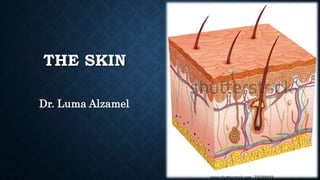
- 1. THE SKIN Dr. Luma Alzamel
- 2. The skin completely covers the body and is continuous with the membranes lining the body orifices. It: • protects the underlying structures from injury and from invasion by microbes. • contains sensory (somatic) nerve endings of pain, temperature and touch. • is involved in the regulation of body temperature.
- 3. Functions of the skin 1. Protection The skin forms a relatively waterproof layer that protects the deeper and more delicate structures. As an important non- specific defense mechanism, it acts as a barrier against: • invasion by microbes. • chemicals. • physical agents, e.g., mild trauma, ultraviolet light. • dehydration. The dermis contains specialized immune cells called Langerhans cells. They phagocytose intruding antigens and travel to lymphoid tissue, where they present antigen to T- lymphocytes, thus stimulating an immune response. Due to the presence of the sensory nerve endings in the skin the body reacts by reflex action to unpleasant or painful stimuli, protecting it from further injury
- 4. 2. Regulation of body temperature The temperature of the body remains constant at about 36.8°C across a wide range of environmental temperatures. In health, variations are usually limited to between 0.5 and 0.75°C, although it is raised slightly in the evening, during exercise and in women just after ovulation. When metabolic rate increases body temperature rises and when it decreases body temperature falls.
- 5. To ensure this constant temperature a balance is maintained between heat produced in the body and heat lost to the environment. Heat production Some of the energy released in the cells during metabolic activity is in the form of heat and the most active organs, chemically and physically, produce the most heat. The principal organs involved are as follows. The muscles. Contraction of skeletal muscles produces a large amount of heat through muscular exercise and Shivering which involves muscle contraction and produces heat when there is the risk of the body temperature falling below normal. The liver is very chemically active, and heat is produced. Metabolic rate and heat production are increased after eating. The digestive organs produce heat during peristalsis and by the chemical reactions involved in digestion.
- 6. Heat loss Most of the heat loss from the body occurs through the skin. Small amounts are lost in expired air, urine and faces. Only the heat lost through the skin can be regulated to maintain a constant body temperature. There is no control overheat lost by the other routes. Heat loss through the skin is affected by the difference between body and environmental temperatures, the amount of the body surface exposed to the air and the type of clothes worn. Air is a poor conductor of heat and when layers of air are trapped in clothing and between the skin and clothing they act as effective insulators against excessive heat loss. For this reason, several layers of lightweight clothes provide more effective insulation against a low environmental temperature than one heavy garment. A balance is maintained between heat production and heat loss. Control is achieved mainly by thermoreceptors in the hypothalamus.
- 7. Mechanisms of heat loss. In evaporation, the body is cooled when heat is used to convert the water in sweat to water vapor. In radiation, exposed parts of the body radiate heat away from the body. In conduction, clothes and other objects in contact with the skin take up heat. In convection, air passing over the exposed parts of the body is heated and rises, cool air replaces it and convection currents are set up. Heat is also lost from the clothes by convection.
- 8. Control of body temperature Nervous control. The temperature regulating center in the hypothalamus is responsive to the temperature of circulating blood. This center controls body temperature through autonomic nerve stimulation of the sweat glands when body temperature rises. The vasomotor center in the medulla oblongata controls the diameter of the small arteries and arterioles, and therefore the amount of blood which circulates in the capillaries in the dermis. The vasomotor center is influenced by the temperature of its blood supply and by nerve impulses from the hypothalamus. When body temperature rises the skin capillaries dilate and the extra blood near the surface increases heat loss by radiation, conduction and convection. The skin is warm and pink in color. When body temperature falls arteriolar constriction conserves heat, and the skin is whiter and feels cool.
- 10. Activity of the sweat glands. When the temperature of the body is increased by 0.25 to 0.5°C the sweat glands are stimulated to secrete sweat, which is conveyed to the surface of the body by ducts. When sweat droplets can be seen on the skin the rate of production is exceeding the rate of evaporation. This is most likely to happen when the environmental air is humid and the temperature high. Loss of heat from the body by unnoticeable evaporation of water through the skin and expired air occurs even when the environmental temperature is low. This is called insensible water loss (around 500 ml per day) and is accompanied by insensible heat loss.
- 11. Effects of vasodilatation. The amount of heat lost from the skin depends to a great extent on the amount of blood in the vessels in the dermis. As heat production increases, the arterioles become dilated and more blood pours into the capillary network in the skin. In addition to increasing the amount of sweat produced the temperature of the skin is raised and there is an increase in the amount of heat lost by radiation, conduction and convection. If the external environmental temperature is low or if heat production is decreased, vasoconstriction is stimulated by sympathetic nerves. This decreases the blood flow near the body surface, conserving heat.
- 12. Fever This is often the result of infection and is caused by release of chemicals (pyrogens) from damaged tissue and the cells involved in inflammation. Pyrogens act on the hypothalamus, which releases prostaglandins that reset the hypothalamic thermostat to a higher temperature. The body responds by activating heat promoting mechanisms, e.g., shivering and vasoconstriction until the new higher temperature is reached. When the thermostat is reset to the normal level, heat- loss mechanisms are activated. There is profuse sweating and vasodilatation accompanied by warm, pink (flushed) skin until body temperature falls to the normal range again.
- 13. Hypothermia This is present when core temperature, e.g., the rectal temperature, is below 35°C. At a rectal temperature below 32°C, compensatory mechanisms to restore body temperature usually fail, e.g., shivering is replaced by muscle rigidity and cramps, vasoconstriction fails to occur and there is lowered blood pressure, pulse and respiration rates. Mental confusion and disorientation occur. Death usually occurs when the temperature falls below 25°C.
- 14. 3. Formation of vitamin D 7-dehydrocholesterol is a lipid-based substance in the skin and ultraviolet light from the sun converts it to vitamin D. This circulates in the blood and is used, with calcium and phosphate, in the formation and maintenance of bone. Any vitamin D in excess of immediate requirements is stored in the liver. 4. Sensation Sensory receptors consist of nerve endings in the dermis that are sensitive to touch, pressure, temperature or pain. Stimulation generates nerve impulses in sensory nerves that are transmitted to the cerebral cortex. Some areas have more sensory receptors than others causing them to be especially sensitive, e.g., the lips and fingertips.
- 16. 5. Absorption This property is limited but substances that can be absorbed include: • some drugs, in transdermal patches, e.g., hormones used as replacement therapy in postmenopausal women, nicotine as an aid to stopping smoking. • some toxic chemicals, e.g., mercury. 6. Excretion The skin is a minor excretory organ for some substances including: • sodium chloride in sweat and excess sweating may lead to abnormally low blood sodium levels. • urea, especially when kidney function is impaired. • aromatic substances, e.g. garlic and other spices.
- 18. Wound healing Conditions required for wound healing Systemic factors. These include good nutritional status and general health. Infection, impaired immunity, poor blood supply and systemic conditions, e.g., diabetes mellitus and cancer, reduce the rate of wound healing. Local factors that facilitate wound healing include: • good blood supply providing oxygen and nutrients and removing waste products • freedom from contamination by, e.g., microbes, foreign bodies, toxic chemicals.
- 19. Primary healing (healing by first intention) This method of healing follows minimal destruction of tissue when the damaged edges of a wound are in close apposition. There are several overlapping stages in the repair process. Inflammation. The cut surfaces become inflamed and blood clot and cell debris fill the gap between them in the first few hours. Phagocytes and fibroblasts migrate into the blood clot: • phagocytes begin to remove the clot and cell debris stimulating fibroblast activity. • fibroblasts secrete collagen fibers which begin to bind the surfaces together.
- 20. Proliferation. There is proliferation of epithelial cells across the wound, through the clot. The epidermis meets and grows upwards until the full thickness is restored. The clot above the new tissue becomes the scab and separates after 3 to 10 days. Granulation tissue, consisting of new capillary buds, phagocytes and fibroblasts, develops, invading the clot and restoring the blood supply to the wound. Fibroblasts continue to secrete collagen fibers as the clot and any bacteria are removed by phagocytosis. Maturation. The granulation tissue is replaced by fibrous scar tissue. Rearrangement of collagen fibers occurs and the strength of the wound increases. In time the scar becomes less vascular, appearing after a few months as a fine line. The channels left when stitches are removed heal by the same process.
- 21. Secondary healing (healing by second intention) This method of healing follows destruction of a large amount of tissue or when the edges of a wound cannot be brought into apposition, e.g., varicose ulcers and pressure sores (decubitus ulcers). The stages of secondary healing are: Inflammation. This develops on the surface of the healthy tissue and separation of necrotic tissue(slough) begins, due mainly to the action of phagocytes in the inflammatory exudate.
- 22. Proliferation. This begins as granulation tissue, consisting of capillary buds, phagocytes and fibroblasts, develops at the base of the cavity. It grows towards the surface, probably stimulated by macrophages. Phagocytes in the plentiful blood supply tend to prevent infection of the wound by ingestion of bacteria after separation of the slough. Some fibroblasts in the wound develop a limited ability to contract, reducing the size of the wound and healing time. When granulation tissue reaches the level of the dermis, epithelial cells at the edges proliferate and grow towards the center.
- 23. Maturation. This occurs as scar tissue replaces granulation tissue, usually over several months until the full thickness of the skin is restored. The fibrous scar tissue is shiny and does not contain sweat glands, hair follicles or sebaceous glands.