1. FUNDUS IN CVS
1. DIABETIC RETINOPATHY
Pathogenesis: Microvascular occlusion, loss of pericytes, proliferation of endothelial cells, perivascular leakage, AV shunts, neovascularisation.
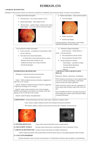
1) Background Diabetic Retinopathy
• Microaneurysms – tiny red spots temporal to fovea
• Retinal haemorrhages – flame shaped, dot blot
• Macular edema – capillary leakage, cystoids macular edema
Hard exudates – waxy yellow lesions with discrete margins
2) Diabetic Maculopathy - edema and hard exudates
• Focal maculopathy
• Diffuse maculopathy
• Ischemic maculopathy
Clinically significant - Retinal edema or hard exudate
within 500 micrometer of centre of macula
3) Pre proliferative Diabetic Retinopathy
• Cotton wool spots – accumulation of neuronal fibres within
the nerve fibre layer
• Intra retinal microvascular abnormalities
- AV Shunts that run from retinal arterioles to venules
- Dilatation and tortuosity, beading of veins
- Peripheral narrowing, silver wiring of arterioles
- Dot blot haemorrhages.
4) Proliferative Diabetic Retinopathy
Arterial occlusion → Retinal hypoxia →
VEGF → Neovascularisation
• Neovascularisation at the disc (NVD)
• Neovascularisation elsewhere (NVE)
5) Advanced Diabetic Eye Disease
- Haemorrhages → preretinal or intragel
- Tractional Retinal detachment
- Tractional retinoschisis
- Rubeosis
HYPERTENSIVE RETINOPATHY
Pathogenesis: Arteriolar narrowing (vasoconstriction)
Fibrosis (involutional sclerosis)
Grade I: Mild to moderate narrowing of arterioles, exaggeration of
light reflex at AV crossings
Grade II: Moderate to marked narrowing of arterioles, exaggeration
of light reflex and changes at AV crossings
Grade III: copper wiring of arterioles, silver wiring of arterioles,
BONNET sign: banking of veins distal to AV crossings. GUNN sign:
tapering of veins on both sides of the crossings
Grade IV: grade III changes with papilloedema
3. RETINAL VASCULAR OCCLUSIVE
DISEASE
Risk factors: Diabetes , Hypertension , Dyslipidemia
CRVO: dilatation and tortuosity of venous segments distal
to the site of occlusion, haemorrhages, retinal edema,
cotton wool spots, disc edema, hyperaemia
CRAO: narrowing of arteries and veins, segmentation of
blood column (cattle trucking), cloudy retina, Cherry red
spot
HoLLEN-HORST PLAQUES: intermittent showering of
minute bright yellow orange crystals (cholesterol emboli)
at arteriolar bifurcations
4. ROTH SPOTS - Retinal haemorrhages with white or pale centres, composed of coagulative fibrin
- Cause: immune complex mediated vasculitis often resulting from bacterial endocarditis
- DD: leukemia ,diabetes, ischaemic events, pernicious anemia, HIV retinopathy, thrombocytopenia
5. LIPAEMIA RETINALIS Creamy white coloured retinal blood vessels in triglyceridemia
6. COCK SCREW VESSELS Increased venous tortuosity due to arteriolisation of veins in Coarctation of Aorta
7. CIRCINATE RETINOPATHY Caused by chronic retinal edema, Eg: Diabetes, Hypertension
Girdle of bright white patches with crenated borders around the macula made up of macrophage aggregates filled with lipids
8. VASCULITIS DISORDERS
Arteriolar attenuation, cotton wool spots, microaneurysms, AV malformations, Ischaemic optic neuropathy, neovascularisation