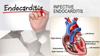
2. INFECTIVE ENDOCARDITIS
DEFINITION:
It is an inflammatory process of the endocardium,
especially the valves.
is a microbial infection of the valves and endothelial
surface of the heart and usually develops in people with
prosthetic heart valves or cardiac structural defects (e.g.
valve disorders)
3. INCIDENCE
•Each year 15,000 to 20,000 new cases are diagnosed.
•Has high morbidity and death rates.
•Infective endocarditis is more common in older people
•high among IV/injection drug users who most commonly develop
infections of the right-sided heart valves
•Hospital-acquired endocarditis occurs most often in patients with
debilitating disease, those with peripherally inserted central catheters
and those receiving haemodialysis or prolonged intravenous or
4. CONTINUE..
• Patients receiving immunosuppressive medications or
corticosteroids may develop fungal endocarditis.
•Invasive procedures, particularly those involving mucosal
surfaces, can cause a bacteremia.
• If a person has some anatomical cardiac defect, bacteremia
can cause bacterial endocarditis.
• The combination of the invasive procedure, the particular
bacterium introduced into the bloodstream, and the cardiac
defect may result in infective endocarditis.
6. Subacute bacterial endocarditis :
develops gradually over several weeks or months
Usually caused by organisms like: Streptococcus viridans
Acute bacterial endocarditis:
Develops over days or weeks with an erratic course and earlier development
of complications
Commonly caused by Staphylococcus aureus.
Native valve endocarditis:
An infection of a previously normal or damaged valve.
7. Prosthetic valve endocarditis:
An infection of a prosthetic valve.
Non bacterial thrombotic endocarditis:
Caused by sterile thrombotic lesions. ( may be in pts with cancer
or other chronic diseases.)
8. RISK FACTORS
• Prosthetic cardiac valves or prosthetic material used for cardiac
valve repair
• History of bacterial endocarditis (even without heart disease)
• Congenital heart disease
• Unrepaired cyanotic congenital heart disease, including patients
with palliative shunts and conduits
9. • Repaired congenital heart disease with residual defects at
the site or adjacent to the site of a prosthetic patch or
device
• Cardiac transplant recipients with valvulopathy
11. PATHOPHYSIOLOGY
Due to etiological factors
A deformity or an injury of the endocardium leads to
accumulation on the endocardium of fibrin and platelets
(clot formation).
Infectious organisms( staph, strepto…)
The infection most frequently results in platelets, fibrin,
blood cells and microorganisms that cluster as
12. The vegetations may embolise to other tissues throughout the
body.
As the clot on the endocardium continues to expand, the infecting
organism is covered by the new clot and concealed from the
body's normal defenses.
The infection may erode through the endocardium into the
underlying structures (e.g. valve leaflets), causing tears or other
deformities of valve leaflets, dehiscence of prosthetic valves,
13.
14. CLINICAL MANIFESTATIONS
•onset is insidious.
•Systemic emboli occur with left-sided heart infective
endocarditis; pulmonary emboli occur with right-sided heart
infective endocarditis
•Fever
•Chills with sweats
•Malaise
•Weakness
•Anorexia
•Weight loss ……
15. • backache
•Splenomegaly
•Flu like symptoms
•symptoms and signs can be non-specific, diagnosis
requires a high index of suspicion. C/M due to
embolization:
Stroke, TIA, aphasia
Myocardial infarction…..
16.
17.
18. clusters of petechiae may be found on the body.
Small, painful nodules (Osler nodes) may be present in
the pads of fingers or toes.
Finger Clubbing
Arthralgia, proteinuria, hematuria
Pulmonary embolus…..
19. Irregular, red or purple, painless, flat macules (Janeway lesions)
may be present on the palms, fingers, hands, soles and toes.
Haemorrhages with pale centres (Roth spots) caused by emboli
may be observed in the fundi of the eyes.
Vision loss
Splinter haemorrhages (i.e. reddish-brown lines and streaks)
Heart failure, which may result from perforation of a valve leaflet,
rupture of chordae, blood flow obstruction due to vegetations, or
intracardiac shunts from dehiscence of prosthetic valves.
20. ASSESSMENT AND DIAGNOSTIC FINDINGS
•History collection
•Physical examination: A heart murmur may be absent initially but
develops in almost all patients. Murmurs that worsen over time
indicate progressive damage from vegetations or perforation of
the valve or the chordate tendineae.
•fever and no obvious source of infection, particularly if a heart
murmur is present.
•Fever is intermittent and may be absent in patients who are
21. •. Blood culture
• CBC : Patients may have elevated white blood cell (WBC)
counts
•patients may be anaemic and have a positive
rheumatoid factor and an elevated erythrocyte
sedimentation rate (ESR) or (-reactive protein.
22. •Microscopic haematuria
•Doppler echocardiography : may assist in the diagnosis by
demonstrating a mass on the valve, prosthetic valve or supporting
structures and by identifying vegetations, abscesses, new
prosthetic valve dehiscence or new regurgitation
•The echocardiogram may reveal the development of heart failure.
•Chest x ray
23. Prevention:
is rare, infective endocarditis may be life-threatening.
A key strategy is primary prevention in high-risk patients (i.e.
those with prosthetic heart valves).
endocarditis prophylaxis should be given for dental and
respiratory procedures
The list of gastrointestinal and genitourinary procedures is
similarly precise and includes procedures that also have a
24. MANAGEMENT
The severity of oral inflammation and infection is a significant factor in
the incidence and degree of bacteraemia.
•Good oral hygiene is probably the most important factor in reducing
the risk of endocarditis in susceptible individuals, and access to high
quality dental care should be facilitated.
• Once a patient is found to have a cardiac anomaly putting them at risk
of endocarditis, the patient should be referred to have their dental
25. •Prophylaxis is recommended only for those invasive
respiratory tract procedures that involve a high risk of
bacteremia. These include tonsillectomy and
adenoidectomy, bronchoscopy with incision or biopsy and
surgery involving bronchial, sinus, nasal or middle ear
mucosa.
•Regular personal and professional oral healthcare and
rinsing with an antiseptic mouthwash for 30 seconds before
26. PHARMACOLOGICAL MANAGEMENT
•Antibiotic:
•penicillin is usually the medication of choice
•Patients are usually instructed to take 2 g of amoxicillin orally 1
hour before the procedure.
• parenterally in a continuous intravenous infusion for 2 to 6
weeks.
***In fungal endocarditis, an antifungal agent, such as
amphotericin B, is the usual treatment.
27. SURGICAL MANAGEMENT
•if the infection does not respond to medications, the patient has a prosthetic heart
valve endocarditis, has a vegetation larger than 1 cm, or develops complications
such as a septal perforation.
•Surgical interventions include:
• valve debridement or excision,
•debridement of vegetations,
• debridement and closure of an abscess and closure of a fistula.
•The aortic valve may be best treated with an autograft. Most patients who
have prosthetic valve endocarditis require valve replacement.
28. NURSING MANAGEMENT
•monitor the patient's temperature; the patient may have fever for
weeks.
•Heart sounds are assessed; a new murmur may indicate
involvement of the valve leaflets.
•monitor for signs and symptoms of systemic embolisation or for
patients with right-sided heart endocarditis,
• monitor for signs and symptoms of pulmonary infarction and
infiltrates.
•assesse signs and symptoms of organ damage such as stroke,
meningitis, heart failure, myocardial infarction, glomerulonephritis
and splenomegaly.
29. •The patient is started on antibiotics as soon as blood cultures have
been obtained.
•Provide the patient and family with emotional support and facilitate
coping strategies during the prolonged course of the infection and
antibiotic treatment required.
•If the patient receives surgical treatment, the nurse should provide
postoperative care and instruction.
•Encourage to have nutritious diet, adequate fluid, and rest.