Digestive system - Anatomy & Physiology - B.Pharm & Pharm.D
•
9 likes•4,174 views

Human Digestive System - Anatomy & Physiology - B.Pharm & Pharm.D - GIT & Its Accessory Organs

Recommended
Recommended
More Related Content
What's hot
What's hot (20)
Similar to Digestive system - Anatomy & Physiology - B.Pharm & Pharm.D
Similar to Digestive system - Anatomy & Physiology - B.Pharm & Pharm.D (20)
More from Kameshwaran Sugavanam
More from Kameshwaran Sugavanam (20)
Recently uploaded
Recently uploaded (20)
Digestive system - Anatomy & Physiology - B.Pharm & Pharm.D
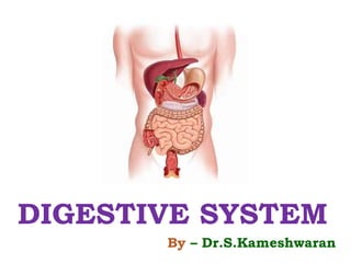
- 1. DIGESTIVE SYSTEM By – Dr.S.Kameshwaran
- 3. Digestive system describes alimentary canal and its accessory organs Digestion “It is a processes that prepare food eaten in the diet for absorption” System begins at the mouth passes through thorax, abdomen, pelvis and ends at the anus. Digestive process gradually breakdown the food eaten until they are in the form suitable for absorption
- 4. ACTIVITIES OF DIGESTIVE SYSTEM 1. Ingestion – TAKING OF FOOD IN TO ALIMENTARY CANAL (Eating/ Drinking) 2. Propulsion: Mixes & moves the content along the alimentary canal 3. Digestion A. Mechanical breakdown – fragment food into smaller particles (Mastication) (Chewing) B. Chemical Digestion of food into small molecules by enzymes present in secretions produced by glands and accessory organs of digestive system Mouth = carbohydrates Stomach = Proteins SI = CH, Proteins, fats, nucleic acids
- 5. 4. Absorption – digested food contents pass through the walls of alimentary canal in to blood and lymph capillaries for circulation and use by body cells 5.Defecation – indigestible un absorpable food substance excreted form ali canal as feces by the process of defecation.
- 6. Anatomy Alimentary canal Also known as Gastrointestinal tract (GI) Long tube through which food passes Commences at mouth terminates at anus Various organs includes: Mouth pharynx esophagus stomach small intestine large intestine
- 8. Accessory digestive organs Various secretion are poured in the alimentary tract Eg: Gastric juice secreted by glands in the lining of stomach Some by glands situated outside the tract Three pairs of salivary glands The pancreas The liver and biliary tracts
- 9. LAYERS OF GIT The GI tract contains four layers: the innermost layer - mucosa, Submucosa, followed by muscularis propria and the outermost layer -adventitia. The structure of these layers varies, in different regions of the digestive system, depending on their function.
- 11. MUCOSA (Mucus membrane): Innermost membrane Contains a lining columnar epithelium, including glandular tissue Glandular tissue secretes mucus – lubricates the inner line, Forms physical barrier that protects them from the damaging effect of digestive enzymes Consist of an underlying layer of loose connective tissue called the lamina propria - which provides vascular support for the epithelium Finally a thin double layer of smooth muscle is often present - the muscularis mucosa for movement of the mucosa.
- 12. SUB MUCOSA: This layer consist of loose connective tissue, contains collagen and some electric fibers Helps in binding of muscle layer to mucosa Contains sympathetic and parasympathetic nerve supply Contains larger blood vessels
- 13. Muscularis propria smooth muscle layer. There are usually two layers; the inner layer is circular, and the outer layer is longitudinal. These layers of smooth muscle are used for peristalsis (rhythmic waves of contraction), Helps to move food down through the gut. Myenteric plexus – the sympathetic and para sympathetic nerve innervation
- 15. Adventia layer (or serosa) It is also called as serosa layer Outermost layer blood vessels, lymphatics and nerves. Made up of loose connective tissue This connective tissue covered by the visceral peritoneum. Which reduces the friction during movements
- 16. Organs of digestive system: Salivary glands Pharynx Esophagus Stomach Small Intestine Large Intestine Rectum Accessory digestive organs: liver, gallbladder, pancreas
- 17. Mouth Oral cavity: mechanical, chemical digestion Mastication: teeth chew food Tongue mixes food + saliva
- 18. The mouth or oral cavity is bounded by muscles and bones Oral cavity is lined throughout with mucous membrane It contains stratified squamous epithelium containing small mucus secreting glands The uvula is a curved fold of muscle covered with mucous membrane Hanging down from the middle of the soft palate
- 19. Teeth: Food is chewed with the help of teeth to breakdown in to smaller particles – mastication Teeth are hard calcified structures that help in mastication Teeths are embadded in the sockets of the mandible and the maxilla Babies are born with two sets deciduous teeth (baby/ Milk teeth) & Permanent teeth (adult teeth)
- 20. DECIDUOUS TEETH There are temporary teeth 10 in each jaw They begin to erupt at about 6 months of age Should all be present by 36 months The permanent teeth begin to replace the deciduous teeth in the 6 th year Consisting 32 teeth is usually completed by the 21 st year (16 in each jaw)
- 21. STRUCTURE: Basic structure of tooth Crown – visible part Root – part with in gum Crown – is covered with enamel (highly mineralised tissue) + dentin Root – is covered with cementum (Hard connective tissue) The part connect the crown & root portion of tooth known as neck
- 23. CROWN: The crown contains enamel & dentin ENAMEL: Hardest and most mineralized part of human body It form outer covering of the tooth Calcium phosphate is the major mineral present DENTIN: It is a hard connective tissue just like a bone It present behind the enamel Accounts for largest portion of tooth It surrounds the pulp (soft CT contains BV & nerve innervating to tooth ROOT Root covered with cementum (Hard connective tissue) All blood vessels and nerves enters in to pulp through the root of teeth
- 24. PERMANENT TEETH: INCISORS – 2-2 CANINE – 1-1 PREMOLARS – 2-2 MOLARS – 3-3 DECIDUOUS TEETH INCISORS – 2 -2 CANINE – 1 - 1 MOLARS -2 – 2 Functions: INCISORS - cutting teeth used for biting of pieces of food CANINES – conical shaped teeth – grasping & tearing the food PREMOLARS – grinding the food (ab in children) (after 9 yrs)
- 25. SALIVARY GLANDS: Oral cavity contains three main pairs of salivary glands Their secretions termed as saliva Which helps to moisten the oral cavity & bolus formation Saliva is the mixture of mucus & serous (watery type) Every day 1.5 lit of saliva produced Types of salivary glands: The parotid glands The submandibular The sublingual glands Apart from that 600-1000 are present in the oral cavity – contribute to small amount of hole saliva production
- 26. PAROTID GLAND: Largest salivary gland Located immediately anterior to the ear on either side It releases serous secretion – via parotid duct - Opens at second upper molar tooth of oral cavity
- 27. THE SUBMANDIBULAR: This is second largest gland – located below the mandible It produce mixed secretions – releases via submandibular duct - Opens in the mandibular region of the oral cavity (adjacent to the frenulum of the tongue)
- 28. The sublingual glands This is the smallest major salivary gland Located in the floor of the mouth It produces mixed secretions It does not have specialized ducts -ductless glands It releases secretions directly in to the floor of mouth via 10-12 small ducts
- 29. COMPOSITION OF SALIVA: Water – 99% Electrolytes (Sod, Pot, Cal, Mag, Bicar, Phosp) Proteins Mucosal glycoprotein Trace of albumin Polypeptides & Oligopeptides Functions of saliva: Chemical digestion of polysaccarides Lubrication of food Cleaning and lubricating mouth Non specific defence – ig & lysosyme combat microbs Taste
- 30. PHARYNX: It is a funnel shaped tube It is made up of skeletal muscle – lined with mucus membrane Divided in to Nasopharynx Oropharynx Laryngopharynx Nasopharynx: Contributes to respiration Oropharunx & Laryngopharynx Contributes to respiratory as well as digestive functions
- 31. Oropharynx & laryngopharynx is common passage way for both respiratory and digestive system food – oral – pharynx – esophagus Pharynx walls 3 layers of muscles: Mucosa – stratified squamous epithelium Middle – fibrous connective tissue- BV, lymph, Nerves Outer layer – number of involuntary muscle – swallowing
- 32. OESOPHAGUS: Also known as food pipe It is a long muscular tube About 25 cm long – 2 cm in dia Lies in the thorax in front of vertebral column Behind trachea and heart Which joins pharynx
- 34. It curves upwards before opening in to the stomach Sharp angle – prevents regurgitation (back flow ) of gastric contents to the oesophagus The upper and lower ends of the esophagus are closed by sphincter Cricopharyngeal/ upper oesphagel sphincter prevents air passing in to the oesophagus
- 35. Anatomy of esophagus: Mucosa Sub mucosa Muscularis propria Functions: Functions of mouth - pharynx - oesophagus Formation of bolus Swallowing (deglutition) A wave of peristalsis is stimulated when the bolus is present in the pharynx – bolus propelled to the stomach via esophagus
- 36. STOAMCH: It is a J shaped dilated portion of alimentary tract It is a hollow, muscular, bag like structure The second phase of digestion takes place in the stomach Stomach size varies with the volume of food it contains 1.5liter for an adult Structure of stomach: Continues with oeophagus at cardiac sphincter Ends with duodenum at pyloric sphincter It has two curvatures Lesser curvature is short Lies on the posterior surface of the stomach It is the downward continuation of posterior wall of oesophagus Just before the pyloric sphincter it curves uwpard o give J shape
- 38. Greater curvature – outer portion of stomach Stomach – divided in to 3 regions FUNDUS – BODY – PYLORUS Fundus: The dome shaped part is formed by the upper curvature of the stomach Body The central region of the stomach Pylorus: It is the lower region of the stomach, that continues in to the duodenum
- 40. At the distal end of pylorus – pyloric sphincter Pyloric sphincter – Guarding the opening between stomach and duodenum When stomach is inactive – sphincter relaxed and open When stomach contains food – sphincter is closed
- 41. WALLS OF THE STOMACH The stomach has 4 layers SEROSA/ VISCERAL PERITONEUM MUSCLE LAYER Outer Longitudinal layer Middle circular layer Inner oblique layer SUBMUCOSA
- 43. GASTRIC JUICE: About 2 liters of gastric juice are secreted daily by specialized secretory glands present in the mucosa Composition of Gastric Juice : Water Mineral salts Mucus – by mucus membrane HCL – by Parietal cells in gastric glands Intrinsic factor – by Parietal cells in gastric glands Inactive enzyme precursor - pepsinogen
- 44. FUNCTIONS OF GASTIC JUICE: WATER: Further liquefies the food swallowed HCL: Acidifies the food and stop the action of salivary amylase Kills ingested microbs Provide acid environment needed for the action of pepsins PEPSINOGENS: Pepsinogen activated to pepsin by HCL & by pepsin already present in the stomach PEPSIN Pepsin is more active at ph between 1.5 - 3.5 digest the proteins
- 45. INTRINSIC FACTOR: It is a protein Important for absorption of vit B12 from ileum (def: pernicious anaemia) MUCUS: Prevents mechanical injury to stomach
- 46. SECRETION OF GASTRIC ACID: HCL in stomach with out food - Fasting juice Secretion reaches max level 1 hour after meal decline to fasting level after 4 hour Acid production is regulated by the parasympathetic nervous sytem 3 PHASES OF GASTRIC ACID SECRETION: CEPHALIC PHASE GASTRIC PHASE INTESTINAL PHASE
- 47. CEPHALIC PHASE: The rate of secretion of gastric juice is high (around 500ml/h) in this phase Flow of juice before food reaching the stomach Occurs due to reflex stimulation of vagus nerves initiated by sight, smell, taste or thought of food Cerebral cortex and appetite centres of hypothalamus sends the neurogenic signal Impulses from hypothalamus reaches the stomach via vagus nerve - and stimulate the secretions of HCL – secreted from parietal cell
- 48. GASTRIC PHASE: Rate of secretion of gastric juice in this phase is lesser (200ml/h) This phase begins as soon as the food enters the stomach G cells in the pylorus & duodenum – secrete gastrine – passes directly in to the circulating blood Gastrine present in blood supplies to stomach – stimulate the gastric glands to produce more gastric juice Gastrine secretion stopped when the pH in pylorus falls less than 3
- 49. INTESTINAL PHASE: Partially digested stomach contents (Chyme) – reach small intestine – secretin & cholecystokinin are produced by endocrine cells of intestinal mucosa They slow down the secretion of Gastric juice & reduce gastric motility Process in more faster if meal contain high fat CH Leaves stomach – 23 hours Protein meal – longer Fatty meal – remain longest
- 51. PEPSIN ROLE IN PROTEIN DIGESTION: Pepsinogen is secreted by the chief cells present in the gastric glands Pepsinogen is converted in to pepsin by HCL acid and previously formed pepsin At pH 3 or less than that the pepsin exerts maximum enzymatic activity It breaks the protein molecule in to smaller peptide chains by acting as catalyst
- 52. FUNCTIONS: Break downs larger molecules in to smaller 2-3 liters of gastric acid secreted per day, high amount secreted in evening Pepsinogen – converted in to pepsin – helps in protein digestion Intrinsic factor produced by parietal cells of stomach is essential for vit B12 absorption
- 53. SMALL INTESTINE: Continue with stomach @ pyloric sphincter Present between the stomach and small intestine 2.5 cm dia, little over 5 meter long Leads in to large intestine @ ileocaecal valve Lies in the abdominal cavity Chemical digestion of food is completed and absorption of most nutrients Absorption of 90% of nutrients from digested food Internal wall is folded to form villi (finger like projections) absorb all the nutrients
- 54. Structurally small intestine can be divided in to 3 parts DUODENUM JEJUNAM ILEUM
- 55. DUODENUM: It is the initial and smallest part of small intestine 25 cm long It is a hollow tube like structure connecting stomach to jejunum Curves around the head of pancreas Majority of the chemical digestion takes place in duodenum Secretions of gall bladder and pancreas merge in a common structure – hepato pancreatic ampulla Enters the duodenum @ duodenal papilla – gaurded by – hepato pancreatic sphincter (Sphincter of oddi)
- 58. JEJUNAM It is a part of small intestine Present between duodenum & ileum 2 meter long ILEUM It is the lower end of the small intestine Continues in to the large intestine to form the caecum 3 meters long, The diameter of intestinal lumen decreases towards the ileum ends at ileocaecal valve Controls flow of material from ileum to caecum (1st part of Large intestine) prevents back flow
- 59. STRUCTURE OF SMALL INTESTINE: Wall of SI – 4 layers Some modifications only in the peritoneum & Mucosa MUCOSA : Surface of SI mucosa is greatly increased by permanent circular folds, VILLI – finger like projections of mucosal layer 0.5 – 1 mm long Coverings consist of columnar epithelial cells with tini micro villi micro villi - 1 micro meter long
- 62. INTESTINAL JUICE: 1-2 liters of intestinal juice secreted/day Present in the form of clear yellow fluid Consist of water, mucus and mineral salts It is slightly alkaline (basic) in nature pH 7.6
- 63. CHEMICAL DIGESTION IN SI When food present in the small intestine Cholecystokinin is produced in the small intestine which promotes the release of Pancreatic enzymes and bile from the gallbladder in to the small intestine Proteins are degraded into small peptides and amino acids Lipids (fats) are degraded into fatty acids and glycerol, Pancreatic lipase & bile - breaks down triglycerides into free fatty acids and monoglycerides Pancreatic amylase breaks down some carbohydrates into oligosaccharides
- 64. Functions: Carbohydrate metabolism Lipid metabolism Protein metabolism Processing of drugs and hormones Excretion of bilirubin Synthesis of bile salts Storage Phagocytosis Activation of vitamin D
- 65. PANCREATIC JUICE: Secreted by exocrine pancreas Which enters the duodenum at the duodenal papilla It consist of Water Mineral salts Enzymes Amylase, lipase, nucleases that digest DNA & RNA INACTIVE ENZYME PRECUROSORS Tripsinogen
- 66. FUNCTIONS: Digestion of proteins Digestion of CH Digestion of fats
- 67. LARGE INTESTINE: It is about 1.5m long Beginning at caecum & terminating at the rectum and anal canal 6.5cm diameter It forms an arch round the coiled up small intestine It is divided in to Caecum Colon Sigmoid colon Rectum
- 68. THE CAECUM: This is first part of large intestine It is a dilated region It has no digestive function Can cause significant problems when inflamed THE COLON: It has 4 parts has same structure and function THE ASCENDING COLON THE TRANSVERSE COLON THE DESCENDING COLON
- 69. THE ASCENDING COLON This passes upwards from he caecum to the level of liver Where it curves to left at hepatic flexure to become transverse colon THE TRANSVERSE COLON: This extends across the abdominal cavity in front of the duodenum & the stomach Near to the spleen it forms splenic flexure and curves downwards to become descending colon THE DESCENDING COLON: It passes down the left side of the abdominal cavity Curves towards the midline It known as sigmoid colon
- 71. THE SIGMOIDCOLON: This part describes as s shaped curve in the pelvic cavity It continues downward to become the rectum THE RECTUM: This is slightly dilated section of the large intestine It is about 13cm length Leads from sigmoid colon Terminates at anal canal THE ANAL CANAL: It is a short passage about 3.8cmlong in adults
- 73. The internal sphincter – contains smooth muscles – under the control of ANS External sphincter – formed by skeletal muscles – its under voluntary control. When individuals wants to defecate – they open it up allowing evacuation of the bowel. STRUCTURE: 4 layers of tissue
- 74. SECRETIONS: Colon mucosa contains number of goblet cells – secrete mucus – lubricate the colon walls CONSTITUENTS OF FEACES: It is a semisolid brown mass Colour due to the presence of stercobilin (heam – bilirubin – enters intestine – excreted in faeces Fibre Dead and live microbes Epithelial cells shed from the walls of the tract Fatty acid Mucus secreted by the epithelial lining of the large intestine Mucus – helps to lubricate the faeces
- 75. FUNCTIONS OF LARGE INTESTINE Absorption – absorption of water by osmosis until food content become semi solid mass Mineralsalts, vitamins and some drugs also absorbed MICROBIAL ACTIVITY: Large intestine contains certain types of bacteria Which synthesis Vitamin k and folic acid Includes Escherichia Coli, Enterobacter aergenes Streptococcus faecalis, Clastridium perfringens They are harmless in humans Become pathogenic if transferred to other part
- 76. Pancreas: It is an elongated digestive gland Pale gray gland 12-15 cm long & 60gms weight Present in the abdominal cavity Consist of broad head a body and a narrow tail Head lies in the curve of duodenum The exocrine pancreas: It contains large number of lobules made up of small acini The walls of it consist of secretory cells Each lobule drained by small duct –unite to form pancreatic duct – open to the duedenum
- 78. Before entering in to deudenum it joins common bile duct to form hepatopancreatic ampulla Opening is controlled by hepato pancreatic sphincter (of oddi) at the duodenal papilla THE ENDOCRINE PANCREAS: Contains specialized cells called pancreatic islets It has no ducts - hormones diffuse directly in to blood Secretes insulin & glucagon – maintains blood glucose level
- 79. LIVER: Largest gland in the body Weighing about 1-2.3 kg Situated in the upper part of the abdominal cavity The liver is enclosed in a thin inelasstic capsule and incompletely covered by a layer of peritoneum Folds of peritoneum form supporting ligaments that attach the liver to the inferior surface of the diaphragm Liver has 4 lobes Obvious Large right lobe Smaller wedge shaped left lobe Two others are caudate & quadrate lobes – in posterior surface
- 81. THE PORTAL FISSURE: The part is posterior surface of the liver where various structures enters and leave the liver Portal vein, hepatic artery, nerve fibres, hepatic ducts, lymph vessels
- 82. STRUCTURE: Lobes of liver made by functional units – lobules Visible to the naked eye Hexagonal in outline formed by cuboidal cells Hepatocytes arranged in a pairs of columns radiating from a central vein
- 83. The spacce between the two pairs of columnar cell is – sinusoid The blood vessels are having incomplete walls - - contents of blood mix and come in to close contact with liver cells Kupffer cells (hepatic macrophages) Ingest and destroy the worn out blood cells & foreign particles present in the blood Liver also secretes bile Bile canaliculi run between the column of liver Each hepatocyte having one side sinusoid and bile canaliculus on other side
- 84. FUNCTIONS OF LIVER: Carbohydrate metabolism Fat metabolism Protein metabolism Breakdown of erythrocytes & defense against microbes Detoxification of drugs and toxic substances Inactivation of hormones Production of heat Secretion of bile storage
- 85. COPOSITION OF BILE: 500 – 1000 ml of bile is secreted by liver Bile consist of Water Mineral salts Mucus Bile pigments - bilirubin Bile salts Cholesterol Functions of bile Fat digestion Excretion of bilirubin
- 86. GALL BLADDER: It is a pear shaped sac attached to the posterior surface of the liver by connective tissue It has a fundus/ expanded end A body or main part and a neck Which is continues with the cystic duct Structure is same like that of GIT with small modifications In peritonium muscle layer & mucus membrane FUNCTIONS OF GALLBLADDER: Reservoir for bile Concentration of the bile increases10 – 15 folds by absorbing the water through the wall of the gallbladder Release of stored bile
- 88. DISEASE OF MOUTH: Mouth ulcers Acute Gingivities (inflammation of gums) Squamous cell carcinoma CONGENITAL DISORDER: Cleft palate and cleft lip DISORDERS OF SALIVARY GLAND: Mumps – viral disease at parotid gland Tumor of salivary glands Sialolithiasis – stones in the salivary duct
- 89. DISORDERS OF THE PHARYNX: Pharyngitis – infected and swellon Tonsillitis – inflammation in tonsils DISORDERS OF OESOPHAGUS: GERD – Gastro Esophageal Reflex Disease Achalasia – esophagus empties slowly DISORDERS OF STOMACH: Gastritis – stomach lining becomes inflamed, irritated, or eroded Gastric Ulcer – condition – sore appear in the inner lining of the stomach
- 90. DISORDERS OF INTESTINE: Appendicitis – Inflammation in appendicitis and filled with pus Crohns disease “Condition in which the GIT become inflammed – progress to all layers of GIT” Symptoms: Chronic diarrhoea Loss in weight Fever Pain and tenderness in abdomen Rectal bleeding
- 91. Disorders of pancreas: Pancreatitis – inflammation in pancreas Zollinger – Ellison Syndrome It is a rare disorder Tumours and ulcer in the digestive system Less than 3 peoples affected in million peoples Due to the tumor increase in gastrin production which increases gastric acid secretion Disorders of Liver: Liver cirrhosis – Loss of liver cells, irreversible scarring of liver cells Hepatitis – liver inflammation Disorders of gall bladder: Gall stones – cholelithiasisi Cholecystitis – inflammation in gallbladder
- 92. Movements of GIT: Also known as Gastro intestinal motility Involuntary mobility of human tubular organs is termend as Motility There are two main movements present in the GIT Propulsion movement & Mixing Movements 1. Propulsion movement Invloved in the movement of chyme through the digestive tract at a rate proportional to the absorption and digestion rate The propulsion movement is called as PERISTALSIS
- 93. MIXING MOVEMENTS: It involved in constant mixing of chyme to expose all the nutritionally important components to the enzyme So the components come in to contact with the lining of intestine where it needs to be absorbed – absorption of nutrients MASTICATION: “IT IS THE PROCESS BY WHICH THE LARGER FOOD PARTICLES BREAKDOWNS IN TO SMALLER ONES BY THE PREMOLAR AND MOLAR TEETH'S” Previously the food is tore and cut by the incisors and canines Temporalis, masseter, medial prerygoid & lateral pterygoid are the muscles involved in the mastication process
- 94. SWALLOWING/ DEGLUTITION “passage of food from mouth to stomach” This process involved in mouth, pharynx & oesophagus There are 3 main phase in swallowing Voluntry phase passage of bolus in to oropharynx Pharyngeal Phase involuntary passage of bolus from pharynx to esophagus Oesophageal Phase Involuntary passage of bolus from esophagus to stomach
- 95. STOMACH MOVEMENTS: Stomach filling Food present in the stomach increases the stomach volume The pressure with in the stomach remain unchanged untill the volume reaches maximum capacity Occur due to the stretching of smooth muscle with out increase in tension
- 96. Mixing stomach contents The food and secretions of stomach glands are mixed to form – chyme Mixing wave (peristalsis) like contraction produced in the stomach body for every 20 seconds towards the pyloric region Fluid part of the Chyme pushed towards the pyloric region while the solid part pushed back towards stomach body This process repeats until food mixes with secretions fully
- 97. STOMACH EMPTYING The food remains in the stomach depends on the type and volume of food After having meal stomach empties with in 3-4 hours Each peristaltic movement pushes small amount of chyme in to the duodenum through pyloric sphincter opening - pyloric pump
- 98. MOVEMENTS OF SMALL INTESTINE: Small intestine carry out mixing and propulsion of chyme – occurs due to the segmental and peristaltic contractions of the smooth muscles Segmental contractions – initiate mixing of the intestinal contents Peristaltic contractions initiate propelling the content further along the digestive tract The rate at which the contraction move is about 1cm/min The contractions faster at proximal end slower at distal end The intestinal motility is regulated by mechanical, chemical stimuli also by
- 99. MOVEMENT OF LARGE INTESTINE: Peristaltic contraction moves the chyme along the ascending colon In transverse colon and descending colon undergo strong peristaltic contraction – called mass movements Which occurs atleast 3-4 times a day A mass movement moves the colon content around 20cms Pushing the content of the colon towards the anus Defecation reflex – stimulated by rectal wall – when it is distended by faeces Which weakly contract rectum - relaxes the
- 100. External anal sphincter – made up of skeletal muscle – controlled by cerebrum – relaxes to expel the faeces. DEFECATION: Voluntary movement to expel faeces Contraction of abdominal muscles – to force the colon contents – expel through anal canal.
- 101. DIGESTION “The process of breakdown of food into smaller molecules that can be absorbed in the blood circulation “ There are two types of digestion Mechanical Digestion – breakdown of food in to smaller molecules Chemical Digestion – Breakdown of chemical covalent bonds in organic molecules (CH,PTN, FAT) The process by which the digested nutrients reaches the blood circulation is called as absorption
- 102. DIGESTION & ABSORPTION OF CARBOHYDRATES: Carbohydrates consist of Polysaccharides, disaccharides & Monosacchardies Digestion invloved in breakdown of Polysaccharides in to smaller chains in to Disaccharides then further breakdow in to Monosacchardies Digestion of CH begins at the oral cavity Partial digestion of starch by salivary amylase Small amount of CH digested in the stomach by gastric amylase & Gelatinase
- 103. Digestion continues in the small intestine by pancreatic amylase Disaccharides are converted in to monosaccharides by the enzyme disaccharidases bound to microvilli of the intestinal epithelium Monosaccharides (glucose & galactose) are taken up into the intestinal epithelium cells via – secondary active transport by sodium ion gradient Monosaccharides (Fructose) – taken up by falicitated diffusion Monosaccharides enters in to the capillaries of intestinal villi are carried via hepatic portal
- 104. DIGESTION & ABSORPTION OF LIPIDS Lipids are insoluble or slightly soluble in water They includes triglycerides, phospholipids, cholesterol, steroids and fat soluble vitamins Triglycerides consist of covelently bound 3 fatty acids and a glycerol molecule Lingual lipase secreted in the oral cavity digest minor amount of lipids Emulsification – conversion of larger lipid droplets in to smaller ones with the help of bile salts Then the lipid molecules are digested by the lipase from pancreas Lipids are digested in to free fatty acids, glycerol, cholesterol and phospholipids
- 105. After the digestion the droplets of fatty acids aggregated by the bile salts to form micelles The micelles attach to the plasma memebranes of intestinal epithelial cells – Fatty acid, glycerol pass in to the cell via simple diffusion With in the cell the fatty acid and glycerol converted in to triglyceride – by protein coating they converted in to chylomicrons – enters in to the circulation
- 106. DIGESTION AND ABSORPTION OF PROTEIN Protein breakdown in to amino acids Actively transported in to various body cells The transport of amino acid is stimulated by Growth hormone & insulin Stomach secrete pepsin – cleaves the covalent bonds of proteins –in to smaller polypeptide chain 10-20 % of ingested protein is digested by gastric pepsin Proteolytic enzyme form pancreas carry out the further digestion PEPTIDASE bound to the microvilli of small intestine breakdown the protein in to dipeptides, tripeptides & amino acids dipeptides, tripeptides enters the intestinal epithelial cells
- 107. More amount of amino acids enters in to the intestinal epithelium when it is in the dipeptides, tripeptides With in the cell dipeptides, tripeptides broken down by dipeptidases and tripeptidases in to amino acids They moves from intestinal epithelial cells in to liver by hepatic portal vein