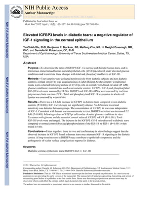
SlideShare a Scribd company logo
1 of 18
Download to read offline
Elevated IGFBP3 levels in diabetic tears: a negative regulator of
IGF-1 signaling in the corneal epithelium
Yu-Chieh Wu, PhD, Benjamin R. Buckner, BS, Meifang Zhu, MS, H. Dwight Cavanagh, MD,
PhD, and Danielle M. Robertson, OD, PhD
Department of Ophthalmology, University of Texas Southwestern Medical Center, Dallas, TX,
USA
Abstract
Purpose—To determine the ratio of IGFBP3:IGF-1 in normal and diabetic human tears, and in
telomerase-immortalized human corneal epithelial cells (hTCEpi) cultured under elevated glucose
conditions and to correlate these changes with total and phosphorylated levels of IGF-1R.
Methods—Tear samples were collected noninvasively from diabetic subjects and non-diabetic
controls; corneal sensitivity was assessed using a Cochet-Bonnet Aesthesiometer. Conditioned
media were collected following culture of hTCEpi cells in normal (5 mM) and elevated (25 mM)
glucose conditions; mannitol was used as an osmotic control. IGFBP3, IGF-1, and phosphorylated
IGF-1R levels were assessed by ELISA. IGFBP3 and IGF-1R mRNA were assessed by real time
polymerase chain reaction (PCR). Total and phosphorylated IGF-1R expression in whole cell
lysates was assessed by western blot.
Results—There was a 2.8-fold increase in IGFBP3 in diabetic tears compared to non-diabetic
controls (P=0.006); IGF-1 levels were not significantly altered. No difference in corneal
sensitivity was detected between groups. The concentration of IGFBP3 in tears was independent
of IGF-1. Consistent with human tear measurements in vivo, IGFBP3 secretion was increased 2.2
fold (P<0.001) following culture of hTCEpi cells under elevated glucose conditions in vitro.
Treatment with glucose and the mannitol control reduced IGFBP3 mRNA (P<0.001). Total
IGF-1R levels were unchanged. The increase in the IGFBP3:IGF-1 ratio detected in diabetic tears
compared to normal controls blocked phosphorylation of the IGF-1R by IGF-1 (P<0.001) when
tested in vitro.
Conclusions—Taken together, these in vivo and confirmatory in vitro findings suggest that the
observed increase in IGFBP3 found in human tears may attenuate IGF-1R signaling in the diabetic
cornea. A long-term increase in IGFBP3 may contribute to epithelial compromise and the
pathogenesis of ocular surface complications reported in diabetes.
Keywords
Diabetes; cornea; epithelium; tears; IGFBP3; IGF-1; IGF-1R
© 2012 Elsevier Inc. All rights reserved.
Corresponding author: Danielle M. Robertson, OD, PhD, Department of Ophthalmology, UT Southwestern Medical Center, 5323
Harry Hines Blvd, Dallas, TX 75390-9057. Tel: 214-648-3416. Danielle.Robertson@utsouthwestern.edu.
Publisher's Disclaimer: This is a PDF file of an unedited manuscript that has been accepted for publication. As a service to our
customers we are providing this early version of the manuscript. The manuscript will undergo copyediting, typesetting, and review of
the resulting proof before it is published in its final citable form. Please note that during the production process errors may be
discovered which could affect the content, and all legal disclaimers that apply to the journal pertain.
The authors have no commercial or proprietary interest in any concept or product discussed in this article.
NIH Public Access
Author Manuscript
Ocul Surf. Author manuscript; available in PMC 2013 April 1.
Published in final edited form as:
Ocul Surf. 2012 April ; 10(2): 100–107. doi:10.1016/j.jtos.2012.01.004.
NIH-PAAuthorManuscriptNIH-PAAuthorManuscriptNIH-PAAuthorManuscript
INTRODUCTION
Diabetic keratopathy can result in substantial and sometimes permanent visual loss
secondary to chronic erosions, scarring, and infectious corneal ulceration. Reported to occur
in more than 70% of patients with diabetes, diabetic keratopathy includes a large spectrum
of conditions, ranging from superficial punctate keratitis to recurrent corneal erosions both
after surgery and de novo, as well as corneal neuropathy.1–4 Alterations in the epithelium of
the diabetic cornea that contribute to the disease phenotype include impaired wound healing
resulting from delayed cellular migration and reduced cellular adhesion and the concomitant
loss of hemidesmosomes, which function to anchor the epithelium to the underlying
basement membrane.5–7 Accompanied by alterations in the basement membrane itself, these
collective changes result in epithelial fragility and disruption of the normal epithelial
barrier.5, 8–12
Insulin-like growth factor binding protein 3, IGFBP3, is an N-linked glycosylated,
phosphorylated, secretory protein with known antiproliferative and pro-apoptotic
functions.13 IGFBP3 belongs to a family of high affinity insulin-like growth factor (IGF)
binding proteins, which function to sequester extracellular IGF-1, preventing IGF-1
activation of the insulin-like growth factor receptor, IGF-1R.14 The IGF-1R is a
glycosylated, transmembrane receptor tyrosine kinase that has vital roles in development
and normal tissue maintenance.15 Both IGFBP3 and IGF-1R have been previously identified
in the corneal epithelium and in cultured corneal epithelial cells16, 17; however, the
functional significance of this localization in mediating homeostatic renewal is unknown. In
addition to mediating IGF-1R signaling, IGFBP3 has also been shown to regulate insulin
resistance18, 19 and apoptosis20–24 in a variety of cell types via IGF-1 independent pathways.
IGFBP3 has also been identified as a hypoxia-responsive protein25, 26 with the potential to
regulate angiogenesis.27–30
Changes in tear production and composition have been associated with diabetes, including
elevated glucose levels,31 an increase in advanced glycation end product modified
proteins,32 and a reduction in reflex tearing.33 In this study, we investigated the expression
levels of IGFBP3 and IGF-1 in human tears of normal and diabetic patients in vivo and
following in vitro culture of telomerase-immortalized human corneal epithelial cells in
elevated glucose. We further investigated IGF-1R expression in normal and high glucose
conditions and phosphorylation status when stimulated at various IGFBP3:IGF-1 ratios.
Significantly, we show for the first time that IGFBP3 is present in human tears and that it is
increased in the tears of patients with diabetes. Moreover, secreted IGFBP3 is similarly
increased following in vitro culture in high glucose. The increase in the IGFBP3:IGF-1 ratio
is associated with a reduction in phosphorylated IGF-1R. Taken together, these findings
suggest that prolonged elevated IGFBP3 expression levels in human tears of diabetics may
contribute to the pathogenesis of the high incidence of corneal complications through the
attenuation of normal IGF-1R-mediated signaling by IGF-1.
METHODS
Study Population
Thirty-three patients, 18 diabetics and 15 non-diabetic normal controls, who met the
inclusion criteria signed an informed consent and were enrolled in this study between June
and August 2011. Inclusion criteria included no recent history of contact lens wear; no use
of topical medications including artificial tears; non-smoking; no use of systemic hormones,
anti-histamines, or anti-depressants; or any previous history of ocular surgery. Diabetic
patients were recruited from the Internal Medicine Diabetic Clinic at UT Southwestern
Medical Center. A diagnosis of diabetes in the patient’s medical chart was required for
Wu et al. Page 2
Ocul Surf. Author manuscript; available in PMC 2013 April 1.
NIH-PAAuthorManuscriptNIH-PAAuthorManuscriptNIH-PAAuthorManuscript
admittance to the study. Normal, non-diabetic patients were recruited from within the
Department of Ophthalmology, UT Southwestern Medical Center. Pertinent medical history
was obtained following consent. Current glycosylated hemoglobin levels were obtained
from medical records. All procedures were approved by the Institutional Review Board at
UT Southwestern Medical Center and were conducted in accordance with the tenets of the
Declaration of Helsinki ethical principles for medical research involving human subjects.
Tear Collection
Each subject underwent one visit at which 10 µl of basal tears were collected sequentially
from the inferior meniscus of the right, then left, eye using a 10 µl glass microcapillary tube
(Drummond, Fisher Scientific, Houston, TX). Samples from right and left eyes were not
pooled but were used to test levels of IGF-1 and IGFBP3, respectively. Care was taken to
minimize reflex tearing. Tear samples were eluted into 1.5 ml Eppendorf tubes and
immediately placed on ice. All samples were collected between 8 a.m. and noon to control
for diurnal fluctuations. Tear samples were stored at −80°C until use. All tear samples were
obtained by a single trained investigator.
Corneal Sensitivity
Corneal sensitivity was measured in the right eye after tear collection using a Cochet-Bonnet
Aesthesiometer (C-BA, Luneau, Paris, France). The C-BA was mounted to a slit lamp base
using a standard lab clamp that allowed for movement in the x, y, z directions. A 0.08 mm
diameter nylon filament applanated the inferior cornea approximately 2 mm above the
inferior limbus. Measurements were initiated with the filament fully extended to 6.0 mm.
Filament length was systematically reduced in 0.5 mm increments until the patient correctly
reported 2 or more of the four stimulus presentations. False presentations were included to
control for incorrect patient responses. All measurements were obtained by a single trained
investigator.
Cell Culture
Human telomerase-immortalized corneal epithelial cells (hTCEpi) were initially isolated
and thereafter routinely maintained in serum-free keratinocyte growth media (KGM-2,
Clonetics-BioWhittaker) containing 5 mM glucose, 0.15 mM calcium and supplemented
with 0.4% bovine pituitary extract, 0.1% human epidermal growth factor, 0.1% insulin,
0.1% hydrocortisone, 0.1% transferrin, 0.1% epinephrine and 0.1% gentamicin sulfate
amphotericin B, as previously described.34 Cells were subcultured on T75 tissue culture
flasks (Falcon Labware; BD Biosciences, Bedford, MA), incubated at 37°C in 5% CO2. and
passaged every 5 days. For high glucose conditions, cells were cultured in serum-free
keratinocyte basal media without supplements for three days on 6-well polystyrene tissue
culture plates (Corning, Lowell, MA). Media contained an additional 20 mM glucose
(Sigma, St. Louis, MO) or 20 mM mannitol (Sigma, St. Louis, MO) as an osmotic control.
Media was changed at day 2. Conditioned media samples were collected at day 2 and day 3
and pooled for later analysis. For all cell culture experiments, assays were performed in
triplicate and repeated three times independently.
ELISA
All samples collected from right and left eyes were used for IGF-1 and IGFBP3 analysis,
respectively. Samples were diluted 1:5 in assay buffer. Conditioned media samples were
concentrated using iCON protein concentrators (Pierce, Rockford, IL). To detect
phosphorylated IGF-1R, 3×10⁵ cultured cells were starved in basal media for 24 hours and
then stimulated by 25 ng/ml of recombinant human IGF-1 (BioVision Inc, Mountain View,
California) with or without recombinant human IGFBP-3 (BioVision Inc, Mountain View,
Wu et al. Page 3
Ocul Surf. Author manuscript; available in PMC 2013 April 1.
NIH-PAAuthorManuscriptNIH-PAAuthorManuscriptNIH-PAAuthorManuscript
California) for 15 minutes at IGFBP3:IGF-1 ratios that simulated the ratios detected in
normal and diabetic human tears. Whole cell lysates were harvested directly in the culture
dish using RIPA buffer containing Halt™ protease and phophatase inhibitor single-use
cocktail (Thermo Fisher, Rockford, IL) on ice for 5 minutes. Total protein concentration was
measured using a Bradford Assay. IGFBP3, IGF-1, and phosphorylated IGF-1R were
detected by ELISA assay (IGFBP3 and IGF-1, R&D Systems, Minneapolis, MN;
phosphorylated IGF-1R, Cell Signaling, Danvers, MA). Media and whole cell lysate
samples were assayed in triplicate. ELISA results of media and whole cell lysate samples
were further normalized to total protein concentration. Each experiment was repeated three
times.
Western Blot
Whole cell lysates were harvested in RIPA buffer as described above. To assess IGF-1R
activation, 3×10⁵ cultured cells were starved in basal media for 24 hours and then stimulated
by 100 ng/ml of recombinant human IGF-1 with or without recombinant human IGFBP-3
for 15 minutes at IGFBP3:IGF-1 ratios identical to those utilized for the ELISA assays.
Supernatant of the lysates were boiled for 5 minutes in 2× SDS-sample buffer (50 mM
Tris·HCl, pH 6.8, 10% glycerol, 4% SDS, 0.01% bromophenol blue, 2% β-
mercaptoethanol), resolved on a TGX™ precast polyacrylamide gel (Bio-rad, Hercules,
CA), and subsequently transferred to an immobilon-P PVDF membrane (Millipore,
Temecula, CA). Membranes were blocked in 5% non-fat milk for 30 minutes at room
temperature and blotted using an antibody against IGF-1R, pIGF-1R, or β-actin overnight at
4°C. Following a 1-hour incubation with a peroxidase-conjugated anti-rabbit or anti-mouse
secondary antibody (Santa Cruz Biotechnology, Santa Cruz, CA), membranes were
visualized using ECL Plus Detection Reagents (Amersham Biosciences, Piscataway, NJ)
and imaged on a Typhoon Variable Mode Imager.
Real-Time RT-Polymerase Chain Reaction (PCR)
Total RNA from hTCEpi cells was extracted with the RNeasy Plus Mini Kit (Qiagen,
Valencia, CA). The synthesis of cDNA was performed by reverse transcription of 1 ug of
total RNA using the QuantiTect RT Kit (Qiagen, Valencia, CA) according to the
manufacturer’s instructions. The expression of β-actin, IGF-1R, and IGFBP-3 was
quantified on an iCycler iQ real-time detection system (Bio-rad, Hercules, CA) using the
QuantiFast SYBR Green PCR Kit (Qiagen, Valencia, CA) according to manufacturer’s
instructions. For each sample, 100 ng of cDNA was subjected to 40 cycles of real-time RT-
PCR in a total volume of 25 µl containing 1µM of each primer sets. Non-template controls
were performed in parallel. Acquisition of fluorescence signals was monitored on an iCycler
and data analysis was performed with the iCycler iQ real-time detection system software
(Bio-rad, Hercules, CA). IGF-1R and IGFBP-3 expression in samples were normalized to β-
actin mRNA expression using the ΔCt method. The primers used in PCR were obtained
from QuantiTect Primer Assay (Qiagen, Valencia, CA) as follows: β-actin probe (Cat.
QT00095431); IGF-1R probe (Cat. QT00005831); IGFBP-3 probe (Cat. QT00072737). All
samples were assayed in triplicate and mean values were calculated. Each experiment was
repeated three times.
Statistical Analysis
Statistical analysis was performed using SigmaStat 3.1 (Systat Software, Inc., San Jose,
CA). For comparisons of differences in age and in protein levels between diabetic and non-
diabetic tears, a student’s t-test was used to determine which groups were significantly
different. For statistical analysis, the diabetic population consisted of patients with either
type 1 or type 2 diabetes. To compare differences in gender between diabetic and non-
diabetic groups, a chi-square test was used. To evaluate a correlation between IGFBP3 and
Wu et al. Page 4
Ocul Surf. Author manuscript; available in PMC 2013 April 1.
NIH-PAAuthorManuscriptNIH-PAAuthorManuscriptNIH-PAAuthorManuscript
IGF-1 levels present in human tears, a Pearson Product Moment correlation was used. For
comparisons in protein levels between conditioned media and cell lysates, a one-way
ANOVA was used with an appropriate post-hoc multiple comparison test. Statistical
significance was set at P<0.05.
RESULTS
Identification of IGFBP3 and IGF-1 in Human Tears
Patient demographics are reported in Table 1. There was no difference in age between
populations (P=0.559); however, due to limited availability of patients who met the
inclusion criteria, there was a difference in gender distribution between groups (P<0.001).
The mean concentration of IGFBP3 in normal non-diabetic human tears was 6.1 ng/ml;
IGF-1 levels were detected at approximately 1.2 ng/ml. In diabetic tears, IGFBP3 was
increased 2.8-fold compared to the level in non-diabetic tears (Figure 1A, p=0.006). When
stratified by disease type, patients with type 2 diabetes had significantly elevated levels of
IGFBP3 in tears compared to non-diabetic controls (P=0.016, data not shown). IGFBP3 was
similarly increased in tears of patients with type 1 diabetes; however, this increase was not
significantly different from controls (P>0.05). There was a trend toward a reduction in
IGF-1 levels in diabetic tears, which was not statistically significant (Figure 1B, P=0.096).
When stratified by type, both type 1 and type 2 showed a slight reduction in IGF-1 in
diabetic tears compared to non-diabetic controls, which was not significant (P=0.072, data
not shown). Changes in IGFBP3 levels were independent of IGF-1 (Figure 1C, p=0.383).
Based upon the UTSW clinical laboratory, HbA1c values of less than 5.8% are considered
normal and 7.0% are considered to be good diabetic control. HbA1c within the diabetic
group (mean of 7.2%) demonstrated moderately well-controlled diabetics, but in whom the
disease was still active. There was no correlation between IGFBP3 in diabetic tears and
HbA1c (r=0.383, P=0.186). No differences in corneal sensitivity were detected between
groups (Figure 2, P= 0.247).
IGFBP3 and IGF-1R Expression and Activation in hTCEpi Cells
After 3 days in culture, 5.6 ng/mg of IGFBP3 was detected in conditioned media under basal
conditions (Figure 3A). In the presence of elevated glucose, there was a 2.2-fold increase in
IGFBP3 compared to the normal glucose control (Figure 3A, P<0.001). There was no
increase in the mannitol control. Western blot for total IGF-1R in whole cell lysates showed
that IGF-1R levels were unchanged in the presence of elevated glucose (Figure 3B). Real
time PCR demonstrated a significant decrease in IGFBP3 mRNA in cells treated with
glucose and the mannitol control compared to non-treated cells (Figure 3C, P<0.001); there
was no change in IGF-1R mRNA between test groups (Figure 3D, p=0.491). In non-
hyperglycemic cells, when stimulated with IGF-1, there was a significant increase in
IGF-1R phosphorylation compared to the non-stimulated control (Figure 4, P<0.001). A
similar increase in receptor phosphorylation was evident when cells were treated with
IGFBP3 and IGF-1 at a ratio that paralleled that seen in the normal tear fluid (P<0.001).
Treatment with IGFBP3 and IGF-1 at the ratio present in diabetic tears completely
attenuated IGF-1R phosphorylation to non-stimulated levels. A schematic illustrating the
relationship between IGFBP3, IGF-1 and IGF-1R activation is presented in Figure 5.
DISCUSSION
This study reports the first identification of IGFBP3 in human tears and demonstrates a
three-fold increase in IGFBP3 in diabetic tears compared to non-diabetic controls. When
stratified by disease type, IGFBP3 was significantly increased in type 2 diabetics. While
type 1 diabetics showed a two-fold increase in tear IGFBP3 levels, this increase was not
Wu et al. Page 5
Ocul Surf. Author manuscript; available in PMC 2013 April 1.
NIH-PAAuthorManuscriptNIH-PAAuthorManuscriptNIH-PAAuthorManuscript
significant. This could be due to the small number of type 1 diabetics recruited in the study
(n=6) or it may be the result of confounding effects from insulin treatment. Importantly, the
change in IGFBP3 identified in this study occurred in the absence of a compensatory
increase in IGF-1 tear levels. While IGFBP3 is a multifunctional protein, the primary role of
extracellular IGFBP3 is to sequester IGF-1 and regulate subsequent IGF-1:IGF-1R
interactions.14 It has been reported that extracellular IGFBP3 binds IGF-1 with a greater
affinity than IGF-1 binding to the IGF-1R.35 Thus, an alteration in the IGFBP3:IGF-1 ratio
through an overall increase in IGFBP3 results in a net reduction in extracellular bioavailable
IGF-1. The impact of reduced free IGF-1 coupled with static IGF-1R levels was
demonstrated in this study through the reduction in the ability of IGF-1 to stimulate IGF-1R
phosphorylation in corneal epithelial cells. Specifically, when tested in vitro at ratios that
were detected in normal human tears, IGFBP3 was not able to inhibit activation of the
receptor by IGF-1; however, IGFBP3 completely abrogated IGF-1 activation of the IGF-1R
when tested at a ratio replicating that found in diabetic tears.
A second potential mechanism by which an increase in IGFBP3 may contribute to ocular
surface alterations is through non-IGF-1R pathways. IGFBP3 has well-established roles in
mediating insulin resistance18, 19, 36 and apoptosis,20–24 and, more recently, it has also been
identified as a regulator of oxidative damage, which is currently regarded as a central
mechanism that underlies the induction of diabetic complications.37, 38 Caspase-mediated
apoptotic pathways are implicated in the pathogenesis of diabetic microvascular changes,39
and IGFPB3 has been reported to potentiate high glucose-induced damage in specific cell
types within the kidney.21 This includes proximal tubular epithelial cells, mesangial cells
and podocytes, where IGFBP3 has been shown to potentiate oxidative stress and stimulate
high glucose-mediated apoptosis.21, 40, 41
Both the attenuation of IGF-1R signaling via IGFBP3 sequestration of IGF-1, as well as
IGFBP3-mediated signaling pathways independent of the IGF-1R, have the potential to
disrupt regulatory mechanisms essential for normal epithelial maintenance and renewal.
Moreover, in the normal corneal epithelium, the IGF-1R has been shown to localize
specifically to E-cadherin complexes at sites of cell-to-cell contact.42 The significance of the
IGF-1R:E-cadherin complex is unknown; however, it likely involves modulation of IGF-1R
signaling. The possible impact of elevated IGFBP3 in human tears and subsequent
abrogation of IGF-1R activation on IGF-1R:E-cadherin signaling may explain in part the
disruption in cellular adhesion that is commonly reported in the diabetic cornea.
Of more importance than the absolute tear concentration in this study is the fold increase in
IGFBP3 in human tears, which was similarly found in conditioned media samples collected
from cultured corneal epithelial cells challenged with high glucose conditions. Growth
factors and other proteins present in the tear fluid have been reported to be derived from the
lacrimal gland, epithelial cells, and blood proteins.43, 44 While the source(s) of IGFBP3 in
the tears has not yet been identified, these data suggest that secretion by corneal epithelial
cells in response to hyperglycemia may account, at least in part, for the presence of IGFBP3
in human tear fluid and support the use of the hTCEpi cell line as an in vitro model to study
these regulatory interactions.
In contrast to protein, IGFBP3 mRNA was significantly reduced following treatment with
glucose and the mannitol control compared to non-treated cells. The simultaneous reduction
in IGFBP3 mRNA by both sugars suggests that IGFBP3 is transcriptionally down-regulated
following osmotic stress in corneal epithelial cells. The specific increase in extracellular
IGFBP3 detected following treatment in high glucose suggests that hyperglycemia regulates
post-transcriptional or post-translational modification of IGFBP3 that may regulate stability
and function. Serum IGFBP3 has been previously identified to undergo non-enzymatic
Wu et al. Page 6
Ocul Surf. Author manuscript; available in PMC 2013 April 1.
NIH-PAAuthorManuscriptNIH-PAAuthorManuscriptNIH-PAAuthorManuscript
glycosylation45, 46; consistent with the hypothesis that IGFBP3 is post-translationally
modified in diabetic tears in response to hyperglycemia, an increase in total glycated
proteins in diabetic tears has been reported.32 Further biochemical studies are necessary to
investigate the potential for hyperglycemic-induced post-translation modification of IGFBP3
and the corresponding impact of these changes on IGFBP3 function.
The absence of any detectable difference in corneal sensitivity using the C-BA in the current
study is not altogether unsurprising. While the C-BA has been reported to be a viable
methodology for obtaining cornea touch thresholds in diabetic eye disease and has been
shown to correlate with severity of neuropathy,47 other studies have suggested that within
the normal population, the C-BA lacks the ability to detect early subtle changes in overall
sensitivity or identify true thresholds due to the limited testing range.48 The mean HbA1c
levels found in the diabetic study group and the absence of any reported treatment for
diabetes-related corneal or retinal complications suggests that these patients are in an earlier
stage of the disease; this is supported by the absence of any significant neuropathy. Data
from one patient were excluded, however, due to a past medical history for surgical
treatment of severe diabetic retinopathy. The excluded patient, a 51-year-old Hispanic male
with type 2 diabetes, presented with an Hb1Ac of 8.5% and a corneal sensitivity of 0.5 mm.
Interestingly, his tested IGFBP3 level was 54 ng/ml, which was outside the standard testing
range of the assay. This represents a 9-fold increase in IGFBP3 over normal tear values and
supports the need for further studies to evaluate the effects of prolonged increased tear
IGFBP3 levels in patients at various stages in the disease process.
The collective findings from this study suggest that the expression of IGF-binding proteins
in tears may modulate IGF-1R signaling in the diabetic cornea. The primary limitations of
this study include a relatively small sample size of type 1 and type 2 diabetics and the
absence of sex-matched controls due to the stringent inclusion criteria. In addition, dry eye
is commonly reported in diabetics and was not included as a test parameter in this study.49
Although patients who were undergoing treatment for dry eye were excluded, a clinical
assessment to confirm the absence of any dry eye symptoms in study subjects was not
performed. Thus, an expanded investigation will be needed to reconfirm and extend the
current findings. Such investigations would include a comprehensive anterior segment
examination to assess ocular surface integrity and tear production and function for potential
confounding effects from asymptomatic dry eye conditions, as well as correlation between
serum and tear glucose levels in patients with both mild and severe disease.
Acknowledgments
Supported in part by NIH Grant R01 EY018219 (DMR), Core Grant EY020799, Training Grant T35 DK066141
(BRB), OneSight Research Foundation, Dallas, Texas (DMR), and a Career Development Award (DMR) and an
unrestricted grant from Research to Prevent Blindness, Inc., New York, New York.
We would like to thank Dr. Jerry Paugh for his expert assistance with the corneal aesthesiometry measurements.
References
1. Kabosova A, Kramerov AA, Aoki AM, et al. Human diabetic corneas preserve wound healing,
basement membrane, integrin and MMP-10 differences from normal corneas in organ culture. Exp
Eye Res. 2003; 77:211–217. [PubMed: 12873452]
2. Schultz RO, Van Horn DL, Peters MA, et al. Diabetic keratopathy. Trans Am Ophthalmol Soc.
1981; 79:189–199.
3. Foulks GN, Thoft RA, Perry HD, et al. Factors related to corneal epithelial complications after
closed vitrectomy in diabetics. Arch Ophthalmol. 1979; 97:1076–1078. [PubMed: 444136]
Wu et al. Page 7
Ocul Surf. Author manuscript; available in PMC 2013 April 1.
NIH-PAAuthorManuscriptNIH-PAAuthorManuscriptNIH-PAAuthorManuscript
4. Lockwood A, Hope-Ross M, Chell P. Neurotrophic keratopathy and diabetes mellitus. Eye. 2006;
20:837–839. [PubMed: 16215544]
5. Azar DT, Spurr-Michaud SJ, Tisdale A, et al. Altered epithelial-basement membrane interactions in
diabetic corneas. Arch Ophthalmol. 1992; 110:537–540. [PubMed: 1532888]
6. Tabatabay CA, Bumbacher M, Baumgartner B, et al. Reduced number of hemidesmosomes in the
corneal epithelium of diabetics with proliferative vitreoretinopathy. Graefes Arch Clin Exp
Ophthalmol. 1988; 226:389–392. [PubMed: 3169590]
7. Wakuta M, Morishige M, Chikama T, et al. Delayed wound closure and phenotypic changes in
corneal epithelium of the spontaneously diabetic Goto-Kakizaki rat. Invest Ophthalmol Vis Sci.
2007; 48:590–596. [PubMed: 17251454]
8. Ljubimov AV, Huang Z-S, Huang GH, et al. Human corneal epithelial basement membrane and
integrin alterations in diabetes and diabetic retinopathy. J Histochem Cytochem. 1998; 46:1033–
1041. [PubMed: 9705969]
9. Gobbels M, Spitznas M, Oldendoerp J. Impairment of corneal epithelial barrier function in
diabetics. Graefes Arch Clin Exp Ophthalmol. 1989; 227:142–144. [PubMed: 2721983]
10. Gekka M, Miyata K, Nagai Y, et al. Corneal epithelial barrier function in diabetic patients. Cornea.
2004; 23:35–37. [PubMed: 14701955]
11. Azar DT, Spurr-Michaud SJ, Tisdale AS, et al. Decreased penetration of anchoring fibrils into the
diabetic stroma. Arch Ophthalmol. 1989; 107:1520–1523. [PubMed: 2803103]
12. Taylor HR, Kimsey RA. Corneal epithelial basement membrane changes in diabetes. Invest
Ophthalmol Vis Sci. 1981; 20:548–553. [PubMed: 7216670]
13. Butt AJ, Williams AC. IGFBP-3 and apoptosis - a license to kill? Apoptosis. 2001; 6:199–205.
[PubMed: 11388669]
14. Baxter RC. Insulin-like growth factor (IGF)-binding proteins: interactions with IGFs and intrinsic
bioactivities. Am J Physiol Endo Metab. 2000; 278:E967–E976.
15. LeRoith D, Werner H, Beitner-Johnson D, et al. Molecular and cellular aspects of the insulin-like
growth factor I receptor. Endocr Rev. 1995; 16:143–163. [PubMed: 7540132]
16. Robertson DM, Ho SI, Hansen BS, et al. Insulin-like growth factor binding protein-3 expression in
the human corneal epithelium. Exp Eye Res. 2007; 85:492–501. [PubMed: 17709104]
17. Rocha EM, Cunha DA, Carneiro EM, et al. Identification of insulin in the tear film and insulin
receptor and IGF-1R on the human ocular surface. Invest Ophthalmol Vis Sci. 2002; 43:963–967.
[PubMed: 11923235]
18. Chan SSY, Twigg SM, Firth SM, et al. Insulin-like growth factor binding protein-3 leads to insulin
resistance in adipocytes. J Clin Endocrinol Metab. 2005; 90:6588–6595. [PubMed: 16189260]
19. Muzumdar RH, Ma X, Fishman S, et al. Central and opposing effects of IGF-1 and IGF-binding
protein-3 on systemic insulin action. Diabetes. 2006; 55:2788–2796. [PubMed: 17003344]
20. Hong J, Zhang G, Dong F, et al. Insulin-like growth factor (IGF)-binding protein-3 mutants that do
not bind IGF-1 and IGF-11 stimulate apoptosis in human prostate cancer cells. J Biol Chem. 2002;
277:10489–10497. [PubMed: 11784719]
21. Yoo E-G, Lee WJ, Kim JH, et al. Insulin-like growth factor-binding protein-3 mediates high
glucose-induced apoptosis by increasing oxidative stress in proximal tubular epithelial cells.
Endocrinology. 2011; 152:3135–3142. [PubMed: 21652730]
22. Lee KW, Ma L, Yan X, et al. Rapid apoptosis induction by IGFBP-3 involves an insulin-like
growth factor-independent nucleomitochondrial translocation of RXRa/Nur77. J Biol Chem. 2005;
280:16942–16948. [PubMed: 15731112]
23. Paharkova-Vatchkova V, Lee KW. Nuclear export and mitochondrial and endoplasmic reticulum
localization of IGF-binding protein 3 regulate its apoptotic properties. Endocr Relat Cancer. 2010;
17:293–302. [PubMed: 20228135]
24. Jia Y, Lee KW, Swerdloff R, et al. Interaction of insulin-like growth factor-binding protein-3 and
BAX in mitochondria promotes germ cell apoptosis. J Biol Chem. 2010; 285:1726–1732.
[PubMed: 19887447]
25. Lee JJ, Natsuizaka M, Ohashi S, et al. Hypoxia activates the cyclooxygenase-2- prostaglandin E
synthase axis. Carcinogenesis. 2010; 31:427–434. [PubMed: 20042640]
Wu et al. Page 8
Ocul Surf. Author manuscript; available in PMC 2013 April 1.
NIH-PAAuthorManuscriptNIH-PAAuthorManuscriptNIH-PAAuthorManuscript
26. Nakamura E, Abreu-e-Lima P, Awakura YI, et al. Clusterin is a secreted marker for a hypoxia-
inducible factor-independent function of the von Hippel-Lindau tumor suppressor protein. Am J
Pathol. 2006; 168:574–584. [PubMed: 16436671]
27. Lofqvist C, Chen J, Connor KM, et al. IGFBP3 suppresses retinopathy through suppression of
oxygen-induced vessel loss and promotion of vascular regrowth. Proc Nat Acad Sci. 2007;
104:10589–10594. [PubMed: 17567756]
28. Granata R, Trovato L, Garbarino G, et al. Dual effects of IGFBP-3 on endothelial cell apoptosis
and survival: involvement of the sphingolipid signaling pathways. FASEB J. 2004; 18:1456–1458.
[PubMed: 15247143]
29. Chang KH, Chan-Ling T, McFarland EL, et al. IGF binding protein-3 regulates hematopoietic stem
cell and endothelial precursor cell function during vascular development. Proc Nat Acad Sci.
2007; 104:10595–10600. [PubMed: 17567755]
30. Kielczewski JL, Calzi L, Shaw LC, et al. Free insulin-like growth factor binding protein-3
(IGFBP-3) reduces retinal vascular permeability in association with a reduction of acid
sphingomyelinase (ASMase). Invest Ophthalmol Vis Sci. 2011; 52:8278–8286. [PubMed:
21931131]
31. Lane JD, Krumholz DM, Sack RA, et al. Tear glucose dynamics in diabetes mellitus. Curr Eye
Res. 2006; 31:895–901. [PubMed: 17114114]
32. Zhao Z, Liu J, Shi B, et al. Advanced glycation end product (AGE) modified proteins in tears of
diabetic patients. Mol Vis. 2010; 16:1576–1584. [PubMed: 20806041]
33. Goebbels M. Tear secretion and tear film function in insulin dependent diabetics. Br J Ophthalmol.
2000; 84:19–21. [PubMed: 10611093]
34. Robertson DM, Li L, Fisher S, et al. Characterization of growth and differentiation in a telomerase-
immortalized human corneal epithelial cell line. Invest Ophthalmol Vis Sci. 2005; 46:470–478.
[PubMed: 15671271]
35. Kelley KM, Oh Y, Gargosky SE, et al. Insulin-like growth factor-binding proteins (IGFBPs) and
their regulatory dynamics. Int J Biochem Cell Biol. 1996; 28:619–737. [PubMed: 8673727]
36. Kim HS, Ali O, Shim M, et al. Insulin-like growth factor binding protein-3 induces insulin
resistance in adipocytes in vitro and in rats in vivo. Pediatr Res. 2007; 61:159–164. [PubMed:
17237715]
37. Brownlee M. The pathobiology of diabetic complications: a unifying mechanism. Diabetes. 2005;
54:1615–1625. [PubMed: 15919781]
38. Rolo AP, Palmeira CM. Diabetes and mitochondrial function: role of hyperglycemia and oxidative
stress. Toxicol Appl Pharmacol. 2006; 212:167–178. [PubMed: 16490224]
39. Mishra R, Emancipator SN, Kern T, et al. High glucose evokes an intrinsic proapoptotic signaling
pathway in mesangial cells. Kidney Int. 2005; 67:82–93. [PubMed: 15610231]
40. Vasylyeva TL, Chen X, Ferry RJ. Insulin-like growth factor binding protein-3 mediates cytokine-
induced mesanglial cell apoptosis. Growth Horm IGF Res. 2005; 15:207–214. [PubMed:
15935983]
41. Vasylyeva TL, Ferry RJ. Novel roles of the IGF-IGFBP axis in etiopathophysioloyg of diabetic
nephropathy. Diabetes Res Clin Pract. 2007; 76:177–186. [PubMed: 17011663]
42. Robertson DM, Zhu M, Wu YC. Cellular distribution of the IGF-1R in corneal epithelial cells. Exp
Eye Res. 2011 Dec 13. [Epub ahead of print].
43. Wilson SE, Liang Q, Kim WJ. Lacrimal gland HGF, KGF, and EGF mRNA levels increase after
corneal epithelial wounding. Invest Ophthalmol Vis Sci. 1999; 40:2185–2190. [PubMed:
10476782]
44. Janssen PT, van Bijsterveld OP. Origin and biosynthesis of human tear fluid proteins. Invest
Ophthalmol Vis Sci. 1983; 24:623–630. [PubMed: 6841010]
45. Cortizo AM, Lee PDK, Cedola NV, et al. Relationship between non-enyzmatic glycosylation and
changes in serum insulin-like growth factor-1 (IGF-1) and IGF-binding protein-3 levels in patients
with type 2 diabetes mellitus. Acta Diabetol. 1998; 35:85–90. [PubMed: 9747960]
46. Cortizo AM, Gagliardino JJ. Changes induced by non-enzymatic glycosylation of IGF-binding
protein-3: effects on its binding properties and on its modulatory effect on IGF-1 mitogenic action.
J Endocrinol. 1995; 144:119–126. [PubMed: 7534327]
Wu et al. Page 9
Ocul Surf. Author manuscript; available in PMC 2013 April 1.
NIH-PAAuthorManuscriptNIH-PAAuthorManuscriptNIH-PAAuthorManuscript
47. Tavakoli M, Kallinikos PA, Efron N, et al. Corneal sensitivity is reduced and relates to the severity
of neuropathy is patients with diabetes. Diab Care. 2007; 30:1895–1897.
48. Golebiowski B, Papas E, Stapleton F. Assessing the sensory function of the ocular surface:
implications for use of a non-contact air jet aesthesiometer versus the Cochet-Bonnet
aesthesiometer. Exp Eye Res. 2011; 92:408–413. [PubMed: 21376718]
49. Manaviat MR, Rashidi M, Afkhami-Ardekani M, et al. Prevalence of dry eye syndrome and
diabetic retinopathy in type 2 diabetic patients. BMC Ophthalmol. 2008; 8:10–14. [PubMed:
18513455]
Wu et al. Page 10
Ocul Surf. Author manuscript; available in PMC 2013 April 1.
NIH-PAAuthorManuscriptNIH-PAAuthorManuscriptNIH-PAAuthorManuscript
Figure 1.
IGFBP3 and IGF-1 expression in human tears. A. IGFBP3 demonstrated a 2.8-fold increase
in expression in diabetic tears compared to normal controls (*p=0.006, Mann-Whitney Rank
Sum test). B. IGF-1 was detected at low levels in normal patients and showed a trend toward
reduced levels in diabetic tears (P=0.096, Mann-Whitney Rank Sum test). Data represented
as mean ± se; n=18 diabetic subjects, 15 non-diabetic controls. C. IGFBP3 levels plotted as
a function of IGF-1 demonstrated that the concentration of IGFBP3 in tears was independent
of IGF-1 (P=0.383, Pearson Product Moment Correlation).
Wu et al. Page 11
Ocul Surf. Author manuscript; available in PMC 2013 April 1.
NIH-PAAuthorManuscriptNIH-PAAuthorManuscriptNIH-PAAuthorManuscript
Figure 2.
Corneal sensitivity measurements. Corneal sensitivity measured in the inferior peripheral
cornea following tear collection using a Cochet-Bonnet Aesthesiometer. Sensitivity
measurements are expressed as mean ± se. No difference was detected between diabetics
and non-diabetic controls (P=0.247, Mann-Whitney Rank Sum test).
Wu et al. Page 12
Ocul Surf. Author manuscript; available in PMC 2013 April 1.
NIH-PAAuthorManuscriptNIH-PAAuthorManuscriptNIH-PAAuthorManuscript
Figure 3.
IGFBP3 and IGF-1R expression in hTCEpi cells. A. IGFBP3 levels in conditioned media
were increased by 2.2-fold under high glucose (25 mM glucose) culture conditions
compared to normal glucose levels (5 mM glucose, P<0.001, One-way ANOVA, Holm-
Sidak post hoc multiple comparison test, n=3). Cells cultured in 20 mM mannitol + 5 mM
glucose control failed to alter IGFBP3 secretion compared to the control. Data represented
as mean ± se. Asterisks indicative of P<0.001. Graph representative of 3 repeated
experiments. B. Western blot for total IGF-1R in hTCEpi cells cultured under normal
glucose (5 mM), high glucose (25 mM), and the mannitol control (5 mM glucose, 20 mM
mannitol). There was no detectable difference in total IGF-1R in any test condition. Βeta-
Wu et al. Page 13
Ocul Surf. Author manuscript; available in PMC 2013 April 1.
NIH-PAAuthorManuscriptNIH-PAAuthorManuscriptNIH-PAAuthorManuscript
actin was used as a loading control. Blot representative of 3 repeated experiments. C and D.
Real-time RT-PCR for IGFBP3 (C) and IGF-1R (D) in hTCEpi cells cultured under normal
glucose (5 mM), high glucose (25 mM), and the mannitol control (5 mM glucose, 20 mM
mannitol). There was no significant difference in IGF-1R mRNA level in any test condition
(P=0.491). IGFBP3 mRNA levels were decreased under high glucose and the mannitol
control culture conditions compared to normal glucose levels (5 mM glucose, P<0.001, One-
way ANOVA, Holm-Sidak post hoc multiple comparison test, n=3). Data represented as
mean ± se. Asterisks indicative of P<0.001. Graphs representative of 3 repeated
experiments.
Wu et al. Page 14
Ocul Surf. Author manuscript; available in PMC 2013 April 1.
NIH-PAAuthorManuscriptNIH-PAAuthorManuscriptNIH-PAAuthorManuscript
Figure 4.
Phosphorylation of the IGF-1R. A. Stimulation of hTCEpi cells with 25 ng/ml IGF-1, 25 ng/
ml IGF-1 + 125 ng/ml IGFBP3 (5× IGFBP3:IGF-1 ratio), or 25 ng/ml IGF-1 + 365 ng/ml
IGFBP3 (14.6× IGFBP3:IGF-1 ratio). Both IGF-1 alone and IGF-1 with IGFBP3 at a 5×
ratio increased phosphorylation of the IGF-1R compared to the non-stimulated control
(P<0.001, One-way ANOVA, Holm-Sidak post hoc multiple comparison test, n=3).
Treatment with IGF-1 and IGFBP3 at a 14.6× ratio completely blocked phosphorylation of
the IGF-1R. Data represented as mean ± se. Asterisks indicative of P<0.001. Data
representative of three repeated experiments. B. Western blot for phosphorylated IGF-1R in
hTCEpi cells under the stimulation with 100 ng/ml IGF-1, 100 ng/ml IGF-1 + 500 ng/ml
Wu et al. Page 15
Ocul Surf. Author manuscript; available in PMC 2013 April 1.
NIH-PAAuthorManuscriptNIH-PAAuthorManuscriptNIH-PAAuthorManuscript
IGFBP3 (5×), or 100 ng/ml IGF-1 + 1.46 µg/ml IGFBP3 (14.6×). Both IGF-1 alone and
IGF-1 with IGFBP3 at a 5× ratio increased phosphorylation of the IGF-1R compared to the
non-stimulated control. Treatment with IGF-1 and IGFBP3 at a 14.6× ratio blocked
phosphorylation of the IGF-1R. Total IGF-1R was used as a loading control. Blot
representative of 3 repeated experiments.
Wu et al. Page 16
Ocul Surf. Author manuscript; available in PMC 2013 April 1.
NIH-PAAuthorManuscriptNIH-PAAuthorManuscriptNIH-PAAuthorManuscript
Figure 5.
Schematic illustrating extracellular modulation of IGF-1R signaling. A. In normal tears, in
the presence of low levels of IGFBP3, free IGF-1 can bind and activate the IGF-1R. B. In
diabetic tears, an increase in IGFBP3 binds free IGF-1, preventing activation and
phosphorylation of the IGF-1R.
Wu et al. Page 17
Ocul Surf. Author manuscript; available in PMC 2013 April 1.
NIH-PAAuthorManuscriptNIH-PAAuthorManuscriptNIH-PAAuthorManuscript
Wu et al. Page 18
Table 1
Patient demographics
Diabetics Non-diabetics
Number of participants 18 (6 type 1; 12 type 2) 15
Age (years)
mean ± sd 48.8 ± 8.5 46.9 ± 10.6
range 31–62 25–63
Gender
male 11 (61%) 5 (33%)
female 7 (39%) 10 (67%)
Race*
Asian 0 4 (27%)
African American 3 (17%) 5 (33%)
Caucasian 12 (67%) 4 (27%)
Hispanic 3 (17%) 2 (13%)
Duration of disease
(years)
mean ± sd 14.8 ± 9.7 NA
range 1 – 31
HbA1c**
mean ± sd 7.2% ± 1.2 Data not available
Retinopathy None diagnosed in None diagnosed in
medical record medical record
*
Sum of mean values greater than 100% due to rounding.
**
Normal HbA1c level at the Aston Ambulatory Care Center Laboratory, UT Southwestern Medical Center is considered ≤ 5.8% ± 1.5.
Ocul Surf. Author manuscript; available in PMC 2013 April 1.

More Related Content

Similar to Diabetes lagrima y cornea

Hunter Syndrome A Case Report
Hunter Syndrome A Case ReportHunter Syndrome A Case Report
Hunter Syndrome A Case Reportijtsrd
 
Bone Morphegenic Protein
Bone Morphegenic ProteinBone Morphegenic Protein
Bone Morphegenic Proteinjfreshour
 
salivary old new dd.pppptx disease and health
salivary old new dd.pppptx disease and healthsalivary old new dd.pppptx disease and health
salivary old new dd.pppptx disease and healthMohamedYElZahar
 
Cystinosis: An “eye opener”
Cystinosis: An “eye opener”Cystinosis: An “eye opener”
Cystinosis: An “eye opener”Apollo Hospitals
 
Effect of gold nanoparticles on postoperative peritoneal adhesions in rats
Effect of gold nanoparticles on postoperative peritoneal adhesions in ratsEffect of gold nanoparticles on postoperative peritoneal adhesions in rats
Effect of gold nanoparticles on postoperative peritoneal adhesions in ratsNanomedicine Journal (NMJ)
 
The role of NSAIDs in periodontal disease progression
The role of NSAIDs in periodontal disease progressionThe role of NSAIDs in periodontal disease progression
The role of NSAIDs in periodontal disease progressionHope Inegbenosun
 
Evolution of biochemical diagnosis
Evolution of biochemical diagnosisEvolution of biochemical diagnosis
Evolution of biochemical diagnosisDr Heena Sharma
 
hyperinsulinemic hypoglycaemia
hyperinsulinemic hypoglycaemiahyperinsulinemic hypoglycaemia
hyperinsulinemic hypoglycaemiaiosrjce
 
Aphios amylon ncs_presentation_111213
Aphios amylon ncs_presentation_111213Aphios amylon ncs_presentation_111213
Aphios amylon ncs_presentation_111213Amylon4747
 
Glaucoma suspect,ocular hypertension, steroid induced glaucoma
Glaucoma suspect,ocular hypertension, steroid induced glaucomaGlaucoma suspect,ocular hypertension, steroid induced glaucoma
Glaucoma suspect,ocular hypertension, steroid induced glaucomaBipin Bista
 
Protein and peptide delivery system
Protein and peptide delivery systemProtein and peptide delivery system
Protein and peptide delivery systemYamini Shah
 
Cannabis and Autism - Dr Christian Bogner, MD
Cannabis and Autism - Dr Christian Bogner, MDCannabis and Autism - Dr Christian Bogner, MD
Cannabis and Autism - Dr Christian Bogner, MDChristian Bogner
 
Aphios amylon ncs_presentation_110113
Aphios amylon ncs_presentation_110113Aphios amylon ncs_presentation_110113
Aphios amylon ncs_presentation_110113Amylon4747
 
Prognostic Value of Serum Amyloid A Protein in Egyptian Infants with Hypoxic ...
Prognostic Value of Serum Amyloid A Protein in Egyptian Infants with Hypoxic ...Prognostic Value of Serum Amyloid A Protein in Egyptian Infants with Hypoxic ...
Prognostic Value of Serum Amyloid A Protein in Egyptian Infants with Hypoxic ...Healthcare and Medical Sciences
 
Adjunctive treatment of chronic
Adjunctive treatment of chronicAdjunctive treatment of chronic
Adjunctive treatment of chronicDr Heena Sharma
 
Can Blackcurrant (Ribes Nigrum L) Help in Control of Ligneous Conjunctivitis?...
Can Blackcurrant (Ribes Nigrum L) Help in Control of Ligneous Conjunctivitis?...Can Blackcurrant (Ribes Nigrum L) Help in Control of Ligneous Conjunctivitis?...
Can Blackcurrant (Ribes Nigrum L) Help in Control of Ligneous Conjunctivitis?...CrimsonpublishersMSOR
 
Acromegaly santosh dhungana
Acromegaly santosh dhunganaAcromegaly santosh dhungana
Acromegaly santosh dhunganaskdBP
 

Similar to Diabetes lagrima y cornea (20)

Hunter Syndrome A Case Report
Hunter Syndrome A Case ReportHunter Syndrome A Case Report
Hunter Syndrome A Case Report
 
Bone Morphegenic Protein
Bone Morphegenic ProteinBone Morphegenic Protein
Bone Morphegenic Protein
 
salivary old new dd.pppptx disease and health
salivary old new dd.pppptx disease and healthsalivary old new dd.pppptx disease and health
salivary old new dd.pppptx disease and health
 
Cystinosis: An “eye opener”
Cystinosis: An “eye opener”Cystinosis: An “eye opener”
Cystinosis: An “eye opener”
 
Effect of gold nanoparticles on postoperative peritoneal adhesions in rats
Effect of gold nanoparticles on postoperative peritoneal adhesions in ratsEffect of gold nanoparticles on postoperative peritoneal adhesions in rats
Effect of gold nanoparticles on postoperative peritoneal adhesions in rats
 
The role of NSAIDs in periodontal disease progression
The role of NSAIDs in periodontal disease progressionThe role of NSAIDs in periodontal disease progression
The role of NSAIDs in periodontal disease progression
 
Evolution of biochemical diagnosis
Evolution of biochemical diagnosisEvolution of biochemical diagnosis
Evolution of biochemical diagnosis
 
hyperinsulinemic hypoglycaemia
hyperinsulinemic hypoglycaemiahyperinsulinemic hypoglycaemia
hyperinsulinemic hypoglycaemia
 
Aphios amylon ncs_presentation_111213
Aphios amylon ncs_presentation_111213Aphios amylon ncs_presentation_111213
Aphios amylon ncs_presentation_111213
 
Effect of sex_hormones _and _prolactin_on _sickle_cell_ erythrocyte_polymeris...
Effect of sex_hormones _and _prolactin_on _sickle_cell_ erythrocyte_polymeris...Effect of sex_hormones _and _prolactin_on _sickle_cell_ erythrocyte_polymeris...
Effect of sex_hormones _and _prolactin_on _sickle_cell_ erythrocyte_polymeris...
 
Glaucoma suspect,ocular hypertension, steroid induced glaucoma
Glaucoma suspect,ocular hypertension, steroid induced glaucomaGlaucoma suspect,ocular hypertension, steroid induced glaucoma
Glaucoma suspect,ocular hypertension, steroid induced glaucoma
 
Effects on Gingiva of Diabetes Mellitus Induced by Streptozotocin in Rats
Effects on Gingiva of Diabetes Mellitus Induced by Streptozotocin in RatsEffects on Gingiva of Diabetes Mellitus Induced by Streptozotocin in Rats
Effects on Gingiva of Diabetes Mellitus Induced by Streptozotocin in Rats
 
Protein and peptide delivery system
Protein and peptide delivery systemProtein and peptide delivery system
Protein and peptide delivery system
 
Cannabis and Autism - Dr Christian Bogner, MD
Cannabis and Autism - Dr Christian Bogner, MDCannabis and Autism - Dr Christian Bogner, MD
Cannabis and Autism - Dr Christian Bogner, MD
 
Aphios amylon ncs_presentation_110113
Aphios amylon ncs_presentation_110113Aphios amylon ncs_presentation_110113
Aphios amylon ncs_presentation_110113
 
Prognostic Value of Serum Amyloid A Protein in Egyptian Infants with Hypoxic ...
Prognostic Value of Serum Amyloid A Protein in Egyptian Infants with Hypoxic ...Prognostic Value of Serum Amyloid A Protein in Egyptian Infants with Hypoxic ...
Prognostic Value of Serum Amyloid A Protein in Egyptian Infants with Hypoxic ...
 
Adjunctive treatment of chronic
Adjunctive treatment of chronicAdjunctive treatment of chronic
Adjunctive treatment of chronic
 
Can Blackcurrant (Ribes Nigrum L) Help in Control of Ligneous Conjunctivitis?...
Can Blackcurrant (Ribes Nigrum L) Help in Control of Ligneous Conjunctivitis?...Can Blackcurrant (Ribes Nigrum L) Help in Control of Ligneous Conjunctivitis?...
Can Blackcurrant (Ribes Nigrum L) Help in Control of Ligneous Conjunctivitis?...
 
Acromegaly santosh dhungana
Acromegaly santosh dhunganaAcromegaly santosh dhungana
Acromegaly santosh dhungana
 
LeAnne Bloedon (Aegerion) Rare Disease Day 2016 Conference
LeAnne Bloedon (Aegerion) Rare Disease Day 2016 Conference LeAnne Bloedon (Aegerion) Rare Disease Day 2016 Conference
LeAnne Bloedon (Aegerion) Rare Disease Day 2016 Conference
 

Recently uploaded

VIP Mumbai Call Girls Hiranandani Gardens Just Call 9920874524 with A/C Room ...
VIP Mumbai Call Girls Hiranandani Gardens Just Call 9920874524 with A/C Room ...VIP Mumbai Call Girls Hiranandani Gardens Just Call 9920874524 with A/C Room ...
VIP Mumbai Call Girls Hiranandani Gardens Just Call 9920874524 with A/C Room ...Garima Khatri
 
Call Girls Ooty Just Call 9907093804 Top Class Call Girl Service Available
Call Girls Ooty Just Call 9907093804 Top Class Call Girl Service AvailableCall Girls Ooty Just Call 9907093804 Top Class Call Girl Service Available
Call Girls Ooty Just Call 9907093804 Top Class Call Girl Service AvailableDipal Arora
 
Call Girls Nagpur Just Call 9907093804 Top Class Call Girl Service Available
Call Girls Nagpur Just Call 9907093804 Top Class Call Girl Service AvailableCall Girls Nagpur Just Call 9907093804 Top Class Call Girl Service Available
Call Girls Nagpur Just Call 9907093804 Top Class Call Girl Service AvailableDipal Arora
 
Call Girls Aurangabad Just Call 9907093804 Top Class Call Girl Service Available
Call Girls Aurangabad Just Call 9907093804 Top Class Call Girl Service AvailableCall Girls Aurangabad Just Call 9907093804 Top Class Call Girl Service Available
Call Girls Aurangabad Just Call 9907093804 Top Class Call Girl Service AvailableDipal Arora
 
Call Girls Service Surat Samaira ❤️🍑 8250192130 👄 Independent Escort Service ...
Call Girls Service Surat Samaira ❤️🍑 8250192130 👄 Independent Escort Service ...Call Girls Service Surat Samaira ❤️🍑 8250192130 👄 Independent Escort Service ...
Call Girls Service Surat Samaira ❤️🍑 8250192130 👄 Independent Escort Service ...CALL GIRLS
 
Call Girls Mumbai Just Call 9907093804 Top Class Call Girl Service Available
Call Girls Mumbai Just Call 9907093804 Top Class Call Girl Service AvailableCall Girls Mumbai Just Call 9907093804 Top Class Call Girl Service Available
Call Girls Mumbai Just Call 9907093804 Top Class Call Girl Service AvailableDipal Arora
 
Bangalore Call Girl Whatsapp Number 100% Complete Your Sexual Needs
Bangalore Call Girl Whatsapp Number 100% Complete Your Sexual NeedsBangalore Call Girl Whatsapp Number 100% Complete Your Sexual Needs
Bangalore Call Girl Whatsapp Number 100% Complete Your Sexual NeedsGfnyt
 
Call Girls Service Jaipur Grishma WhatsApp ❤8445551418 VIP Call Girls Jaipur
Call Girls Service Jaipur Grishma WhatsApp ❤8445551418 VIP Call Girls JaipurCall Girls Service Jaipur Grishma WhatsApp ❤8445551418 VIP Call Girls Jaipur
Call Girls Service Jaipur Grishma WhatsApp ❤8445551418 VIP Call Girls Jaipurparulsinha
 
VIP Service Call Girls Sindhi Colony 📳 7877925207 For 18+ VIP Call Girl At Th...
VIP Service Call Girls Sindhi Colony 📳 7877925207 For 18+ VIP Call Girl At Th...VIP Service Call Girls Sindhi Colony 📳 7877925207 For 18+ VIP Call Girl At Th...
VIP Service Call Girls Sindhi Colony 📳 7877925207 For 18+ VIP Call Girl At Th...jageshsingh5554
 
The Most Attractive Hyderabad Call Girls Kothapet 𖠋 6297143586 𖠋 Will You Mis...
The Most Attractive Hyderabad Call Girls Kothapet 𖠋 6297143586 𖠋 Will You Mis...The Most Attractive Hyderabad Call Girls Kothapet 𖠋 6297143586 𖠋 Will You Mis...
The Most Attractive Hyderabad Call Girls Kothapet 𖠋 6297143586 𖠋 Will You Mis...chandars293
 
Call Girls Bangalore Just Call 9907093804 Top Class Call Girl Service Available
Call Girls Bangalore Just Call 9907093804 Top Class Call Girl Service AvailableCall Girls Bangalore Just Call 9907093804 Top Class Call Girl Service Available
Call Girls Bangalore Just Call 9907093804 Top Class Call Girl Service AvailableDipal Arora
 
Call Girls Siliguri Just Call 9907093804 Top Class Call Girl Service Available
Call Girls Siliguri Just Call 9907093804 Top Class Call Girl Service AvailableCall Girls Siliguri Just Call 9907093804 Top Class Call Girl Service Available
Call Girls Siliguri Just Call 9907093804 Top Class Call Girl Service AvailableDipal Arora
 
Book Paid Powai Call Girls Mumbai 𖠋 9930245274 𖠋Low Budget Full Independent H...
Book Paid Powai Call Girls Mumbai 𖠋 9930245274 𖠋Low Budget Full Independent H...Book Paid Powai Call Girls Mumbai 𖠋 9930245274 𖠋Low Budget Full Independent H...
Book Paid Powai Call Girls Mumbai 𖠋 9930245274 𖠋Low Budget Full Independent H...Call Girls in Nagpur High Profile
 
(👑VVIP ISHAAN ) Russian Call Girls Service Navi Mumbai🖕9920874524🖕Independent...
(👑VVIP ISHAAN ) Russian Call Girls Service Navi Mumbai🖕9920874524🖕Independent...(👑VVIP ISHAAN ) Russian Call Girls Service Navi Mumbai🖕9920874524🖕Independent...
(👑VVIP ISHAAN ) Russian Call Girls Service Navi Mumbai🖕9920874524🖕Independent...Taniya Sharma
 
Call Girls Bareilly Just Call 9907093804 Top Class Call Girl Service Available
Call Girls Bareilly Just Call 9907093804 Top Class Call Girl Service AvailableCall Girls Bareilly Just Call 9907093804 Top Class Call Girl Service Available
Call Girls Bareilly Just Call 9907093804 Top Class Call Girl Service AvailableDipal Arora
 
Top Quality Call Girl Service Kalyanpur 6378878445 Available Call Girls Any Time
Top Quality Call Girl Service Kalyanpur 6378878445 Available Call Girls Any TimeTop Quality Call Girl Service Kalyanpur 6378878445 Available Call Girls Any Time
Top Quality Call Girl Service Kalyanpur 6378878445 Available Call Girls Any TimeCall Girls Delhi
 
Call Girls Jabalpur Just Call 9907093804 Top Class Call Girl Service Available
Call Girls Jabalpur Just Call 9907093804 Top Class Call Girl Service AvailableCall Girls Jabalpur Just Call 9907093804 Top Class Call Girl Service Available
Call Girls Jabalpur Just Call 9907093804 Top Class Call Girl Service AvailableDipal Arora
 
Lucknow Call girls - 8800925952 - 24x7 service with hotel room
Lucknow Call girls - 8800925952 - 24x7 service with hotel roomLucknow Call girls - 8800925952 - 24x7 service with hotel room
Lucknow Call girls - 8800925952 - 24x7 service with hotel roomdiscovermytutordmt
 
Call Girls Kochi Just Call 9907093804 Top Class Call Girl Service Available
Call Girls Kochi Just Call 9907093804 Top Class Call Girl Service AvailableCall Girls Kochi Just Call 9907093804 Top Class Call Girl Service Available
Call Girls Kochi Just Call 9907093804 Top Class Call Girl Service AvailableDipal Arora
 
Russian Escorts Girls Nehru Place ZINATHI 🔝9711199012 ☪ 24/7 Call Girls Delhi
Russian Escorts Girls  Nehru Place ZINATHI 🔝9711199012 ☪ 24/7 Call Girls DelhiRussian Escorts Girls  Nehru Place ZINATHI 🔝9711199012 ☪ 24/7 Call Girls Delhi
Russian Escorts Girls Nehru Place ZINATHI 🔝9711199012 ☪ 24/7 Call Girls DelhiAlinaDevecerski
 

Recently uploaded (20)

VIP Mumbai Call Girls Hiranandani Gardens Just Call 9920874524 with A/C Room ...
VIP Mumbai Call Girls Hiranandani Gardens Just Call 9920874524 with A/C Room ...VIP Mumbai Call Girls Hiranandani Gardens Just Call 9920874524 with A/C Room ...
VIP Mumbai Call Girls Hiranandani Gardens Just Call 9920874524 with A/C Room ...
 
Call Girls Ooty Just Call 9907093804 Top Class Call Girl Service Available
Call Girls Ooty Just Call 9907093804 Top Class Call Girl Service AvailableCall Girls Ooty Just Call 9907093804 Top Class Call Girl Service Available
Call Girls Ooty Just Call 9907093804 Top Class Call Girl Service Available
 
Call Girls Nagpur Just Call 9907093804 Top Class Call Girl Service Available
Call Girls Nagpur Just Call 9907093804 Top Class Call Girl Service AvailableCall Girls Nagpur Just Call 9907093804 Top Class Call Girl Service Available
Call Girls Nagpur Just Call 9907093804 Top Class Call Girl Service Available
 
Call Girls Aurangabad Just Call 9907093804 Top Class Call Girl Service Available
Call Girls Aurangabad Just Call 9907093804 Top Class Call Girl Service AvailableCall Girls Aurangabad Just Call 9907093804 Top Class Call Girl Service Available
Call Girls Aurangabad Just Call 9907093804 Top Class Call Girl Service Available
 
Call Girls Service Surat Samaira ❤️🍑 8250192130 👄 Independent Escort Service ...
Call Girls Service Surat Samaira ❤️🍑 8250192130 👄 Independent Escort Service ...Call Girls Service Surat Samaira ❤️🍑 8250192130 👄 Independent Escort Service ...
Call Girls Service Surat Samaira ❤️🍑 8250192130 👄 Independent Escort Service ...
 
Call Girls Mumbai Just Call 9907093804 Top Class Call Girl Service Available
Call Girls Mumbai Just Call 9907093804 Top Class Call Girl Service AvailableCall Girls Mumbai Just Call 9907093804 Top Class Call Girl Service Available
Call Girls Mumbai Just Call 9907093804 Top Class Call Girl Service Available
 
Bangalore Call Girl Whatsapp Number 100% Complete Your Sexual Needs
Bangalore Call Girl Whatsapp Number 100% Complete Your Sexual NeedsBangalore Call Girl Whatsapp Number 100% Complete Your Sexual Needs
Bangalore Call Girl Whatsapp Number 100% Complete Your Sexual Needs
 
Call Girls Service Jaipur Grishma WhatsApp ❤8445551418 VIP Call Girls Jaipur
Call Girls Service Jaipur Grishma WhatsApp ❤8445551418 VIP Call Girls JaipurCall Girls Service Jaipur Grishma WhatsApp ❤8445551418 VIP Call Girls Jaipur
Call Girls Service Jaipur Grishma WhatsApp ❤8445551418 VIP Call Girls Jaipur
 
VIP Service Call Girls Sindhi Colony 📳 7877925207 For 18+ VIP Call Girl At Th...
VIP Service Call Girls Sindhi Colony 📳 7877925207 For 18+ VIP Call Girl At Th...VIP Service Call Girls Sindhi Colony 📳 7877925207 For 18+ VIP Call Girl At Th...
VIP Service Call Girls Sindhi Colony 📳 7877925207 For 18+ VIP Call Girl At Th...
 
The Most Attractive Hyderabad Call Girls Kothapet 𖠋 6297143586 𖠋 Will You Mis...
The Most Attractive Hyderabad Call Girls Kothapet 𖠋 6297143586 𖠋 Will You Mis...The Most Attractive Hyderabad Call Girls Kothapet 𖠋 6297143586 𖠋 Will You Mis...
The Most Attractive Hyderabad Call Girls Kothapet 𖠋 6297143586 𖠋 Will You Mis...
 
Call Girls Bangalore Just Call 9907093804 Top Class Call Girl Service Available
Call Girls Bangalore Just Call 9907093804 Top Class Call Girl Service AvailableCall Girls Bangalore Just Call 9907093804 Top Class Call Girl Service Available
Call Girls Bangalore Just Call 9907093804 Top Class Call Girl Service Available
 
Call Girls Siliguri Just Call 9907093804 Top Class Call Girl Service Available
Call Girls Siliguri Just Call 9907093804 Top Class Call Girl Service AvailableCall Girls Siliguri Just Call 9907093804 Top Class Call Girl Service Available
Call Girls Siliguri Just Call 9907093804 Top Class Call Girl Service Available
 
Book Paid Powai Call Girls Mumbai 𖠋 9930245274 𖠋Low Budget Full Independent H...
Book Paid Powai Call Girls Mumbai 𖠋 9930245274 𖠋Low Budget Full Independent H...Book Paid Powai Call Girls Mumbai 𖠋 9930245274 𖠋Low Budget Full Independent H...
Book Paid Powai Call Girls Mumbai 𖠋 9930245274 𖠋Low Budget Full Independent H...
 
(👑VVIP ISHAAN ) Russian Call Girls Service Navi Mumbai🖕9920874524🖕Independent...
(👑VVIP ISHAAN ) Russian Call Girls Service Navi Mumbai🖕9920874524🖕Independent...(👑VVIP ISHAAN ) Russian Call Girls Service Navi Mumbai🖕9920874524🖕Independent...
(👑VVIP ISHAAN ) Russian Call Girls Service Navi Mumbai🖕9920874524🖕Independent...
 
Call Girls Bareilly Just Call 9907093804 Top Class Call Girl Service Available
Call Girls Bareilly Just Call 9907093804 Top Class Call Girl Service AvailableCall Girls Bareilly Just Call 9907093804 Top Class Call Girl Service Available
Call Girls Bareilly Just Call 9907093804 Top Class Call Girl Service Available
 
Top Quality Call Girl Service Kalyanpur 6378878445 Available Call Girls Any Time
Top Quality Call Girl Service Kalyanpur 6378878445 Available Call Girls Any TimeTop Quality Call Girl Service Kalyanpur 6378878445 Available Call Girls Any Time
Top Quality Call Girl Service Kalyanpur 6378878445 Available Call Girls Any Time
 
Call Girls Jabalpur Just Call 9907093804 Top Class Call Girl Service Available
Call Girls Jabalpur Just Call 9907093804 Top Class Call Girl Service AvailableCall Girls Jabalpur Just Call 9907093804 Top Class Call Girl Service Available
Call Girls Jabalpur Just Call 9907093804 Top Class Call Girl Service Available
 
Lucknow Call girls - 8800925952 - 24x7 service with hotel room
Lucknow Call girls - 8800925952 - 24x7 service with hotel roomLucknow Call girls - 8800925952 - 24x7 service with hotel room
Lucknow Call girls - 8800925952 - 24x7 service with hotel room
 
Call Girls Kochi Just Call 9907093804 Top Class Call Girl Service Available
Call Girls Kochi Just Call 9907093804 Top Class Call Girl Service AvailableCall Girls Kochi Just Call 9907093804 Top Class Call Girl Service Available
Call Girls Kochi Just Call 9907093804 Top Class Call Girl Service Available
 
Russian Escorts Girls Nehru Place ZINATHI 🔝9711199012 ☪ 24/7 Call Girls Delhi
Russian Escorts Girls  Nehru Place ZINATHI 🔝9711199012 ☪ 24/7 Call Girls DelhiRussian Escorts Girls  Nehru Place ZINATHI 🔝9711199012 ☪ 24/7 Call Girls Delhi
Russian Escorts Girls Nehru Place ZINATHI 🔝9711199012 ☪ 24/7 Call Girls Delhi
 

Diabetes lagrima y cornea

  • 1. Elevated IGFBP3 levels in diabetic tears: a negative regulator of IGF-1 signaling in the corneal epithelium Yu-Chieh Wu, PhD, Benjamin R. Buckner, BS, Meifang Zhu, MS, H. Dwight Cavanagh, MD, PhD, and Danielle M. Robertson, OD, PhD Department of Ophthalmology, University of Texas Southwestern Medical Center, Dallas, TX, USA Abstract Purpose—To determine the ratio of IGFBP3:IGF-1 in normal and diabetic human tears, and in telomerase-immortalized human corneal epithelial cells (hTCEpi) cultured under elevated glucose conditions and to correlate these changes with total and phosphorylated levels of IGF-1R. Methods—Tear samples were collected noninvasively from diabetic subjects and non-diabetic controls; corneal sensitivity was assessed using a Cochet-Bonnet Aesthesiometer. Conditioned media were collected following culture of hTCEpi cells in normal (5 mM) and elevated (25 mM) glucose conditions; mannitol was used as an osmotic control. IGFBP3, IGF-1, and phosphorylated IGF-1R levels were assessed by ELISA. IGFBP3 and IGF-1R mRNA were assessed by real time polymerase chain reaction (PCR). Total and phosphorylated IGF-1R expression in whole cell lysates was assessed by western blot. Results—There was a 2.8-fold increase in IGFBP3 in diabetic tears compared to non-diabetic controls (P=0.006); IGF-1 levels were not significantly altered. No difference in corneal sensitivity was detected between groups. The concentration of IGFBP3 in tears was independent of IGF-1. Consistent with human tear measurements in vivo, IGFBP3 secretion was increased 2.2 fold (P<0.001) following culture of hTCEpi cells under elevated glucose conditions in vitro. Treatment with glucose and the mannitol control reduced IGFBP3 mRNA (P<0.001). Total IGF-1R levels were unchanged. The increase in the IGFBP3:IGF-1 ratio detected in diabetic tears compared to normal controls blocked phosphorylation of the IGF-1R by IGF-1 (P<0.001) when tested in vitro. Conclusions—Taken together, these in vivo and confirmatory in vitro findings suggest that the observed increase in IGFBP3 found in human tears may attenuate IGF-1R signaling in the diabetic cornea. A long-term increase in IGFBP3 may contribute to epithelial compromise and the pathogenesis of ocular surface complications reported in diabetes. Keywords Diabetes; cornea; epithelium; tears; IGFBP3; IGF-1; IGF-1R © 2012 Elsevier Inc. All rights reserved. Corresponding author: Danielle M. Robertson, OD, PhD, Department of Ophthalmology, UT Southwestern Medical Center, 5323 Harry Hines Blvd, Dallas, TX 75390-9057. Tel: 214-648-3416. Danielle.Robertson@utsouthwestern.edu. Publisher's Disclaimer: This is a PDF file of an unedited manuscript that has been accepted for publication. As a service to our customers we are providing this early version of the manuscript. The manuscript will undergo copyediting, typesetting, and review of the resulting proof before it is published in its final citable form. Please note that during the production process errors may be discovered which could affect the content, and all legal disclaimers that apply to the journal pertain. The authors have no commercial or proprietary interest in any concept or product discussed in this article. NIH Public Access Author Manuscript Ocul Surf. Author manuscript; available in PMC 2013 April 1. Published in final edited form as: Ocul Surf. 2012 April ; 10(2): 100–107. doi:10.1016/j.jtos.2012.01.004. NIH-PAAuthorManuscriptNIH-PAAuthorManuscriptNIH-PAAuthorManuscript
  • 2. INTRODUCTION Diabetic keratopathy can result in substantial and sometimes permanent visual loss secondary to chronic erosions, scarring, and infectious corneal ulceration. Reported to occur in more than 70% of patients with diabetes, diabetic keratopathy includes a large spectrum of conditions, ranging from superficial punctate keratitis to recurrent corneal erosions both after surgery and de novo, as well as corneal neuropathy.1–4 Alterations in the epithelium of the diabetic cornea that contribute to the disease phenotype include impaired wound healing resulting from delayed cellular migration and reduced cellular adhesion and the concomitant loss of hemidesmosomes, which function to anchor the epithelium to the underlying basement membrane.5–7 Accompanied by alterations in the basement membrane itself, these collective changes result in epithelial fragility and disruption of the normal epithelial barrier.5, 8–12 Insulin-like growth factor binding protein 3, IGFBP3, is an N-linked glycosylated, phosphorylated, secretory protein with known antiproliferative and pro-apoptotic functions.13 IGFBP3 belongs to a family of high affinity insulin-like growth factor (IGF) binding proteins, which function to sequester extracellular IGF-1, preventing IGF-1 activation of the insulin-like growth factor receptor, IGF-1R.14 The IGF-1R is a glycosylated, transmembrane receptor tyrosine kinase that has vital roles in development and normal tissue maintenance.15 Both IGFBP3 and IGF-1R have been previously identified in the corneal epithelium and in cultured corneal epithelial cells16, 17; however, the functional significance of this localization in mediating homeostatic renewal is unknown. In addition to mediating IGF-1R signaling, IGFBP3 has also been shown to regulate insulin resistance18, 19 and apoptosis20–24 in a variety of cell types via IGF-1 independent pathways. IGFBP3 has also been identified as a hypoxia-responsive protein25, 26 with the potential to regulate angiogenesis.27–30 Changes in tear production and composition have been associated with diabetes, including elevated glucose levels,31 an increase in advanced glycation end product modified proteins,32 and a reduction in reflex tearing.33 In this study, we investigated the expression levels of IGFBP3 and IGF-1 in human tears of normal and diabetic patients in vivo and following in vitro culture of telomerase-immortalized human corneal epithelial cells in elevated glucose. We further investigated IGF-1R expression in normal and high glucose conditions and phosphorylation status when stimulated at various IGFBP3:IGF-1 ratios. Significantly, we show for the first time that IGFBP3 is present in human tears and that it is increased in the tears of patients with diabetes. Moreover, secreted IGFBP3 is similarly increased following in vitro culture in high glucose. The increase in the IGFBP3:IGF-1 ratio is associated with a reduction in phosphorylated IGF-1R. Taken together, these findings suggest that prolonged elevated IGFBP3 expression levels in human tears of diabetics may contribute to the pathogenesis of the high incidence of corneal complications through the attenuation of normal IGF-1R-mediated signaling by IGF-1. METHODS Study Population Thirty-three patients, 18 diabetics and 15 non-diabetic normal controls, who met the inclusion criteria signed an informed consent and were enrolled in this study between June and August 2011. Inclusion criteria included no recent history of contact lens wear; no use of topical medications including artificial tears; non-smoking; no use of systemic hormones, anti-histamines, or anti-depressants; or any previous history of ocular surgery. Diabetic patients were recruited from the Internal Medicine Diabetic Clinic at UT Southwestern Medical Center. A diagnosis of diabetes in the patient’s medical chart was required for Wu et al. Page 2 Ocul Surf. Author manuscript; available in PMC 2013 April 1. NIH-PAAuthorManuscriptNIH-PAAuthorManuscriptNIH-PAAuthorManuscript
  • 3. admittance to the study. Normal, non-diabetic patients were recruited from within the Department of Ophthalmology, UT Southwestern Medical Center. Pertinent medical history was obtained following consent. Current glycosylated hemoglobin levels were obtained from medical records. All procedures were approved by the Institutional Review Board at UT Southwestern Medical Center and were conducted in accordance with the tenets of the Declaration of Helsinki ethical principles for medical research involving human subjects. Tear Collection Each subject underwent one visit at which 10 µl of basal tears were collected sequentially from the inferior meniscus of the right, then left, eye using a 10 µl glass microcapillary tube (Drummond, Fisher Scientific, Houston, TX). Samples from right and left eyes were not pooled but were used to test levels of IGF-1 and IGFBP3, respectively. Care was taken to minimize reflex tearing. Tear samples were eluted into 1.5 ml Eppendorf tubes and immediately placed on ice. All samples were collected between 8 a.m. and noon to control for diurnal fluctuations. Tear samples were stored at −80°C until use. All tear samples were obtained by a single trained investigator. Corneal Sensitivity Corneal sensitivity was measured in the right eye after tear collection using a Cochet-Bonnet Aesthesiometer (C-BA, Luneau, Paris, France). The C-BA was mounted to a slit lamp base using a standard lab clamp that allowed for movement in the x, y, z directions. A 0.08 mm diameter nylon filament applanated the inferior cornea approximately 2 mm above the inferior limbus. Measurements were initiated with the filament fully extended to 6.0 mm. Filament length was systematically reduced in 0.5 mm increments until the patient correctly reported 2 or more of the four stimulus presentations. False presentations were included to control for incorrect patient responses. All measurements were obtained by a single trained investigator. Cell Culture Human telomerase-immortalized corneal epithelial cells (hTCEpi) were initially isolated and thereafter routinely maintained in serum-free keratinocyte growth media (KGM-2, Clonetics-BioWhittaker) containing 5 mM glucose, 0.15 mM calcium and supplemented with 0.4% bovine pituitary extract, 0.1% human epidermal growth factor, 0.1% insulin, 0.1% hydrocortisone, 0.1% transferrin, 0.1% epinephrine and 0.1% gentamicin sulfate amphotericin B, as previously described.34 Cells were subcultured on T75 tissue culture flasks (Falcon Labware; BD Biosciences, Bedford, MA), incubated at 37°C in 5% CO2. and passaged every 5 days. For high glucose conditions, cells were cultured in serum-free keratinocyte basal media without supplements for three days on 6-well polystyrene tissue culture plates (Corning, Lowell, MA). Media contained an additional 20 mM glucose (Sigma, St. Louis, MO) or 20 mM mannitol (Sigma, St. Louis, MO) as an osmotic control. Media was changed at day 2. Conditioned media samples were collected at day 2 and day 3 and pooled for later analysis. For all cell culture experiments, assays were performed in triplicate and repeated three times independently. ELISA All samples collected from right and left eyes were used for IGF-1 and IGFBP3 analysis, respectively. Samples were diluted 1:5 in assay buffer. Conditioned media samples were concentrated using iCON protein concentrators (Pierce, Rockford, IL). To detect phosphorylated IGF-1R, 3×10⁵ cultured cells were starved in basal media for 24 hours and then stimulated by 25 ng/ml of recombinant human IGF-1 (BioVision Inc, Mountain View, California) with or without recombinant human IGFBP-3 (BioVision Inc, Mountain View, Wu et al. Page 3 Ocul Surf. Author manuscript; available in PMC 2013 April 1. NIH-PAAuthorManuscriptNIH-PAAuthorManuscriptNIH-PAAuthorManuscript
  • 4. California) for 15 minutes at IGFBP3:IGF-1 ratios that simulated the ratios detected in normal and diabetic human tears. Whole cell lysates were harvested directly in the culture dish using RIPA buffer containing Halt™ protease and phophatase inhibitor single-use cocktail (Thermo Fisher, Rockford, IL) on ice for 5 minutes. Total protein concentration was measured using a Bradford Assay. IGFBP3, IGF-1, and phosphorylated IGF-1R were detected by ELISA assay (IGFBP3 and IGF-1, R&D Systems, Minneapolis, MN; phosphorylated IGF-1R, Cell Signaling, Danvers, MA). Media and whole cell lysate samples were assayed in triplicate. ELISA results of media and whole cell lysate samples were further normalized to total protein concentration. Each experiment was repeated three times. Western Blot Whole cell lysates were harvested in RIPA buffer as described above. To assess IGF-1R activation, 3×10⁵ cultured cells were starved in basal media for 24 hours and then stimulated by 100 ng/ml of recombinant human IGF-1 with or without recombinant human IGFBP-3 for 15 minutes at IGFBP3:IGF-1 ratios identical to those utilized for the ELISA assays. Supernatant of the lysates were boiled for 5 minutes in 2× SDS-sample buffer (50 mM Tris·HCl, pH 6.8, 10% glycerol, 4% SDS, 0.01% bromophenol blue, 2% β- mercaptoethanol), resolved on a TGX™ precast polyacrylamide gel (Bio-rad, Hercules, CA), and subsequently transferred to an immobilon-P PVDF membrane (Millipore, Temecula, CA). Membranes were blocked in 5% non-fat milk for 30 minutes at room temperature and blotted using an antibody against IGF-1R, pIGF-1R, or β-actin overnight at 4°C. Following a 1-hour incubation with a peroxidase-conjugated anti-rabbit or anti-mouse secondary antibody (Santa Cruz Biotechnology, Santa Cruz, CA), membranes were visualized using ECL Plus Detection Reagents (Amersham Biosciences, Piscataway, NJ) and imaged on a Typhoon Variable Mode Imager. Real-Time RT-Polymerase Chain Reaction (PCR) Total RNA from hTCEpi cells was extracted with the RNeasy Plus Mini Kit (Qiagen, Valencia, CA). The synthesis of cDNA was performed by reverse transcription of 1 ug of total RNA using the QuantiTect RT Kit (Qiagen, Valencia, CA) according to the manufacturer’s instructions. The expression of β-actin, IGF-1R, and IGFBP-3 was quantified on an iCycler iQ real-time detection system (Bio-rad, Hercules, CA) using the QuantiFast SYBR Green PCR Kit (Qiagen, Valencia, CA) according to manufacturer’s instructions. For each sample, 100 ng of cDNA was subjected to 40 cycles of real-time RT- PCR in a total volume of 25 µl containing 1µM of each primer sets. Non-template controls were performed in parallel. Acquisition of fluorescence signals was monitored on an iCycler and data analysis was performed with the iCycler iQ real-time detection system software (Bio-rad, Hercules, CA). IGF-1R and IGFBP-3 expression in samples were normalized to β- actin mRNA expression using the ΔCt method. The primers used in PCR were obtained from QuantiTect Primer Assay (Qiagen, Valencia, CA) as follows: β-actin probe (Cat. QT00095431); IGF-1R probe (Cat. QT00005831); IGFBP-3 probe (Cat. QT00072737). All samples were assayed in triplicate and mean values were calculated. Each experiment was repeated three times. Statistical Analysis Statistical analysis was performed using SigmaStat 3.1 (Systat Software, Inc., San Jose, CA). For comparisons of differences in age and in protein levels between diabetic and non- diabetic tears, a student’s t-test was used to determine which groups were significantly different. For statistical analysis, the diabetic population consisted of patients with either type 1 or type 2 diabetes. To compare differences in gender between diabetic and non- diabetic groups, a chi-square test was used. To evaluate a correlation between IGFBP3 and Wu et al. Page 4 Ocul Surf. Author manuscript; available in PMC 2013 April 1. NIH-PAAuthorManuscriptNIH-PAAuthorManuscriptNIH-PAAuthorManuscript
  • 5. IGF-1 levels present in human tears, a Pearson Product Moment correlation was used. For comparisons in protein levels between conditioned media and cell lysates, a one-way ANOVA was used with an appropriate post-hoc multiple comparison test. Statistical significance was set at P<0.05. RESULTS Identification of IGFBP3 and IGF-1 in Human Tears Patient demographics are reported in Table 1. There was no difference in age between populations (P=0.559); however, due to limited availability of patients who met the inclusion criteria, there was a difference in gender distribution between groups (P<0.001). The mean concentration of IGFBP3 in normal non-diabetic human tears was 6.1 ng/ml; IGF-1 levels were detected at approximately 1.2 ng/ml. In diabetic tears, IGFBP3 was increased 2.8-fold compared to the level in non-diabetic tears (Figure 1A, p=0.006). When stratified by disease type, patients with type 2 diabetes had significantly elevated levels of IGFBP3 in tears compared to non-diabetic controls (P=0.016, data not shown). IGFBP3 was similarly increased in tears of patients with type 1 diabetes; however, this increase was not significantly different from controls (P>0.05). There was a trend toward a reduction in IGF-1 levels in diabetic tears, which was not statistically significant (Figure 1B, P=0.096). When stratified by type, both type 1 and type 2 showed a slight reduction in IGF-1 in diabetic tears compared to non-diabetic controls, which was not significant (P=0.072, data not shown). Changes in IGFBP3 levels were independent of IGF-1 (Figure 1C, p=0.383). Based upon the UTSW clinical laboratory, HbA1c values of less than 5.8% are considered normal and 7.0% are considered to be good diabetic control. HbA1c within the diabetic group (mean of 7.2%) demonstrated moderately well-controlled diabetics, but in whom the disease was still active. There was no correlation between IGFBP3 in diabetic tears and HbA1c (r=0.383, P=0.186). No differences in corneal sensitivity were detected between groups (Figure 2, P= 0.247). IGFBP3 and IGF-1R Expression and Activation in hTCEpi Cells After 3 days in culture, 5.6 ng/mg of IGFBP3 was detected in conditioned media under basal conditions (Figure 3A). In the presence of elevated glucose, there was a 2.2-fold increase in IGFBP3 compared to the normal glucose control (Figure 3A, P<0.001). There was no increase in the mannitol control. Western blot for total IGF-1R in whole cell lysates showed that IGF-1R levels were unchanged in the presence of elevated glucose (Figure 3B). Real time PCR demonstrated a significant decrease in IGFBP3 mRNA in cells treated with glucose and the mannitol control compared to non-treated cells (Figure 3C, P<0.001); there was no change in IGF-1R mRNA between test groups (Figure 3D, p=0.491). In non- hyperglycemic cells, when stimulated with IGF-1, there was a significant increase in IGF-1R phosphorylation compared to the non-stimulated control (Figure 4, P<0.001). A similar increase in receptor phosphorylation was evident when cells were treated with IGFBP3 and IGF-1 at a ratio that paralleled that seen in the normal tear fluid (P<0.001). Treatment with IGFBP3 and IGF-1 at the ratio present in diabetic tears completely attenuated IGF-1R phosphorylation to non-stimulated levels. A schematic illustrating the relationship between IGFBP3, IGF-1 and IGF-1R activation is presented in Figure 5. DISCUSSION This study reports the first identification of IGFBP3 in human tears and demonstrates a three-fold increase in IGFBP3 in diabetic tears compared to non-diabetic controls. When stratified by disease type, IGFBP3 was significantly increased in type 2 diabetics. While type 1 diabetics showed a two-fold increase in tear IGFBP3 levels, this increase was not Wu et al. Page 5 Ocul Surf. Author manuscript; available in PMC 2013 April 1. NIH-PAAuthorManuscriptNIH-PAAuthorManuscriptNIH-PAAuthorManuscript
  • 6. significant. This could be due to the small number of type 1 diabetics recruited in the study (n=6) or it may be the result of confounding effects from insulin treatment. Importantly, the change in IGFBP3 identified in this study occurred in the absence of a compensatory increase in IGF-1 tear levels. While IGFBP3 is a multifunctional protein, the primary role of extracellular IGFBP3 is to sequester IGF-1 and regulate subsequent IGF-1:IGF-1R interactions.14 It has been reported that extracellular IGFBP3 binds IGF-1 with a greater affinity than IGF-1 binding to the IGF-1R.35 Thus, an alteration in the IGFBP3:IGF-1 ratio through an overall increase in IGFBP3 results in a net reduction in extracellular bioavailable IGF-1. The impact of reduced free IGF-1 coupled with static IGF-1R levels was demonstrated in this study through the reduction in the ability of IGF-1 to stimulate IGF-1R phosphorylation in corneal epithelial cells. Specifically, when tested in vitro at ratios that were detected in normal human tears, IGFBP3 was not able to inhibit activation of the receptor by IGF-1; however, IGFBP3 completely abrogated IGF-1 activation of the IGF-1R when tested at a ratio replicating that found in diabetic tears. A second potential mechanism by which an increase in IGFBP3 may contribute to ocular surface alterations is through non-IGF-1R pathways. IGFBP3 has well-established roles in mediating insulin resistance18, 19, 36 and apoptosis,20–24 and, more recently, it has also been identified as a regulator of oxidative damage, which is currently regarded as a central mechanism that underlies the induction of diabetic complications.37, 38 Caspase-mediated apoptotic pathways are implicated in the pathogenesis of diabetic microvascular changes,39 and IGFPB3 has been reported to potentiate high glucose-induced damage in specific cell types within the kidney.21 This includes proximal tubular epithelial cells, mesangial cells and podocytes, where IGFBP3 has been shown to potentiate oxidative stress and stimulate high glucose-mediated apoptosis.21, 40, 41 Both the attenuation of IGF-1R signaling via IGFBP3 sequestration of IGF-1, as well as IGFBP3-mediated signaling pathways independent of the IGF-1R, have the potential to disrupt regulatory mechanisms essential for normal epithelial maintenance and renewal. Moreover, in the normal corneal epithelium, the IGF-1R has been shown to localize specifically to E-cadherin complexes at sites of cell-to-cell contact.42 The significance of the IGF-1R:E-cadherin complex is unknown; however, it likely involves modulation of IGF-1R signaling. The possible impact of elevated IGFBP3 in human tears and subsequent abrogation of IGF-1R activation on IGF-1R:E-cadherin signaling may explain in part the disruption in cellular adhesion that is commonly reported in the diabetic cornea. Of more importance than the absolute tear concentration in this study is the fold increase in IGFBP3 in human tears, which was similarly found in conditioned media samples collected from cultured corneal epithelial cells challenged with high glucose conditions. Growth factors and other proteins present in the tear fluid have been reported to be derived from the lacrimal gland, epithelial cells, and blood proteins.43, 44 While the source(s) of IGFBP3 in the tears has not yet been identified, these data suggest that secretion by corneal epithelial cells in response to hyperglycemia may account, at least in part, for the presence of IGFBP3 in human tear fluid and support the use of the hTCEpi cell line as an in vitro model to study these regulatory interactions. In contrast to protein, IGFBP3 mRNA was significantly reduced following treatment with glucose and the mannitol control compared to non-treated cells. The simultaneous reduction in IGFBP3 mRNA by both sugars suggests that IGFBP3 is transcriptionally down-regulated following osmotic stress in corneal epithelial cells. The specific increase in extracellular IGFBP3 detected following treatment in high glucose suggests that hyperglycemia regulates post-transcriptional or post-translational modification of IGFBP3 that may regulate stability and function. Serum IGFBP3 has been previously identified to undergo non-enzymatic Wu et al. Page 6 Ocul Surf. Author manuscript; available in PMC 2013 April 1. NIH-PAAuthorManuscriptNIH-PAAuthorManuscriptNIH-PAAuthorManuscript
  • 7. glycosylation45, 46; consistent with the hypothesis that IGFBP3 is post-translationally modified in diabetic tears in response to hyperglycemia, an increase in total glycated proteins in diabetic tears has been reported.32 Further biochemical studies are necessary to investigate the potential for hyperglycemic-induced post-translation modification of IGFBP3 and the corresponding impact of these changes on IGFBP3 function. The absence of any detectable difference in corneal sensitivity using the C-BA in the current study is not altogether unsurprising. While the C-BA has been reported to be a viable methodology for obtaining cornea touch thresholds in diabetic eye disease and has been shown to correlate with severity of neuropathy,47 other studies have suggested that within the normal population, the C-BA lacks the ability to detect early subtle changes in overall sensitivity or identify true thresholds due to the limited testing range.48 The mean HbA1c levels found in the diabetic study group and the absence of any reported treatment for diabetes-related corneal or retinal complications suggests that these patients are in an earlier stage of the disease; this is supported by the absence of any significant neuropathy. Data from one patient were excluded, however, due to a past medical history for surgical treatment of severe diabetic retinopathy. The excluded patient, a 51-year-old Hispanic male with type 2 diabetes, presented with an Hb1Ac of 8.5% and a corneal sensitivity of 0.5 mm. Interestingly, his tested IGFBP3 level was 54 ng/ml, which was outside the standard testing range of the assay. This represents a 9-fold increase in IGFBP3 over normal tear values and supports the need for further studies to evaluate the effects of prolonged increased tear IGFBP3 levels in patients at various stages in the disease process. The collective findings from this study suggest that the expression of IGF-binding proteins in tears may modulate IGF-1R signaling in the diabetic cornea. The primary limitations of this study include a relatively small sample size of type 1 and type 2 diabetics and the absence of sex-matched controls due to the stringent inclusion criteria. In addition, dry eye is commonly reported in diabetics and was not included as a test parameter in this study.49 Although patients who were undergoing treatment for dry eye were excluded, a clinical assessment to confirm the absence of any dry eye symptoms in study subjects was not performed. Thus, an expanded investigation will be needed to reconfirm and extend the current findings. Such investigations would include a comprehensive anterior segment examination to assess ocular surface integrity and tear production and function for potential confounding effects from asymptomatic dry eye conditions, as well as correlation between serum and tear glucose levels in patients with both mild and severe disease. Acknowledgments Supported in part by NIH Grant R01 EY018219 (DMR), Core Grant EY020799, Training Grant T35 DK066141 (BRB), OneSight Research Foundation, Dallas, Texas (DMR), and a Career Development Award (DMR) and an unrestricted grant from Research to Prevent Blindness, Inc., New York, New York. We would like to thank Dr. Jerry Paugh for his expert assistance with the corneal aesthesiometry measurements. References 1. Kabosova A, Kramerov AA, Aoki AM, et al. Human diabetic corneas preserve wound healing, basement membrane, integrin and MMP-10 differences from normal corneas in organ culture. Exp Eye Res. 2003; 77:211–217. [PubMed: 12873452] 2. Schultz RO, Van Horn DL, Peters MA, et al. Diabetic keratopathy. Trans Am Ophthalmol Soc. 1981; 79:189–199. 3. Foulks GN, Thoft RA, Perry HD, et al. Factors related to corneal epithelial complications after closed vitrectomy in diabetics. Arch Ophthalmol. 1979; 97:1076–1078. [PubMed: 444136] Wu et al. Page 7 Ocul Surf. Author manuscript; available in PMC 2013 April 1. NIH-PAAuthorManuscriptNIH-PAAuthorManuscriptNIH-PAAuthorManuscript
  • 8. 4. Lockwood A, Hope-Ross M, Chell P. Neurotrophic keratopathy and diabetes mellitus. Eye. 2006; 20:837–839. [PubMed: 16215544] 5. Azar DT, Spurr-Michaud SJ, Tisdale A, et al. Altered epithelial-basement membrane interactions in diabetic corneas. Arch Ophthalmol. 1992; 110:537–540. [PubMed: 1532888] 6. Tabatabay CA, Bumbacher M, Baumgartner B, et al. Reduced number of hemidesmosomes in the corneal epithelium of diabetics with proliferative vitreoretinopathy. Graefes Arch Clin Exp Ophthalmol. 1988; 226:389–392. [PubMed: 3169590] 7. Wakuta M, Morishige M, Chikama T, et al. Delayed wound closure and phenotypic changes in corneal epithelium of the spontaneously diabetic Goto-Kakizaki rat. Invest Ophthalmol Vis Sci. 2007; 48:590–596. [PubMed: 17251454] 8. Ljubimov AV, Huang Z-S, Huang GH, et al. Human corneal epithelial basement membrane and integrin alterations in diabetes and diabetic retinopathy. J Histochem Cytochem. 1998; 46:1033– 1041. [PubMed: 9705969] 9. Gobbels M, Spitznas M, Oldendoerp J. Impairment of corneal epithelial barrier function in diabetics. Graefes Arch Clin Exp Ophthalmol. 1989; 227:142–144. [PubMed: 2721983] 10. Gekka M, Miyata K, Nagai Y, et al. Corneal epithelial barrier function in diabetic patients. Cornea. 2004; 23:35–37. [PubMed: 14701955] 11. Azar DT, Spurr-Michaud SJ, Tisdale AS, et al. Decreased penetration of anchoring fibrils into the diabetic stroma. Arch Ophthalmol. 1989; 107:1520–1523. [PubMed: 2803103] 12. Taylor HR, Kimsey RA. Corneal epithelial basement membrane changes in diabetes. Invest Ophthalmol Vis Sci. 1981; 20:548–553. [PubMed: 7216670] 13. Butt AJ, Williams AC. IGFBP-3 and apoptosis - a license to kill? Apoptosis. 2001; 6:199–205. [PubMed: 11388669] 14. Baxter RC. Insulin-like growth factor (IGF)-binding proteins: interactions with IGFs and intrinsic bioactivities. Am J Physiol Endo Metab. 2000; 278:E967–E976. 15. LeRoith D, Werner H, Beitner-Johnson D, et al. Molecular and cellular aspects of the insulin-like growth factor I receptor. Endocr Rev. 1995; 16:143–163. [PubMed: 7540132] 16. Robertson DM, Ho SI, Hansen BS, et al. Insulin-like growth factor binding protein-3 expression in the human corneal epithelium. Exp Eye Res. 2007; 85:492–501. [PubMed: 17709104] 17. Rocha EM, Cunha DA, Carneiro EM, et al. Identification of insulin in the tear film and insulin receptor and IGF-1R on the human ocular surface. Invest Ophthalmol Vis Sci. 2002; 43:963–967. [PubMed: 11923235] 18. Chan SSY, Twigg SM, Firth SM, et al. Insulin-like growth factor binding protein-3 leads to insulin resistance in adipocytes. J Clin Endocrinol Metab. 2005; 90:6588–6595. [PubMed: 16189260] 19. Muzumdar RH, Ma X, Fishman S, et al. Central and opposing effects of IGF-1 and IGF-binding protein-3 on systemic insulin action. Diabetes. 2006; 55:2788–2796. [PubMed: 17003344] 20. Hong J, Zhang G, Dong F, et al. Insulin-like growth factor (IGF)-binding protein-3 mutants that do not bind IGF-1 and IGF-11 stimulate apoptosis in human prostate cancer cells. J Biol Chem. 2002; 277:10489–10497. [PubMed: 11784719] 21. Yoo E-G, Lee WJ, Kim JH, et al. Insulin-like growth factor-binding protein-3 mediates high glucose-induced apoptosis by increasing oxidative stress in proximal tubular epithelial cells. Endocrinology. 2011; 152:3135–3142. [PubMed: 21652730] 22. Lee KW, Ma L, Yan X, et al. Rapid apoptosis induction by IGFBP-3 involves an insulin-like growth factor-independent nucleomitochondrial translocation of RXRa/Nur77. J Biol Chem. 2005; 280:16942–16948. [PubMed: 15731112] 23. Paharkova-Vatchkova V, Lee KW. Nuclear export and mitochondrial and endoplasmic reticulum localization of IGF-binding protein 3 regulate its apoptotic properties. Endocr Relat Cancer. 2010; 17:293–302. [PubMed: 20228135] 24. Jia Y, Lee KW, Swerdloff R, et al. Interaction of insulin-like growth factor-binding protein-3 and BAX in mitochondria promotes germ cell apoptosis. J Biol Chem. 2010; 285:1726–1732. [PubMed: 19887447] 25. Lee JJ, Natsuizaka M, Ohashi S, et al. Hypoxia activates the cyclooxygenase-2- prostaglandin E synthase axis. Carcinogenesis. 2010; 31:427–434. [PubMed: 20042640] Wu et al. Page 8 Ocul Surf. Author manuscript; available in PMC 2013 April 1. NIH-PAAuthorManuscriptNIH-PAAuthorManuscriptNIH-PAAuthorManuscript
  • 9. 26. Nakamura E, Abreu-e-Lima P, Awakura YI, et al. Clusterin is a secreted marker for a hypoxia- inducible factor-independent function of the von Hippel-Lindau tumor suppressor protein. Am J Pathol. 2006; 168:574–584. [PubMed: 16436671] 27. Lofqvist C, Chen J, Connor KM, et al. IGFBP3 suppresses retinopathy through suppression of oxygen-induced vessel loss and promotion of vascular regrowth. Proc Nat Acad Sci. 2007; 104:10589–10594. [PubMed: 17567756] 28. Granata R, Trovato L, Garbarino G, et al. Dual effects of IGFBP-3 on endothelial cell apoptosis and survival: involvement of the sphingolipid signaling pathways. FASEB J. 2004; 18:1456–1458. [PubMed: 15247143] 29. Chang KH, Chan-Ling T, McFarland EL, et al. IGF binding protein-3 regulates hematopoietic stem cell and endothelial precursor cell function during vascular development. Proc Nat Acad Sci. 2007; 104:10595–10600. [PubMed: 17567755] 30. Kielczewski JL, Calzi L, Shaw LC, et al. Free insulin-like growth factor binding protein-3 (IGFBP-3) reduces retinal vascular permeability in association with a reduction of acid sphingomyelinase (ASMase). Invest Ophthalmol Vis Sci. 2011; 52:8278–8286. [PubMed: 21931131] 31. Lane JD, Krumholz DM, Sack RA, et al. Tear glucose dynamics in diabetes mellitus. Curr Eye Res. 2006; 31:895–901. [PubMed: 17114114] 32. Zhao Z, Liu J, Shi B, et al. Advanced glycation end product (AGE) modified proteins in tears of diabetic patients. Mol Vis. 2010; 16:1576–1584. [PubMed: 20806041] 33. Goebbels M. Tear secretion and tear film function in insulin dependent diabetics. Br J Ophthalmol. 2000; 84:19–21. [PubMed: 10611093] 34. Robertson DM, Li L, Fisher S, et al. Characterization of growth and differentiation in a telomerase- immortalized human corneal epithelial cell line. Invest Ophthalmol Vis Sci. 2005; 46:470–478. [PubMed: 15671271] 35. Kelley KM, Oh Y, Gargosky SE, et al. Insulin-like growth factor-binding proteins (IGFBPs) and their regulatory dynamics. Int J Biochem Cell Biol. 1996; 28:619–737. [PubMed: 8673727] 36. Kim HS, Ali O, Shim M, et al. Insulin-like growth factor binding protein-3 induces insulin resistance in adipocytes in vitro and in rats in vivo. Pediatr Res. 2007; 61:159–164. [PubMed: 17237715] 37. Brownlee M. The pathobiology of diabetic complications: a unifying mechanism. Diabetes. 2005; 54:1615–1625. [PubMed: 15919781] 38. Rolo AP, Palmeira CM. Diabetes and mitochondrial function: role of hyperglycemia and oxidative stress. Toxicol Appl Pharmacol. 2006; 212:167–178. [PubMed: 16490224] 39. Mishra R, Emancipator SN, Kern T, et al. High glucose evokes an intrinsic proapoptotic signaling pathway in mesangial cells. Kidney Int. 2005; 67:82–93. [PubMed: 15610231] 40. Vasylyeva TL, Chen X, Ferry RJ. Insulin-like growth factor binding protein-3 mediates cytokine- induced mesanglial cell apoptosis. Growth Horm IGF Res. 2005; 15:207–214. [PubMed: 15935983] 41. Vasylyeva TL, Ferry RJ. Novel roles of the IGF-IGFBP axis in etiopathophysioloyg of diabetic nephropathy. Diabetes Res Clin Pract. 2007; 76:177–186. [PubMed: 17011663] 42. Robertson DM, Zhu M, Wu YC. Cellular distribution of the IGF-1R in corneal epithelial cells. Exp Eye Res. 2011 Dec 13. [Epub ahead of print]. 43. Wilson SE, Liang Q, Kim WJ. Lacrimal gland HGF, KGF, and EGF mRNA levels increase after corneal epithelial wounding. Invest Ophthalmol Vis Sci. 1999; 40:2185–2190. [PubMed: 10476782] 44. Janssen PT, van Bijsterveld OP. Origin and biosynthesis of human tear fluid proteins. Invest Ophthalmol Vis Sci. 1983; 24:623–630. [PubMed: 6841010] 45. Cortizo AM, Lee PDK, Cedola NV, et al. Relationship between non-enyzmatic glycosylation and changes in serum insulin-like growth factor-1 (IGF-1) and IGF-binding protein-3 levels in patients with type 2 diabetes mellitus. Acta Diabetol. 1998; 35:85–90. [PubMed: 9747960] 46. Cortizo AM, Gagliardino JJ. Changes induced by non-enzymatic glycosylation of IGF-binding protein-3: effects on its binding properties and on its modulatory effect on IGF-1 mitogenic action. J Endocrinol. 1995; 144:119–126. [PubMed: 7534327] Wu et al. Page 9 Ocul Surf. Author manuscript; available in PMC 2013 April 1. NIH-PAAuthorManuscriptNIH-PAAuthorManuscriptNIH-PAAuthorManuscript
  • 10. 47. Tavakoli M, Kallinikos PA, Efron N, et al. Corneal sensitivity is reduced and relates to the severity of neuropathy is patients with diabetes. Diab Care. 2007; 30:1895–1897. 48. Golebiowski B, Papas E, Stapleton F. Assessing the sensory function of the ocular surface: implications for use of a non-contact air jet aesthesiometer versus the Cochet-Bonnet aesthesiometer. Exp Eye Res. 2011; 92:408–413. [PubMed: 21376718] 49. Manaviat MR, Rashidi M, Afkhami-Ardekani M, et al. Prevalence of dry eye syndrome and diabetic retinopathy in type 2 diabetic patients. BMC Ophthalmol. 2008; 8:10–14. [PubMed: 18513455] Wu et al. Page 10 Ocul Surf. Author manuscript; available in PMC 2013 April 1. NIH-PAAuthorManuscriptNIH-PAAuthorManuscriptNIH-PAAuthorManuscript
  • 11. Figure 1. IGFBP3 and IGF-1 expression in human tears. A. IGFBP3 demonstrated a 2.8-fold increase in expression in diabetic tears compared to normal controls (*p=0.006, Mann-Whitney Rank Sum test). B. IGF-1 was detected at low levels in normal patients and showed a trend toward reduced levels in diabetic tears (P=0.096, Mann-Whitney Rank Sum test). Data represented as mean ± se; n=18 diabetic subjects, 15 non-diabetic controls. C. IGFBP3 levels plotted as a function of IGF-1 demonstrated that the concentration of IGFBP3 in tears was independent of IGF-1 (P=0.383, Pearson Product Moment Correlation). Wu et al. Page 11 Ocul Surf. Author manuscript; available in PMC 2013 April 1. NIH-PAAuthorManuscriptNIH-PAAuthorManuscriptNIH-PAAuthorManuscript
  • 12. Figure 2. Corneal sensitivity measurements. Corneal sensitivity measured in the inferior peripheral cornea following tear collection using a Cochet-Bonnet Aesthesiometer. Sensitivity measurements are expressed as mean ± se. No difference was detected between diabetics and non-diabetic controls (P=0.247, Mann-Whitney Rank Sum test). Wu et al. Page 12 Ocul Surf. Author manuscript; available in PMC 2013 April 1. NIH-PAAuthorManuscriptNIH-PAAuthorManuscriptNIH-PAAuthorManuscript
  • 13. Figure 3. IGFBP3 and IGF-1R expression in hTCEpi cells. A. IGFBP3 levels in conditioned media were increased by 2.2-fold under high glucose (25 mM glucose) culture conditions compared to normal glucose levels (5 mM glucose, P<0.001, One-way ANOVA, Holm- Sidak post hoc multiple comparison test, n=3). Cells cultured in 20 mM mannitol + 5 mM glucose control failed to alter IGFBP3 secretion compared to the control. Data represented as mean ± se. Asterisks indicative of P<0.001. Graph representative of 3 repeated experiments. B. Western blot for total IGF-1R in hTCEpi cells cultured under normal glucose (5 mM), high glucose (25 mM), and the mannitol control (5 mM glucose, 20 mM mannitol). There was no detectable difference in total IGF-1R in any test condition. Βeta- Wu et al. Page 13 Ocul Surf. Author manuscript; available in PMC 2013 April 1. NIH-PAAuthorManuscriptNIH-PAAuthorManuscriptNIH-PAAuthorManuscript
  • 14. actin was used as a loading control. Blot representative of 3 repeated experiments. C and D. Real-time RT-PCR for IGFBP3 (C) and IGF-1R (D) in hTCEpi cells cultured under normal glucose (5 mM), high glucose (25 mM), and the mannitol control (5 mM glucose, 20 mM mannitol). There was no significant difference in IGF-1R mRNA level in any test condition (P=0.491). IGFBP3 mRNA levels were decreased under high glucose and the mannitol control culture conditions compared to normal glucose levels (5 mM glucose, P<0.001, One- way ANOVA, Holm-Sidak post hoc multiple comparison test, n=3). Data represented as mean ± se. Asterisks indicative of P<0.001. Graphs representative of 3 repeated experiments. Wu et al. Page 14 Ocul Surf. Author manuscript; available in PMC 2013 April 1. NIH-PAAuthorManuscriptNIH-PAAuthorManuscriptNIH-PAAuthorManuscript
  • 15. Figure 4. Phosphorylation of the IGF-1R. A. Stimulation of hTCEpi cells with 25 ng/ml IGF-1, 25 ng/ ml IGF-1 + 125 ng/ml IGFBP3 (5× IGFBP3:IGF-1 ratio), or 25 ng/ml IGF-1 + 365 ng/ml IGFBP3 (14.6× IGFBP3:IGF-1 ratio). Both IGF-1 alone and IGF-1 with IGFBP3 at a 5× ratio increased phosphorylation of the IGF-1R compared to the non-stimulated control (P<0.001, One-way ANOVA, Holm-Sidak post hoc multiple comparison test, n=3). Treatment with IGF-1 and IGFBP3 at a 14.6× ratio completely blocked phosphorylation of the IGF-1R. Data represented as mean ± se. Asterisks indicative of P<0.001. Data representative of three repeated experiments. B. Western blot for phosphorylated IGF-1R in hTCEpi cells under the stimulation with 100 ng/ml IGF-1, 100 ng/ml IGF-1 + 500 ng/ml Wu et al. Page 15 Ocul Surf. Author manuscript; available in PMC 2013 April 1. NIH-PAAuthorManuscriptNIH-PAAuthorManuscriptNIH-PAAuthorManuscript
  • 16. IGFBP3 (5×), or 100 ng/ml IGF-1 + 1.46 µg/ml IGFBP3 (14.6×). Both IGF-1 alone and IGF-1 with IGFBP3 at a 5× ratio increased phosphorylation of the IGF-1R compared to the non-stimulated control. Treatment with IGF-1 and IGFBP3 at a 14.6× ratio blocked phosphorylation of the IGF-1R. Total IGF-1R was used as a loading control. Blot representative of 3 repeated experiments. Wu et al. Page 16 Ocul Surf. Author manuscript; available in PMC 2013 April 1. NIH-PAAuthorManuscriptNIH-PAAuthorManuscriptNIH-PAAuthorManuscript
  • 17. Figure 5. Schematic illustrating extracellular modulation of IGF-1R signaling. A. In normal tears, in the presence of low levels of IGFBP3, free IGF-1 can bind and activate the IGF-1R. B. In diabetic tears, an increase in IGFBP3 binds free IGF-1, preventing activation and phosphorylation of the IGF-1R. Wu et al. Page 17 Ocul Surf. Author manuscript; available in PMC 2013 April 1. NIH-PAAuthorManuscriptNIH-PAAuthorManuscriptNIH-PAAuthorManuscript
  • 18. NIH-PAAuthorManuscriptNIH-PAAuthorManuscriptNIH-PAAuthorManuscript Wu et al. Page 18 Table 1 Patient demographics Diabetics Non-diabetics Number of participants 18 (6 type 1; 12 type 2) 15 Age (years) mean ± sd 48.8 ± 8.5 46.9 ± 10.6 range 31–62 25–63 Gender male 11 (61%) 5 (33%) female 7 (39%) 10 (67%) Race* Asian 0 4 (27%) African American 3 (17%) 5 (33%) Caucasian 12 (67%) 4 (27%) Hispanic 3 (17%) 2 (13%) Duration of disease (years) mean ± sd 14.8 ± 9.7 NA range 1 – 31 HbA1c** mean ± sd 7.2% ± 1.2 Data not available Retinopathy None diagnosed in None diagnosed in medical record medical record * Sum of mean values greater than 100% due to rounding. ** Normal HbA1c level at the Aston Ambulatory Care Center Laboratory, UT Southwestern Medical Center is considered ≤ 5.8% ± 1.5. Ocul Surf. Author manuscript; available in PMC 2013 April 1.