Vascular rings & slings
•Download as PPTX, PDF•
2 likes•943 views

vascular arch compression ... 4 year retrospective study

Recommended
More Related Content
What's hot
What's hot (20)
Similar to Vascular rings & slings
Similar to Vascular rings & slings (20)
More from Jyotindra Singh
More from Jyotindra Singh (20)
Recently uploaded
Recently uploaded (20)
Vascular rings & slings
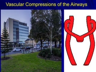
- 1. Vascular Compressions of the Airways
- 2. Introduction Aortic arch anomalies producing tracheo esophageal constriction account for 1 to 2% of all congenital heart disease The compression can result in symptoms characterized by upper airway symptoms or dysphagia. Vascular rings are classified according to embryologic, pathologic, and radiographic criteria 2
- 3. Group I-Complete vascular ring Double aortic arch Right aortic arch with retroesophageal component Left aortic arch and right descending aorta with right ligamentum arteriosum or patent ductus arteriosus Cervical aortic arch complex
- 4. Double Aortic Arch Both arches patent Symmetrical origin Atretic L arch distal to the origin of L SCA Atretic segment between L CCA and L SCA
- 5. Group II -Incomplete vascular ring Left aortic arch and retroesophageal right Subclavian artery Tracheal compression by Innominate or left common carotid artery Ductus arteriosus sling Malrotation of heart with patent ductus arteriosus GROUP III- Pulmonary artery Sling
- 6. 4 years Retrospective Review of Complex Vasculo compressive airway patients at POW 6 TYPE OF ANOMALY n ( % ) DAA ( Double Aortic Arch ) 4 ( 20 ) RALL( Right aortic arch with persistent left ligament) 11 ( 55) ASA/RDA ( Aberrant right subclavian artery /left aortic arch with right descending aorta ) 2 ( 10) RA / RLIA ( Right arch with a retroesophageal left innominate artery) 1 ( 5 ) PS ( Pulmonary sling ) 2 ( 10 )
- 7. COMPLEX AIRWAY PATIENTS 4 11 2 1 2 0 2 4 6 8 10 12 DAA RALL ASA/RDA RA/RLIA PS
- 8. Clinical Presentation 8 STRIDOR 18 ( 90%) NOISY BREATHING 10 ( 50%) BRASSY COUGH 12 ( 60%) CHOKING EPISODES 10 ( 50%) WHEEZES ( misdiagnosed asthma ) 15 ( 75%) INTERRUPTED FEEDING 13 ( 65 %) RECURRENT CHEST INFECTION 8 ( 40 %) ATTACKS OF CYANOSIS 9 ( 45%) RECURRENT VOMITTING 4 ( 20%) APNEA 3 ( 15 % )
- 9. Case – Double Aortic Arch 3 mth old Hx: - Normal antenatal scans - Stridor since 2/52 of age - 2 x choking episodes in last 2 weeks LBO – Severe Tracheomalacia CTA – Double Aortic Arch – Left dominant arch with Right sided descending aorta. 2D Echo – Small PFO/ Tiny PDA 9
- 12. Case 2 9.5 yr old female. Symptom- Dysphagia – Increased in last 6 months. Gastroscopy -Compression of the oesophagus with pulsations from aorta noted from 25-30cm. Bronchoscopy -Segment of almost complete tracheal rings - small segment of trachealis, on right side Barium swallow - There is a fairly mild indentation of the right side of the oesophagus related to the right aortic arch. There is a prominent posterior impression on the upper portion of the intrathoracic oesophagus which is likely due to the known diverticulum of Kommerell. CT Scan - Right aortic arch - Komerrel diverticulum - Aberrant subclavian artery 12
- 13. Type II vascular rings (right aortic arch with retroesophageal ligamentum arteriosum) account for 45% of complete vascular rings. The majority of these defects have left descending aortas. A remnant or stump of the left fourth arch (Kommerell's diverticulum) may persist. The left subclavian artery arises aberrantly behind the esophagus from the right-sided arch or from Kommerell's diverticulum. Near this point the left-sided ligamentum emanates and joins the left pulmonary artery to create a complete ring about the trachea and esophagus. 13
- 14. Aberrant subclavian is seen crossing behind the oesophagus Dilated oesophagus is apparent infront of the crossing vessel Dysphagia Lusoria
- 15. CASE 3 8 week old baby transferred to SCH post resp. arrest. Chest Xray – Hyperinflation of right lung with complete collapse of left lung. LBO – Significant Tracheobronchomalacia Echo – Moderate ASD , Aberrant RSCA, Interrupted IVC with azygous continuation. CT Scan - Collapsed left lung and mediastinal shift to left. The right main bronchus is narrowed from the carina to its branching point. Surgical discussion – Aortopexy vs Posterior tracheopexy. Timing of ASD repair. 15
- 16. Posterior Tracheopexy Marked hyperinflation of right lung with complete collapse of left.Interrupted IVC with large azygous vein Large azygous vein seen in relation with thoracic duct. Trachea, right MB and left MB, oesophagus with thoracic duct and anterior spinal ligament all dissected and displayed16Right Thoracotomy Approach
- 17. negative pressure during respiration. Post Op PREOP – Marked hyperinflation Immediate POSTOP – Improved aeration Patient is Extubated. Trachea midline. Of Right lung with mediastinal shift. Of left lung Weaned to CPAP - High flows – Room air. 2 months Postop – Closure of Ostium secundum ASD + Aortopexy for anterior tracheal collapse
- 18. CASE 4 3 Month old infant Case of Double aortic arch with significant tracheobronchomalacia. Left thoracotomy - Division of ductus+ plication of kommerels diverticulum. Initial improvement but had to be reintubated 10 days later. Anterior aortopexy via a superior hemisternotomy. Remained intubated and ventilated with ongoing evidence of more distal tracheobronchomalacia. Underwent Posterior tracheopexy. Extubated 6th postop day – CPAP- High Flows – Room air.
- 19. SURGICAL APPROACH & PRINCIPLE Surgical approach is determined by which arch is dominant. When the right arch is dominant (75%)- left thoracotomy. When the left arch is dominant (18%)- right thoracotomy. When the arches are balanced (7%), - left thoracotomy is preferred. If the patient has a significant intracardiac lesion-median sternotomy. MRI / CT scan shows Right posterior arch is smaller- Right thoracotomy
- 20. MultiDisciplinary Approach To have a robust system for managing airway patients one needs to work closely with - Gen Paediatric - ENT - Gen Paediatric Surgery - Anethesist/ Intensivist - Respiratory - Cardiology - Radiology - Sleep Team
- 21. Thank You