SlideShare a Scribd company logo
1 of 109
Lymphedema following breast cancer: The importance of
surgical methods and obesity
Rebecca J. Tsai, PhDa,*, Leslie K. Dennis, PhDa,b, Charles F.
Lynch, MD, PhDa, Linda G.
Snetselaar, RD, PhD, LDa, Gideon K.D. Zamba, PhDc, and
Carol Scott-Conner, MD, PhD,
MBAd
aDepartment of Epidemiology, College of Public Health,
University of Iowa, Iowa City, IA, USA.
bDivision of Epidemiology and Biostatistics, College of Public
Health, University of Arizona,
Tucson, AZ, USA.
cDepartment of Biostatistics, College of Public Health,
University of Iowa, Iowa City, IA, USA.
dDepartment of Surgery, College of Medicine, University of
Iowa, Iowa City, IA, USA.
Abstract
Background: Breast cancer-related arm lymphedema is a serious
complication that can
adversely affect quality of life. Identifying risk factors that
contribute to the development of
lymphedema is vital for identifying avenues for prevention. The
aim of this study was to examine
the association between the development of arm lymphedema
and both treatment and personal
(e.g., obesity) risk factors.
Methods: Women diagnosed with breast cancer in Iowa during
2004 and followed through 2010,
who met eligibility criteria, were asked to complete a short
computer assisted telephone interview
about chronic conditions, arm activities, demographics, and
lymphedema status. Lymphedema was
characterized by a reported physician-diagnosis, a difference
between arms in the circumference
(> 2cm), or the presence of multiple self-reported arm
symptoms (at least two of five major arm
symptoms, and at least four total arm symptoms). Relative risks
(RR) were estimated using
logistic regression.
Results: Arm lymphedema was identified in 102 of 522
participants (19.5%). Participants treated
by both axillary dissection and radiation therapy were more
likely to have arm lymphedema than
treated by either alone. Women with advanced cancer stage,
positive nodes, and larger tumors
along with a body mass index > 40 were also more likely to
develop lymphedema. Arm activity
level was not associated with lymphedema.
*Correspondence and Reprints to: Rebecca Tsai, National
Institute for Occupational Safety and Health, 4676 Columbia
Parkway,
R-17, Cincinnati, OH 45226. [email protected] Phone:
(513)841-4398. Fax: (513) 841-4489.
Authorship contribution
All authors contributed to the conception, design, drafting,
revision, and the final review of this manuscript.
Competing interest
Conflicts of Interest and Source of Funding: This study was
funded by the National Cancer Institute Grant Number:
5R03CA130031.
All authors do not declare any conflict of interest.
HHS Public Access
Author manuscript
Front Womens Health. Author manuscript; available in PMC
2018 December 14.
Published in final edited form as:
Front Womens Health. 2018 June ; 3(2): .
A
u
th
o
r M
a
n
u
scrip
t
A
u
th
o
r M
a
n
u
scrip
t
A
u
th
o
r M
a
n
u
scrip
t
A
u
th
o
r M
a
n
u
scrip
t
Conclusions: Surgical methods, cancer characteristics and
obesity were found to contribute to
the development of arm lymphedema. Vigorous arm activity
post-surgery was not found to
increase the risk of arm lymphedema.
Keywords
arm activity; arm lymphedema; body mass index; breast cancer
comorbidity; surgery
Introduction
In the United States, breast cancer is the most common cancer
excluding non-melanoma
skin cancers among women [1]. It is estimated that 266,120
women will be diagnosed with
breast cancer in 2018, 90% of whom will survive from breast
cancer at least five years [2, 3].
Lymphedema of the arm (here forward referred to as
lymphedema) is believed to be a
treatment complication that adversely affects breast cancer
survivors. However, there is
conflicting information regarding which treatments are risk
factors and limited research on
other risk factors for lymphedema. Lymphedema causes the
accumulation of fluid (swelling)
in the arm and 15–20% of breast cancer survivors are expected
to develop this condition in
their lifetimes [4]. Lymphedema is a progressive disease; if not
treated and controlled, severe
pain and disability can result.
Lymphedema research evaluating treatment or personal risk
factors has yielded conflicting
results. Guidelines that warned breast cancer survivors against
vigorous or repetitive exercise
[5] are now being challenged by recent evidence disputing the
previously reported harm of
vigorous arm activities [6–11].
This study looked at the association between the development of
lymphedema and treatment
and personal (e.g. obesity, arm activity) risk factors among a
cohort of women diagnosed
with breast cancer in Iowa during 2004 and followed through
2010 for symptoms of
lymphedema. This study attempted to examine arm exercise in
multiple ways.
Materials and Methods
Breast cancer cases were identified through the Iowa Cancer
Registry (ICR). The ICR is a
population-based registry that is part of the National Cancer
Institute’s Surveillance
Epidemiology and End Results (SEER) program. A total of 2164
breast cancer cases were
diagnosed among Iowa residents during 2004. Ineligible
subjects included 9 males, 236
women over age 80 at breast cancer diagnosis, and 145 cases
known to be deceased. We
excluded breast cancer cases who had a previous or subsequent
cancer diagnosis (N=323), or
had more than one primary tumor at time of initial breast cancer
diagnosis (N=174) except
for in-situ cervical cancer or non-melanoma skin cancer. Due to
low 5-year survival, stage
IV breast cancer cases (N=76) were also excluded. A total of
1,201 met our inclusion
criteria. The interview was completed by 522 (43.5%) eligible
women with a participation
rate among those we were able to contact of 50.6% (522/1,020).
Participants that were
unstaged (N=17) were not included in the staging analysis as
only stages I to III were
compared, but were included in analyses for treatment and
socio-demographic factors. The
Institutional Review Board at the University of Iowa has
approved this study.
Tsai et al. Page 2
Front Womens Health. Author manuscript; available in PMC
2018 December 14.
A
u
th
o
r M
a
n
u
scrip
t
A
u
th
o
r M
a
n
u
scrip
t
A
u
th
o
r M
a
n
u
scrip
t
A
u
th
o
r M
a
n
u
scrip
t
Recruitment
Physicians of subjects were first contacted to see if there were
any reasons why the woman
should not be approached for this study. Physician consent was
assumed if the physician did
not contact the ICR within three weeks, per ICRs standard
passive consent policy.
Thereafter, an invitation letter with elements of consent (as
required by the Institutional
Review Board at the University of Iowa) was sent to each
woman. Two weeks after mailing
the letters, a trained interviewer called the subjects. Subjects
received up to 10 call attempts
on different days of the week and at different times of the day.
Subjects were traced for
addresses or phone numbers through internet sources as needed.
The ICR provided information for demographic, disease- and
treatment-related factors.
These included date of birth, date of breast cancer diagnosis,
laterality of cancer, tumor size,
cancer stage, number of lymph nodes examined, scope of lymph
node dissection, number of
positive lymph nodes found, number of lymph nodes removed,
date and type of first-course
therapy (surgery, chemotherapy, radiation and hormone
therapy), and surgery type.
Participant interview
The interview was designed to collect information not available
through the ICR records. We
used cognitive interviewing and piloting to develop the
questionnaire. Rewording and
reformatting of questions were done to clarify and facilitate the
interviewing process.
Computer-assisted telephone interviewing (CATI) was used to
allow for data checks during
the interview to minimize data entry errors. The average time of
interview was 17 minutes.
Demographic information collected included marital status,
highest level of education, hand
dominance, and self-reported height and weight to calculate
body mass index (BMI) at time
of diagnosis. Radiation therapy to the axilla was also self-
reported.
Self-reported lymphedema was collected through the CATI in
three different ways. First,
subjects were asked if they were ever diagnosed by a physician
with lymphedema. If
diagnosed with lymphedema, they were also asked whether or
not it had resolved. Second,
they were asked if they experienced 13 specific arm/hand
symptoms within the last three
months (Table 1). Third, they were asked to measure the arm
circumference of both arms at
two different locations (one hand width above and below the
elbow crease). Subjects were
also asked if they used specific methods at least once a week to
treat or prevent
lymphedema. Additional information was collected on arm
infection, chronic conditions
diagnosed prior to breast cancer diagnosis/and or arm
lymphedema diagnosis (e.g., high
blood pressure, high cholesterol, heart attack, coronary heart
disease, stroke, congestive
heart failure, emphysema, chronic bronchitis, asthma, thyroid
condition, liver condition,
kidney failure, osteoporosis, diabetes, and arthritis), airplane
trips taken the year after breast
cancer diagnosis, lifting heavy objects, and physical therapy.
A portion of the interview focused on specific arm activities
(swimming, playing tennis,
weightlifting, and gardening) and overall arm activity levels.
Overall arm activities were
broken down into four combinations based on the positioning
(above or below the shoulders)
and the intensity (vigorous or moderate) of the arm activity.
Each subject was asked to
estimate the number of hours per week for each combination of
arm activity during 1) the
Tsai et al. Page 3
Front Womens Health. Author manuscript; available in PMC
2018 December 14.
A
u
th
o
r M
a
n
u
scrip
t
A
u
th
o
r M
a
n
u
scrip
t
A
u
th
o
r M
a
n
u
scrip
t
A
u
th
o
r M
a
n
u
scrip
t
past year, 2) one year prior to breast cancer diagnosis, and 3)
one year after the subject was
able to resume routine household activities. The frequency and
the intensity of arm activities
were later combined into low, medium and high levels. High
level was defined as doing
vigorous arm activities for more than two hours per week. Low
level was defined as doing
vigorous arm activities for less than one hour per week and
doing moderate arm activities for
two or less hours per week.
Lymphedema categorization
Lymphedema was characterized in 3 different ways; 1)
physician-diagnosed, not resolved, 2)
the circumference of the affected arm was greater than 2cm
larger than the other arm (either
above or below the elbow crease), or 3) the presence of multiple
self-reported arm
symptoms. For arm symptoms, a woman must have reported at
least two of five major arm
symptoms (shirt sleeve felt tight, arm felt swollen, heavy, tense
or hard) and at least four
total arm symptoms (major symptoms plus arm felt numb, stiff,
or painful, rash on arm,
other arm symptoms, cannot see knuckles or veins on hand, or
rings felt tight). The arm
symptoms definition was determined based on the experience of
our expert panel. In this
report a woman was considered to have lymphedema if she had
a positive indication of
lymphedema based on any of the three assessment criteria. The
distribution of lymphedema
status based on these 3 criteria is reported in Table 1.
Reliability and representativeness
We examined reliability of the telephone interview among 19
subjects with lymphedema and
20 subjects without lymphedema (based on the initial
interview). The second interview was
approximately 6 weeks after the initial interview. Kappa
coefficients ranged between 0.4–0.8
for most items, which indicated fair to good agreement.
No significant differences between participants and non-
participants were found for disease
characteristics and breast cancer treatments, indicating that the
study results may be
generalized to breast cancer cases diagnosed in Iowa during
2004.
Statistical analysis
Univariate relative risk estimates (RRs) with 95% confidence
intervals (95% CI) were
calculated using unconditional logistic regression. Potential
confounders were identified
prior to analysis based on biologic plausibility. Estimates were
adjusted for confounders that
conferred a 10% or greater change from the crude RR. For
factors of interest in which less
than 20 subjects indicated they had the condition, confounders
that presented a >20%
change from the crude RR were adjusted for in the final model.
RESULTS
Cumulative incidence of lymphedema
Arm lymphedema subsequent to breast cancer treatment was
identified in 102 (19.5%)
participants. The time between initial breast cancer treatment
and onset of arm symptoms or
physician-diagnosed lymphedema are graphed in Figure 1. The
majority of lymphedema
Tsai et al. Page 4
Front Womens Health. Author manuscript; available in PMC
2018 December 14.
A
u
th
o
r M
a
n
u
scrip
t
A
u
th
o
r M
a
n
u
scrip
t
A
u
th
o
r M
a
n
u
scrip
t
A
u
th
o
r M
a
n
u
scrip
t
cases was persistent cases, and was diagnosed within two years
after the initial breast cancer
treatment.
Participants’ characteristics
At the time of interview, the average age of participants was 63
years and the mean BMI was
28.8 kg/m2. One-third (30.5%) of participants were college
graduates and 70% were
married. Neither education level nor marital status was
associated with lymphedema (Table
2). Subjects who were under 50 at the time of interview were
more likely to develop
lymphedema than subjects aged 75+ years (RR=2.95, 95% CI:
1.25, 6.98). Participants with
a BMI ≥30 (35.9%) were more likely to develop lymphedema
(RR=2.15, 95% CI: 1.35,
3.42) than those with a BMI <30. An increasing trend in the
RRs was observed as BMI
increased over 30 (Table 2).
Breast cancer disease and treatment
For cancer characteristics, 87% of participants were classified
as having stage I or II breast
cancer and the mean tumor size was 19mm. In regards to breast
cancer surgical treatments,
57% of women were treated with lumpectomy and 34% with
sentinel node biopsy with an
average of 8 nodes removed. Only 30.5% were detected with
positive nodes and no trend
was seen with increasing number of positive nodes (data not
shown). Participants with > 10
lymph nodes removed were found to have an increased risk of
developing lymphedema in
the presence of radiation therapy. However this effect was
reduced after adjustment for
axillary dissection. Our results observed a trend of increasing
risk as an increasing number
of nodes was removed. Radiation therapy was received by 63%
of women, and among those
who received radiation, 30 stated that radiation was directed to
the axillary area as well as
the breast. Over half of the participants had chemotherapy
and/or hormonal therapy as part
of their breast cancer treatment.
Lymphedema was associated with stage III cancer (RR=2.23,
95% CI: 1.09, 4.55), tumors ≥
30mm (RR=2.76, 95% CI: 1.16, 6.58), and the presence of
positive nodes (RR=1.88, 95%
CI: 1.13, 3.13) (Table 3). Axillary dissection and radiation were
found to interact (p=0.01).
The combination of both axillary dissection and radiation
therapy showed a slightly stronger
association with lymphedema (RR=2.61, 95% CI: 1.27, 5.39)
than either axillary dissection
(RR=2.21, 95% CI: 1.32, 3.68) or radiation alone (RR=1.29,
95% CI: 0.81, 2.04). Radiation
directed to the axillary area (RR=1.10, 95% CI: 0.62–1.93) and
other treatment factors were
not associated with lymphedema (Table 3).
Chronic conditions
Lymphedema was associated with chronic bronchitis (RR=3.45,
95% CI: 1.24, 9.63). A
borderline increased risk for developing lymphedema was seen
among participants who
were diagnosed with osteoarthritis/ rheumatoid arthritis
(RR=1.57, 95% CI: 0.93, 2.67)
and/or kidney failure (RR=4.70, 95% CI: 0.89, 24.85). No
association was found with high
blood pressure, diabetes or other conditions reported after
adjustment for age, BMI, and the
interaction of axillary dissection and radiation.
Tsai et al. Page 5
Front Womens Health. Author manuscript; available in PMC
2018 December 14.
A
u
th
o
r M
a
n
u
scrip
t
A
u
th
o
r M
a
n
u
scrip
t
A
u
th
o
r M
a
n
u
scrip
t
A
u
th
o
r M
a
n
u
scrip
t
Arm activity and other personal factors
No associations were found between lymphedema and specific
arm activities including
swimming, playing tennis, weightlifting or gardening. When
analyses were restricted to
participants who had the same level of arm activity before and
after breast cancer diagnosis,
no association between lymphedema and arm activity level
either above or below the
shoulders was found (Table 4). Surgery on dominant side
(RR=1.49, 95% CI: 0.95–2.32),
and air travel (RR=0.98, 95% CI: 0.63–1.52) were not
associated with lymphedema in this
study. An association was seen between infection and
lymphedema (RR =8.51, 95% CI:
3.07, 23.61). However, all but one participant developed arm
infection after lymphedema
diagnosis.
Discussion
The prevalence of arm lymphedema in women diagnosed with
breast cancer in Iowa in 2004
was 19.5% five-years after diagnosis, similar to results reported
from previous studies [4, 12,
13]. This study, similar to other studies [14–19], found that
BMI was associated with the
development of lymphedema among these women. The
association with increased BMI was
evident both for the study definition of lymphedema and when
defined only as physician-
diagnosed cases. This suggests that the association seen was not
an artifact of measurement
error in lymphedema. Obesity, because of larger tissue volume
and higher fat content, may
have contributed to lymphedema development through increased
difficulty of performing
surgery or required alternative treatment techniques [20, 21]. In
addition, obesity may
increase lymphatic stress by exacerbating the inflammatory
response or prolonging the
surgical healing time [22]. Moreover, the increased amount of
adipose tissue may act as a
reservoir for lymphatic fluids [20]. Furthermore, one small
study found that weight loss was
correlated with a significant reduction in arm volume [23].
Obesity is also linked to chronic
conditions such as high blood pressure and diabetes, which may
further impair a lethargic
lymphatic system by disrupting fluid balance.
An increase in lymphedema risk was observed when both
axillary dissection and radiation
therapy were performed. A number of studies [24–29] have
suggested that the addition of
radiation therapy to axillary dissection increases the risk of
lymphedema. Radiation after
axillary dissection may have induced additional fibrosis that
could compress or block
lymphatic vessels. Participants in this study who had radiation
and no axillary dissection
were generally diagnosed with early stage breast cancer (stage I
or II) and had less invasive
treatments. Conversely, women who receive both axillary
dissection and radiation therapy
tended to be stage III and thus were treated more aggressively.
This may further explain the
interaction observed between axillary dissection and radiation
therapy.
Overall, published reports have both supported [16, 17, 29–32]
and refuted [21, 33, 34] that
increasing number of nodes excised is linked to arm
lymphedema risk. This study did not
find such an association after adjusting for axillary dissection.
Axillary dissection was
identified as a confounder because it was speculated that the
association with the
development of lymphedema may have been attributed to the
intactness of the lymphatic
network in the axilla rather than how many nodes were removed
[30]. Axillary dissection is
generally indicated in the presence of positive nodes and leads
to an increased number of
Tsai et al. Page 6
Front Womens Health. Author manuscript; available in PMC
2018 December 14.
A
u
th
o
r M
a
n
u
scrip
t
A
u
th
o
r M
a
n
u
scrip
t
A
u
th
o
r M
a
n
u
scrip
t
A
u
th
o
r M
a
n
u
scrip
t
nodes excised. Axillary dissection, a procedure that disrupts the
lymphatic network,
remained associated with lymphedema even after adjusting for
the number of lymph nodes
removed.
While breast cancer treatments are major contributors to
lymphedema, the association
between lymphedema and advance stages of cancer, positive
nodes, or large tumors persisted
even after adjusting for axillary dissection. It is possible that
advanced disease or larger
tumors may disrupt or damage regional lymphatics.
The presence of most chronic conditions did not influence the
subsequent development of
lymphedema. While it was speculated that conditions such as
high blood pressure and
diabetes may exacerbate a damaged lymphatic system due to
increased hydrostatic pressure
[35], we did not find such an association in this study.
Medications taken to control high
blood pressure [14], may have negated the effect of increased
hydrostatic pressure. Both
chronic bronchitis and kidney conditions were linked to the
development of lymphedema.
Kidney failure may be associated with fluid retention that may
cause edema [35], thus
further complicating an already delicate lymphatic system. It is
also possible that these
subjects may have a surveillance bias for physician-diagnosed
lymphedema due to visiting
the doctor for these other conditions. This study’s findings are
inconclusive since <20
subjects were diagnosed with these conditions. Arthritis and
autoimmune diseases can
contribute to lymphedema through inflammation to the joints,
blood or lymph vessels, which
may be reflected in the borderline association we saw.
While most of the previously published studies did not find an
association between age and
lymphedema, we, similar to Geller et al.[14], found younger age
was associated with
developing lymphedema. It has been suggested that younger
women may have advanced
cancer which required more invasive treatments [36]. This study
found that younger women
under the age of 50 were more likely to have positive nodes
(41% versus 19%) or be
diagnosed at a higher stage after adjustment for confounders.
They were also more likely to
have axillary dissection. Moreover, younger women are more
active outside of the home and
may be more likely to notice the effects of mild lymphedema
[37]. Also, older women tend
to have extensive co-morbidity and might pay less attention to
arm symptoms. Hence, arm
symptoms related to lymphedema may have been under-reported
by older women [37, 38].
While this study attempted to capture arm lymphedema in both
an objective and subjective
way (based on arm symptoms experienced within the last three
months), under-reporting of
arm symptoms (subjective method) is an issue because
subclinical cases of lymphedema
may be missed.
Specific activities were not found to be associated with
lymphedema. Among women who
weight lifted, an increased amount of time spent weightlifting
above the heart was not found
to be associated with lymphedema. The results from this study
were similar to another study
in that none of the specific activities or overall arm activity
level showed increased risk for
lymphedema [39]. Arm lymphedema can lead to a reduction of
arm activity level. In an
attempt to avoid this bias, only subjects that reported no change
in arm activity level both
above and below the shoulders a year after resuming household
activities (as compared to a
year before breast cancer diagnosis) were included in the
overall arm activity level analysis.
Tsai et al. Page 7
Front Womens Health. Author manuscript; available in PMC
2018 December 14.
A
u
th
o
r M
a
n
u
scrip
t
A
u
th
o
r M
a
n
u
scrip
t
A
u
th
o
r M
a
n
u
scrip
t
A
u
th
o
r M
a
n
u
scrip
t
With this restriction, arm activity level was not associated with
lymphedema. However
without this restriction, we found low level of arm activities
below the shoulders was
associated with lymphedema (RR=2.40, 95% CI: 1.38, 4.20)
(data not shown). We believe
that this may be a reflection of decreased level of arm activity
due to the presence of
lymphedema. Overall, our findings on arm activity do not
support an association with post-
operative arm exercise and arm lymphedema.
Although air travel (41.6% of our subjects) has been speculated
by both clinicians and breast
cancer survivors to be a potential risk factor for lymphedema,
such an association was not
observed in our study or the study by Kilbreath et al [40]. It is
probable that having
lymphedema puts breast cancer survivors at risk for getting an
arm infection due to
decreased lymphatic circulation.
Strengths
This study was conducted using a population-based cohort of
breast cancer survivors five to
six years after breast cancer diagnosis, thereby avoiding
erroneous inclusion of acute
lymphedema cases. Participants reporting physician-diagnosed
lymphedema were
additionally asked if their condition has since resolved to
decrease misclassification.
Furthermore, objective and subjective assessments were applied
to capture subclinical cases.
Thirty-two percent of subjects reporting resolved lymphedema
were later identified to have
lymphedema through subclinical means. In addition, obtaining
lymphedema status five or
more years after breast cancer diagnosis allowed us to observe
the long-term risk from
treatments, as many studies have short follow-up times of 1–2
years after diagnosis or
treatments.
Limitations
The biggest limitation was that all of our measures of
lymphedema were self-reported. Due
to caller identification and increased usage of cell phones, we
were unable to reach as many
subjects as we anticipated and 181 eligible women could not be
traced from ICR
information.
Conclusion
Among this cohort of breast cancer survivors, we found
lymphedema to have a prevalence of
19.5% five years after diagnosis, with most developing
lymphedema within the first 2 years
after surgery. In particular, women with a high BMI were found
to be at risk for developing
lymphedema, suggesting that obesity may further promote
inflammation which can lead to
lymphatic impairment. Younger age was also associated with
lymphedema development.
The combination of axillary dissection and radiation therapy
doubled the risk of developing
lymphedema. Low level of arm activity was not found to be
associated with lymphedema.
Acknowledgments
Funding
This study was funded by the National Cancer Institute Grant
Number: 5R03CA130031 (PI: Rebecca Tsai, PhD).
Tsai et al. Page 8
Front Womens Health. Author manuscript; available in PMC
2018 December 14.
A
u
th
o
r M
a
n
u
scrip
t
A
u
th
o
r M
a
n
u
scrip
t
A
u
th
o
r M
a
n
u
scrip
t
A
u
th
o
r M
a
n
u
scrip
t
Abbreviations:
BMI body mass index
CATI Computer-assisted telephone interviewing
ICR Iowa Cancer Registry
RR Relative Risk
CI Confidence Interval
REFERENCES
1. Siegel R, Ma J, Zou Z, Jemal A (2014) Cancer statistics,
2014. CA: a cancer journal for clinicians
64:9–29. [PubMed: 24399786]
2. Surveillance Epidemiology and End Results, 2018 SEER Stat
fact sheets: breast. Available at: http://
seer.cancer.gov/statfacts/html/breast.html [Accessed April 26
2018].
3. Noone AM HN, Krapcho M, Miller D, Brest A, Yu M, Ruhl J,
Tatalovich Z, Mariotto A, Lewis DR,
Chen HS, Feuer EJ, Cronin KA (eds), SEER Cancer Statistics
Review, 1975–2015, National Cancer
Institute Bethesda, MD, https://seer.cancer.gov/csr/1975_2015/,
based on November 2017 SEER
data submission, posted to the SEER web site, 4 2018.
4. Velanovich V, Szymanski W (1999) Quality of life of breast
cancer patients with lymphedema. Am J
Surg 177:184–7; discussion 8. [PubMed: 10219851]
5. Kent H (1996) Breast-cancer survivors begin to challenge
exercise taboos. CMAJ 155:969–71.
[PubMed: 8837548]
6. Harris SR, Niesen-Vertommen SL (2000) Challenging the
myth of exercise-induced lymphedema
following breast cancer: a series of case reports. J Surg Oncol
74:95–8; discussion 8–9. [PubMed:
10914817]
7. McKenzie DC, Kalda AL (2003) Effect of upper extremity
exercise on secondary lymphedema in
breast cancer patients: a pilot study. J Clin Oncol 21:463–6.
[PubMed: 12560436]
8. Lee TS, Kilbreath SL, Sullivan G, Refshauge KM, Beith JM,
et al. (2009) Factors that affect
intention to avoid strenuous arm activity after breast cancer
surgery. Oncol Nurs Forum 36:454–62.
[PubMed: 19581236]
9. Harris SR (2012) “We’re all in the same boat”: a review of
the benefits of dragon boat racing for
women living with breast cancer. Evidence-based
complementary and alternative medicine : eCAM
2012:167651. [PubMed: 22811743]
10. Baumann FT, Reike A, Reimer V, Schumann M, Hallek M,
et al. (2018) Effects of physical
exercise on breast cancer-related secondary lymphedema: a
systematic review. Breast cancer
research and treatment.
11. Bloomquist K, Oturai P, Steele ML, Adamsen L, Moller T,
et al. (2018) Heavy-Load Lifting: Acute
Response in Breast Cancer Survivors at Risk for Lymphedema.
Med Sci Sports Exerc 50:187–95.
[PubMed: 28991039]
12. Petrek JA, Heelan MC (1998) Incidence of breast
carcinoma-related lymphedema. Cancer
83:2776–81. [PubMed: 9874397]
13. Sakorafas GH, Peros G, Cataliotti L, Vlastos G (2006)
Lymphedema following axillary lymph
node dissection for breast cancer. Surg Oncol 15:153–65.
[PubMed: 17187979]
14. Geller BM, Vacek PM, O’Brien P, Secker-Walker RH
(2003) Factors associated with arm swelling
after breast cancer surgery. J Women’s Health 12:921–30.
15. Helyer LK, Varnic M, Le LW, Leong W, McCready D
(2010) Obesity is a risk factor for
developing postoperative lymphedema in breast cancer patients.
Breast J 16:48–54. [PubMed:
19889169]
16. Clark B, Sitzia J, Harlow W (2005) Incidence and risk of
arm oedema following treatment for
breast cancer: a three-year follow-up study. Q J Med 98:343–8.
Tsai et al. Page 9
Front Womens Health. Author manuscript; available in PMC
2018 December 14.
A
u
th
o
r M
a
n
u
scrip
t
A
u
th
o
r M
a
n
u
scrip
t
A
u
th
o
r M
a
n
u
scrip
t
A
u
th
o
r M
a
n
u
scrip
t
http://seer.cancer.gov/statfacts/html/breast.html
https://seer.cancer.gov/csr/1975_2015/
17. Dominick SA, Madlensky L, Natarajan L, Pierce JP (2013)
Risk factors associated with breast
cancer-related lymphedema in the WHEL Study. Journal of
cancer survivorship : research and
practice 7:115–23. [PubMed: 23212606]
18. DiSipio T, Rye S, Newman B, Hayes S (2013) Incidence of
unilateral arm lymphoedema after
breast cancer: a systematic review and meta-analysis. Lancet
Oncol 14:500–15. [PubMed:
23540561]
19. Ribeiro Pereira ACP, Koifman RJ, Bergmann A (2017)
Incidence and risk factors of lymphedema
after breast cancer treatment: 10 years of follow-up. Breast
36:67–73. [PubMed: 28992556]
20. Meric F, Buchholz TA, Mirza NQ, Vlastos G, Ames FC, et
al. (2002) Long-term complications
associated with breast-conservation surgery and radiotherapy.
Ann Surg Oncol 9:543–9. [PubMed:
12095969]
21. Goffman TE, Laronga C, Wilson L, Elkins D (2004)
Lymphedema of the arm and breast in
irradiated breast cancer patients: risks in an era of dramatically
changing axillary surgery. Breast J
10:405–11. [PubMed: 15327493]
22. Ahmed RL, Schmitz KH, Prizment AE, Folsom AR (2011)
Risk factors for lymphedema in breast
cancer survivors, the Iowa Women’s Health Study. Breast
Cancer Res Treat 130:981–91.
[PubMed: 21761159]
23. Shaw C, Mortimer P, Judd PA (2007) Randomized
controlled trial comparing a low-fat diet with a
weight-reduction diet in breast cancer-related lymphedema.
Cancer 109:1949–56. [PubMed:
17393377]
24. Kissin MW, Querci della Rovere G, Easton D, Westbury G
(1986) Risk of lymphoedema following
the treatment of breast cancer. Br J Surg 73:580–4. [PubMed:
3730795]
25. Schunemann H, Willich N (1998) Lymphoedema of the arm
after primary treatment of breast
cancer. Anticancer Res 18:2235–6. [PubMed: 9703792]
26. Herd-Smith A, Russo A, Muraca MG, Del Turco MR,
Cardona G (2001) Prognostic factors for
lymphedema after primary treatment of breast carcinoma.
Cancer 92:1783–7. [PubMed:
11745250]
27. Kwan W, Jackson J, Weir LM, Dingee C, McGregor G, et al.
(2002) Chronic arm morbidity after
curative breast cancer treatment: prevalence and impact on
quality of life. J Clin Oncol 20:4242–8.
[PubMed: 12377968]
28. Keramopoulos A, Tsionou C, Minaretzis D, Michalas S,
Aravantinos D (1993) Arm morbidity
following treatment of breast cancer with total axillary
dissection: a multivariated approach.
Oncology 50:445–9. [PubMed: 8233285]
29. Hinrichs CS, Watroba NL, Rezaishiraz H, Giese W, Hurd T,
et al. (2004) Lymphedema secondary
to postmastectomy radiation: incidence and risk factors. Ann
Surg Oncol 11:573–80. [PubMed:
15172932]
30. Goldberg JI, Wiechmann LI, Riedel ER, Morrow M, Van
Zee KJ (2010) Morbidity of sentinel node
biopsy in breast cancer: the relationship between the number of
excised lymph nodes and
lymphedema. Ann Surg Oncol 17:3278–86. [PubMed:
20574774]
31. Petrek JA, Senie RT, Peters M, Rosen PP (2001)
Lymphedema in a cohort of breast carcinoma
survivors 20 years after diagnosis. Cancer 92:1368–77.
[PubMed: 11745212]
32. Zou L, Liu FH, Shen PP, Hu Y, Liu XQ, et al. (2018) The
incidence and risk factors of related
lymphedema for breast cancer survivors post-operation: a 2-year
follow-up prospective cohort
study. Breast Cancer 25:309–14. [PubMed: 29397555]
33. Yen TW, Fan X, Sparapani R, Laud PW, Walker AP, et al.
(2009) A contemporary, population-
based study of lymphedema risk factors in older women with
breast cancer. Ann Surg Oncol
16:979–88. [PubMed: 19194754]
34. Beaulac SM, McNair LA, Scott TE, LaMorte WW, Kavanah
MT (2002) Lymphedema and quality
of life in survivors of early-stage breast cancer. Arch Surg
137:1253–7. [PubMed: 12413312]
35. Paulus BM, Ali S, Zia AA, Munir A, Davis RC, Jr, et al.
(2008) Causes and consequences of
systemic venous hypertension. Am J Med Sci 336:489–97.
[PubMed: 19092322]
36. Paskett ED, Naughton MJ, McCoy TP, Case LD, Abbott JM
(2007) The epidemiology of arm and
hand swelling in premenopausal breast cancer survivors. Cancer
Epidemiol Biomarkers Prev
16:775–82. [PubMed: 17416770]
Tsai et al. Page 10
Front Womens Health. Author manuscript; available in PMC
2018 December 14.
A
u
th
o
r M
a
n
u
scrip
t
A
u
th
o
r M
a
n
u
scrip
t
A
u
th
o
r M
a
n
u
scrip
t
A
u
th
o
r M
a
n
u
scrip
t
37. Armer J, Fu MR (2005) Age differences in post-breast
cancer lymphedema signs and symptoms.
Cancer Nurs 28:200–7. [PubMed: 15915063]
38. Warmuth MA, Bowen G, Prosnitz LR, Chu L, Broadwater G,
et al. (1998) Complications of
axillary lymph node dissection for carcinoma of the breast: a
report based on a patient survey.
Cancer 83:1362–8. [PubMed: 9762937]
39. Ahmed RL, Thomas W, Yee D, Schmitz KH (2006)
Randomized controlled trial of weight training
and lymphedema in breast cancer survivors. J Clin Oncol
24:2765–72. [PubMed: 16702582]
40. Kilbreath SL, Ward LC, Lane K, McNeely M, Dylke ES, et
al. (2010) Effect of air travel on
lymphedema risk in women with history of breast cancer. Breast
Cancer Res Treat 120:649–54.
[PubMed: 20180016]
Tsai et al. Page 11
Front Womens Health. Author manuscript; available in PMC
2018 December 14.
A
u
th
o
r M
a
n
u
scrip
t
A
u
th
o
r M
a
n
u
scrip
t
A
u
th
o
r M
a
n
u
scrip
t
A
u
th
o
r M
a
n
u
scrip
t
Figure 1.
Cumulative Incidence of Arm Lymphedema diagnosed by a
physician or by having 4 +
symptoms among breast cancer survivors in Iowa, 2004
Tsai et al. Page 12
Front Womens Health. Author manuscript; available in PMC
2018 December 14.
A
u
th
o
r M
a
n
u
scrip
t
A
u
th
o
r M
a
n
u
scrip
t
A
u
th
o
r M
a
n
u
scrip
t
A
u
th
o
r M
a
n
u
scrip
t
A
u
th
o
r M
a
n
u
scrip
t
A
u
th
o
r M
a
n
u
scrip
t
A
u
th
o
r M
a
n
u
scrip
t
A
u
th
o
r M
a
n
u
scrip
t
Tsai et al. Page 13
Table 1.
Distribution of self-reported lymphedema assessments among
study participants diagnosed with breast cancer
in Iowa, 2004.
Participants %
N=522
Physician-diagnosed lymphedema
Unresolved 44 8.4
Resolved 28 5.4
Not diagnosed 450 86.2
Arm measurement
>2cm difference between arms 49 9.4
≤ 2 cm difference between arms 351 67.2
Missing 122 23.4
Arm symptoms*
Subjective indication of arm lymphedema 45 8.6
No subjective indication of arm lymphedema 477 91.4
Arm lymphedema based on any of the above
Arm lymphedema 102 19.5
No arm lymphedema 420 80.5
*
Subjective indication is present if subject has at least 2 major
symptoms (shirt sleeve felt tight, arm felt swollen, heavy, tense
or hard) and at least
4 total symptoms (major arm symptoms plus arm felt numb,
stiff, or painful, rash on arm, other arm symptoms, cannot see
knuckles or veins on
hand, or rings felt tight)
Front Womens Health. Author manuscript; available in PMC
2018 December 14.
A
u
th
o
r M
a
n
u
scrip
t
A
u
th
o
r M
a
n
u
scrip
t
A
u
th
o
r M
a
n
u
scrip
t
A
u
th
o
r M
a
n
u
scrip
t
Tsai et al. Page 14
Table 2.
Relative risk of demographic factors and lymphedema among
subjects diagnosed with breast cancer in Iowa,
2004
Lymphedema Adjusted
Yes No RR CI
Age at interview
75–85 12 (11.8) 86 (20.5) Ref *
70–74 14 (13.7) 56 (13.3) 1.85 0.75, 4.55
65–69 16 (15.7) 51 (12.1) 1.77 0.72, 4.34
60–64 13 (12.8) 61 (14.5) 1.29 0.52, 3.18
55–59 13 (12.8) 60 (14.3) 1.40 0.56, 3.49
50–54 14 (13.7) 57 (13.6) 1.61 0.66, 3.94
25–49 20 (19.6) 49 (11.7) 2.95 1.25, 6.98
Trend RR comparing age 75–85 to 25–49 1.85 0.93, 3.70
Education
≤ High school 45 (44.5) 192 (46.0)
Ref
†
Some college 25 (24.8) 98 (23.5) 0.90 0.50, 1.63
≥ College 31 (30.7) 127 (30.5) 0.94 0.53, 1.65
Trend RR comparing ≥ College to ≤ High school 0.93 0.53, 1.64
Married
No 33 (33.0) 124 (29.5)
Ref
‡
Yes 67 (67.0) 296 (70.5) 0.76 0.46, 1.23
BMI (kg/m2)
<18.5 0 4 (1.0) N/A
18.5–24.9 22 (21.8) 138 (33.1) Ref c
25–29.9 28 (27.7) 140 (33.6) 1.20 0.64, 2.23
30–34.9 20 (19.8) 83 (19.9) 1.50 0.76, 2.98
35–39.9 13 (12.9) 35 (8.4) 2.51 1.12, 5.65
40+ 18 (17.8) 17 (4.1) 5.58 2.45, 12.7
Trend RR comparing 40+ to 18.5–24.9 7.43 3.03, 18.2
RR= relative risk; CI = confidence interval; BMI= body mass
index.
Note: Total number of subjects may not add up to 522 due to
missing data
*
Adjusted for BMI, axillary dissection, number of lymph nodes
removed, any radiation, and any radiation x axillary dissection
†
Adjusted for BMI, age, axillary dissection, any radiation, and
any radiation x axillary dissection
‡
Adjusted for age, axillary dissection, any radiation, and any
radiation x axillary dissection
Front Womens Health. Author manuscript; available in PMC
2018 December 14.
A
u
th
o
r M
a
n
u
scrip
t
A
u
th
o
r M
a
n
u
scrip
t
A
u
th
o
r M
a
n
u
scrip
t
A
u
th
o
r M
a
n
u
scrip
t
Tsai et al. Page 15
Table 3.
Relative Risk of Lymphedema by Breast Cancer Treatment and
Disease Factors Among Subjects Diagnosed
with Breast Cancer in Iowa, 2004
Lymphedema Adjusted
Yes No RR CI
Stage
Stage I 42 (42.0) 235 (58.2) Ref *
Stage II 38 (38.0) 139 (34.4) 1.13 0.66, 1.92
Stage III 20 (20.0) 30 (7.4) 2.23 1.09, 4.55
Trend RR comparing Stage III to Stage I 1.99 0.99, 3.98
Primary tumor size (mm)
0–9 15 (15.8) 106 (26.6)
Ref
†
10–14 17 (17.9) 88 (22.1) 1.26 0.57, 2.75
15–29 39 (41.1) 158 (39.6) 1.60 0.78, 3.34
30+ 24 (25.3) 47 (11.8) 2.76 1.16, 6.58
Trend RR comparing 30+ to 0–9 2.65 1.14, 6.13
Surgery
Lumpectomy 54 (52.9) 243 (58.1)
Ref
‡
Mastectomy 13 (12.8) 56 (13.4) 1.09 0.39, 3.04
Modified radical mastectomy 35 (34.3) 119 (28.5) 1.53 0.75,
3.15
# of lymph node removed
0–2 17 (16.8) 126 (30.4) Ref *
3–6 21 (20.8) 108 (26.0) 1.10 0.50, 2.39
7–10 18 (17.8) 57 (13.73) 1.45 0.56, 3.78
>10 45 (44.6) 124 (29.9) 1.60 0.66, 3.84
Trend RR comparing >10 to 0–2 1.64 0.74, 3.61
Positive nodes
No 54 (52.9) 300 (73.5) Ref *
Yes 48 (47.1) 108(26.5) 1.88 1.13, 3.13
Radiation to the axilla
§
No 60 (65.2) 272 (61.8) Ref *
Yes 32 (34.8) 107 (28.2) 1.10 0.62, 1.93
Chemotherapy
No 40 (40.0) 208 (49.9) Ref *
Yes 60 (60.0) 209 (50.1) 1.01 0.61, 1.67
Hormones therapy
No 47 (47.0) 183 (44.6) Ref *
Yes 53 (53.0) 227 (55.4) 0.92 0.58, 1.46
RR= relative risk; CI = confidence interval; BMI= body mass
index
Note: Total number of subjects may not add up to 522 due to
missing data
Front Womens Health. Author manuscript; available in PMC
2018 December 14.
A
u
th
o
r M
a
n
u
scrip
t
A
u
th
o
r M
a
n
u
scrip
t
A
u
th
o
r M
a
n
u
scrip
t
A
u
th
o
r M
a
n
u
scrip
t
Tsai et al. Page 16
*
Adjusted for age, axillary dissection, any radiation, and any
radiation x axillary dissection
†
Adjusted for age, axillary dissection, chemotherapy, number of
lymph nodes removed, any radiation, and any radiation x
axillary dissection
‡
Adjusted for age, any radiation to axilla, axillary dissection,
any radiation, and any radiation x axillary dissection
§
Among subjects who received radiation therapy
Front Womens Health. Author manuscript; available in PMC
2018 December 14.
A
u
th
o
r M
a
n
u
scrip
t
A
u
th
o
r M
a
n
u
scrip
t
A
u
th
o
r M
a
n
u
scrip
t
A
u
th
o
r M
a
n
u
scrip
t
Tsai et al. Page 17
Table 4.
Relative Risk of Arm Activities and Lymphedema Among
Subjects Diagnosed with Breast Cancer in Iowa,
2004, Restricted to Women Who Reported no Change in Arm
Activity Level Before and After Breast Cancer
Diagnosis
Lymphedema Crude Adjusted
Arm activities Yes No RR CI RR CI
Above the shoulders: *
High 9 (20) 35 (13.3) Ref
Ref
†
Medium 14 (31.1) 113 (43) 0.48 0.19, 1.21 0.51 0.19, 1.40
Low 22 (48.9) 115 (43.7) 0.74 0.31, 1.76 0.78 0.29, 2.07
Below the shoulders: *
High 18 (40) 126 (47.9) Ref
Ref
†
Medium 17 (37.8) 92 (35) 1.29 0.63, 2.65 1.38 0.63, 3.01
Low 10 (22.2) 45 (17.1) 1.56 0.67, 3.62 1.59 0.60, 4.24
RR= relative risk; CI = confidence interval.
*
Only included subjects that reported no change in arm activity
level from before breast cancer diagnosis to after resuming
household activities
(N=308)
†
Adjusted for BMI, age, axillary dissection, any radiation, and
any radiation x axillary dissection
Front Womens Health. Author manuscript; available in PMC
2018 December 14.
AbstractIntroductionMaterials and
MethodsRecruitmentParticipant interviewLymphedema
categorizationReliability and representativenessStatistical
analysisRESULTSCumulative incidence of
lymphedemaParticipants’ characteristicsBreast cancer disease
and treatmentChronic conditionsArm activity and other personal
factorsDiscussionStrengthsLimitationsConclusionReferencesFig
ure 1.Table 1.Table 2.Table 3.Table 4.
1
Running head: ARTICLE CRITIQUE
8
EBP GRANT PROPOSAL
Chapter 11: Lymphedema
The purpose of this paper is to discuss lymphedema and
critique an article on the topic. Lymphedema is when there is an
obstruction in the lymphatic vessels that cause the tissues in the
extremities to swell (Hubert & Vanmeter, 2018). This also
allows for accumulation of lymph in the tissues as well.
Commonly, this disorder is congenital and may involve the
lymph nodes along with the vessels (Hubert & Vanmeter, 2018).
It can also be caused by blockage of the lymph vessels due to
parasitic worms (Hubert & Vanmeter, 2018). When the lymph
states to build up in the body, the more the extremity swells
(Huber & Vanmeter, 2018). As lymphedema continues to
progress over time, the extremity becomes enlarged, firm and
painful (Hubert & Vanmeter, 2018). Lymphedema can be
chronic as well, which leads to frequent infection (Hubert &
Vanmeter, 2018). According to Johns Hopkins Medicine (2020),
lymphedema can occur after cancer surgery when lymph nodes
are removed.
Authors Rebecca J. Tsai, Leslie K. Dennis, Charles F.
Lynch, Linda G. Snetselaar, Gideon K. D. Zamba, Carol Scott-
Conner published an article on lymphedema after breast cancer
entitled “Lymphedema following breast cancer: The importance
of surgical methods and obesity” Published to Front Women’s
Health in 2018. The purpose of the article is to discuss the
research that the authors have conducted on the association
between developing lymphedema after cancer surgery and
personal risk factors (Tsai et al., 2018). This research will allow
for them to better understand if there are certain factors that
make a person more at risk for developing lymphedema after
cancer surgery. The literature was drawn from a systemic
approach. This is because it focuses on a specific question and
critically appraises all relevant research. The review focuses on
cause and effect meaning how does one issue effect the other. In
this case, the authors are correlating developing lymphedema
after surgery and certain risk factors that can affect this. Tsai et
al. (2018) identify concern that the measures of the study are
from subject self-reporting. The authors feel that there can be
issues with self-reporting because the patients do not always
participate until the end of the study.
The aim of the study is to understand if there is a
correlation between arm lymphedema and certain personal risk
factors (Tasi et al., 2018). To achieve an accurate study, the
authors used a population-based cohort design. This allows for
better understanding of the research for this specific population
of women. The sample was obtained in Iowa from 2004 to 2010.
Tsai et al. (2018) states that the women who met the criteria,
completed a short telephone interview about their lymphedema
status, arm activities, demographics and chronic conditions that
they currently have. The patients were confirmed to have
lymphedema from physician reports and the presence of at least
four major arm symptoms that can occur with the disease (Tsai
et al., 2018). There were 522 participants in the study, which
seems to be an adequate size for a population-based cohort
design.
The study showed that lymphedema was identified in 102
of the 522 patients (Tsai et al., 2018). The results showed that
people who had radiation and some dissection if the axilla were
more at risk for lymphedema after breast cancer surgery (Tsai et
al., 2018). Tsai et al. (2018) states that the women who were the
most likely to have lymphedema had a body mass index above
40. This shows that obesity, the characteristics of the cancer
and the methods used in surgery were all major factors in the
patient developing lymphedema (Tsai et al., 2018). The authors
found that obesity promotes the inflammation of the body which
leads to issues with lymphedema. The authors also found that
women who were younger had a higher chance of developing
lymphedema as well. The authors are satisfied with the results
and do not mention a need for further testing (Tsai et al., 2018).
The article is greatly recommended as a nurse and a future
advanced practice nurse. This article is easy to comprehend and
has pertinent information that can be used in practice. The
information can be useful when assessing patients who have had
breast cancer surgery. Knowing and understanding the risk
factors for developing lymphedema after breast cancer surgery
can allow for preventative care for the patients who are most at
risk. It may not be completely curable, but there are certain
steps that can be taken to reduce the symptoms or even keep it
from starting in the first placed (Johns Hopkins Medicine,
2020). It is apparent that advanced practice nurses should
understand the signs and symptoms of developing lymphedema
so that it can be treated adequately.
References
Hubert, R. & VanMeter, K. C. (2018). Gould's pathophysiology
for the health professions. St. Louis, MO: Elsevier Saunders.
Johns Hopkins Medicine. (2020). Breast cancer: lymphedema
after treatment. Retrieved from
https://www.hopkinsmedicine.org/health/conditions-and-
diseases/breast-cancer/breast-cancer-lymphedema-after-
treatment
Tsai R. J., Dennis, L. K., Lynch, C. F., Snetselaar, L. G.,
Zamba, G. K. D., Scott-Conner, C. (2018). Lymphedema
following breast cancer: the importance of surgical methods and
obesity. Front Women’s Health, 3(2), 1-17.
References
Lastname, C. (2008). Title of the source without caps except
Proper Nouns or: First word after colon. The Journal or
Publication Italicized and Capped, Vol#(Issue#), Page numbers.
Lastname, O. (2010). Online journal using DOI or digital
object identifier. Main Online Journal Name, Vol#(Issue#), 159-
192. doi: 10.1000/182
Lastname, W. (2009). If there is no DOI use the URL of the
main website referenced. Article Without DOI Reference,
Vol#(Issue#), 166-212. Retrieved from
http://www.mainwebsite.org
Journal of
Clinical Medicine
Article
Optimizing Hydroxyurea Treatment for Sickle Cell
Disease Patients: The Pharmacokinetic Approach
Charlotte Nazon 1, Amelia-Naomi Sabo 2,3, Guillaume Becker
3,4, Jean-Marc Lessinger 2,
Véronique Kemmel 2,3,* and Catherine Paillard 1,5,*
1 Hôpitaux Universitaires de Strasbourg, Centre de compétence
pour les maladies constitutionnelles du
globule rouge et de l’érythropoïèse, Service d’hématologie
oncologie pédiatrique, Avenue Molière,
67200 Strasbourg, France; [email protected]
2 Laboratoire de Pharmacologie et Toxicologie
Neurocardiovasculaire, Faculté de Médecine, 11 rue Humann,
67085 Strasbourg, France; [email protected] (A.-N.S.);
[email protected] (J.-M.L.)
3 Hôpitaux Universitaires de Strasbourg, Hôpital de
Hautepierre, Laboratoire de Biochimie et Biologie
Moléculaire, Avenue Molière, 67200 Strasbourg, France;
[email protected]
4 Hôpitaux Universitaires de Strasbourg, Service de la
Pharmacie, Avenue Molière, 67200 Strasbourg, France
5 Laboratoire d’ImmunoRhumatologie Moléculaire, INSERM
UMR_S 1109, LabEx Transplantex, Fédération
de Médecine Translationnelle de Strasbourg, 4 rue Kirschleger,
67085 Strasbourg Cedex, France
* Correspondence: [email protected] (V.K.); [email protected]
(C.P.);
Tel.: +33-(0)-3-88-12-75-33 (V.K.); +33-(0)-3-88-12-88-23
(C.P.)
Received: 21 August 2019; Accepted: 11 October 2019;
Published: 16 October 2019
����������
�������
Abstract: Background: Hydroxyurea (HU) is a FDA- and EMA-
approved drug that earned an
important place in the treatment of patients with severe sickle
cell anemia (SCA) by showing its
efficacy in many studies. This medication is still underused due
to fears of physicians and families
and must be optimized. Methods: We analyzed our population
and identified HU pharmacokinetic
(PK) parameters in order to adapt treatment in the future.
Working with a pediatric population,
we searched for the most indicative sampling time to reduce the
number of samples needed. Results:
Nine children treated by HU for severe SCA were included for
this PK study. HU quantification
was made using a validated gas chromatography/mass
spectrometry (GC/MS) method. Biological
parameters (of effectiveness and compliance) and clinical data
were collected. None of the nine
children reached the therapeutic target defined by Dong et al. as
an area under the curve (AUC)
= 115 h.mg/L; four patients were suspected to be non-
compliant. Only two patients had an HbF
over 20%. The 2 h sample was predictive of the medication
exposure (r2 = 0.887). Conclusions: It is
urgent to be more efficient in the treatment of SCA, and
pharmacokinetics can be an important asset
in SCA patients.
Keywords: sickle cell disease; sickle cell anemia; hydroxyurea;
pharmacokinetics
1. Introduction
Sickle cell anemia (SCA) is one of the most common inherited
diseases in the world. It affects more
than 300,000 infants born annually worldwide, and the
epidemiologic projections show a growing
tendency for the years to come (30% increase by 2050) [1]. It is
an autosomal recessive disease affecting
the red blood cells due to a point missense mutation on the
hemoglobin beta chain. This mutation
leads to a polymerization of the hemoglobin (Hb), which causes
an increased density, dehydration,
and deformation of red blood cells, forming the sickle cells [2].
Clinical manifestations of SCA include hemolytic anemia, vaso-
occlusive crisis (VOC), and bacterial
susceptibility and can have an impact on many organs [3–9].
J. Clin. Med. 2019, 8, 1701; doi:10.3390/jcm8101701
www.mdpi.com/journal/jcm
http://www.mdpi.com/journal/jcm
http://www.mdpi.com
https://orcid.org/0000-0003-4680-4293
http://dx.doi.org/10.3390/jcm8101701
http://www.mdpi.com/journal/jcm
https://www.mdpi.com/2077-
0383/8/10/1701?type=check_update&version=2
J. Clin. Med. 2019, 8, 1701 2 of 10
Hydroxyurea (HU) is a FDA- and EMA-approved drug that
earned an important place in the
treatment of patients with severe SCA. It has shown its efficacy
in multiple studies by reducing the
morbi-mortality and frequency of VOC, transfusions, and
hospitalizations for those patients [10–16].
Furthermore, HU treatment is associated with improvement in
hemoglobin concentration illustrated
by increasing mean corpuscular volume (MCV) and Hemoglobin
F (HbF) levels [17]. One way to
adjust HU dose, mostly used in American centers, is to
introduce HU at 15–20 mg/kg/day and to make
a dose escalation until a mild myelosuppression tolerated by the
patient is obtained, which indicates
that the maximum tolerated dose (MTD) has been reached [18].
The dose escalation of HU depends on
three hematological parameters: the neutrophil count (1.5–3
G/L), the reticulocyte count (100–200 G/L),
and the platelet count (>80 G/L) [17]. When the MTD is
reached, the risk/benefit balance is optimal for
the patient [19]. Despite the fact that the escalation to MTD has
proven to be the best way to dose HU,
many European centers use a fixed-dose strategy (20
mg/kg/day).
The dose escalation method presents some drawbacks: it
requires frequent outpatient visits and
laboratory tests and usually takes 6 to 12 months; the treatment
is usually suboptimal during that
time [20–22]. The fact that there is a period during which the
treatment is not providing any clinical
improvement, and that frequent laboratory tests and medical
consultations are needed, does not help
patient adherence, which is already low in this population
[23,24]. It has been highlighted by Brandow
et al., who showed that the patients who refused HU gave the
following reasons: fear of cancer and
other side effects in the majority followed by not wanting to
take medication, not wanting to have
required laboratory monitoring, or not thinking the medication
would work [23]. Regarding toxicities,
even if we now know that the myelosuppression is transient
[18,25], that the azoospermia caused by
HU could be reversible [26], and that there is no evidence it has
a genotoxic or a leukemogenic effect [15],
the reticence among physicians and families is still strong,
leading to an underuse of this treatment [23].
Studies showed that the escalation to MTD did not add any
toxicities [27]. Organ damage begins early
in life, worsens over time, and is irreversible. Accordingly,
early optimal treatment in young patients
who have not yet developed serious or irreversible organ
damage is a necessity.
Another difficulty remains in pharmacokinetic variations of HU.
Indeed, for a same dose of
HU, drug exposure may vary five times in adults and three times
in children [25]. There are major
inter-individual variations regarding absorption, profiles,
distribution, and clearance of HU [28].
Logically, HU is not efficacious at the same dosage for
everyone: In a study of Dong et al., posology at
MTD ranges from 14.2 to 35.5 mg/kg/day [21]. It makes
standard patient care impossible.
For these reasons, a personalized dose optimization process that
can rapidly identify MTD for
individual SCA patients is highly desirable. The main goal of
this study was to analyze our population
and identify the pharmacokinetic parameters of HU to be able to
adapt and optimize their treatment in
the future. By doing so, we intend to have a model we can use
to adapt the dose of HU and reach the
MTD for an earlier clinical benefit for our patients more
quickly. Working with a pediatric population,
we wanted to find the most indicative sampling time to reduce
the number of blood collections needed.
2. Experimental Section
2.1. Patients
We included, prospectively, all the patients with SCA of <20
years of age, treated by HU attending
our hospital (Hôpitaux Universitaires de Strasbourg, France),
for a follow-up consultation between
February and May 2018. More than 100 patients with SCA (SS
or SC) are followed in our hospital,
including 27 treated by HU. In our hospital, HU is introduced at
15 mg/kg/day and normally increased
to reach MTD following hematological criteria: the neutrophil
count (1.5–3 G/L), the reticulocyte count
(100–200 G/L), and the platelet count (>80 G/L). Although,
most of the time, if there is no clinical
manifestation of SCA, no dose adjustment is done.
J. Clin. Med. 2019, 8, 1701 3 of 10
All patients and/or parents/guardians provided written informed
consent before enrollment in
the study. This study received approval from the Institutional
review board of Strasbourg University
Hospital (DRCI 2018-project n◦6112) and the French data
protection authority (CNIL-n◦2215437).
2.2. Study Design
Plasma samples were collected at the following times: pre-dose
and at 10 min, 20 min, 1 h, 2 h,
4 h, and 6 h after oral HU administration at the patient’s usual
dose. Whole blood samples were
transported and/or stored at 2–8 ◦C for a maximum of four h
before centrifugation, and then aliquoted
plasma was rapidly frozen at −20 ◦C.
Demographic information and standard laboratory parameters
were collected: Neutrophil,
reticulocyte, and platelet counts were measured in turn to
identify the individual tolerability of the
treatment, hemoglobin, MCV, and the percentage of HbF to
monitor the clinical efficacy of HU.
We evaluated the adherence of our patients by asking them
about the frequency of missed doses
over the past six months. Genotypic data were collected,
looking for associated alpha thalassemia
and haplotype.
2.3. GC-MS
The dosage of HU was made by our hospital’s Biochemistry and
Molecular Biology Laboratory
using a gas chromatography coupled with mass spectrometry
(GC-MS) technique (version 2.0,
CHU Strasbourg, Strasbourg, France), which can be used
routinely. A Thermo Scientific™ ISQ Series
(Thermo Fisher Scientific, Courtaboeuf, France) was used for
the analysis. Chromatography was
performed on a low polarity RTX-5-MS (30 m × 0.25 mm × 0.25
µm) capillary column, and helium was
the carrier gas. The total analysis time of a sample was 13 min.
Data were obtained from the Thermo
Xcalibur© software (© 2012 Thermo Fisher Scientific Inc.,
version 3.1, Courtaboeuf, France).
It is a sensitive and specific method validated by our local
laboratory that is feasible with a low
quantity of plasma (50 µL). The measurement of this method
ranges from 0.79 to 100 mg/L with a
detection limit of 0.28 mg/L.
2.4. Modelling and Statistical Analysis
The plasma concentration–time data was analyzed by a non-
compartmental method to obtain
a concentration–time curve. The pharmacokinetics’ parameters
were defined for each patient: the
maximum plasma concentration (Cmax), the time to reach the
Cmax (Tmax), and the area under the
curve (AUC). The Cmax were identified by a graphical analysis.
The mean, standard deviation, and median were calculated using
Microsoft Excel. The simulated
AUC0–6h were calculated by the linear log trapezoidal rule
(version 6, GraphPad Prism, San Diego,
CA, USA).
2.5. Optimal Sampling
Exploration of the relationship between HU concentration–time
and exposition was made.
The significant linear correlations were defined by the
determination coefficient r2 > 0.5. AUC was
tested by a non-compartmental method. Data analysis was made
using the GraphPad Prism® program
(version 6, GraphPad Prism, San Diego, CA, USA).
3. Results
3.1. Characteristics of Patients
Nine patients were included, and their characteristics are shown
in Table 1. These patients were
on HU treatment for multiple VOC or acute chest syndrome
(ACS). Most of them were treated by HU
for more than four years, and their daily doses ranged from 12.9
to 24.6 mg/kg/day.
J. Clin. Med. 2019, 8, 1701 4 of 10
Table 1. Patient characteristics.
Demographic Characteristics
Sex ratio M/F 0.8 (4/5)
Age
Mean ± standard deviation 14.4 (±3.7)
Median 16.5
Weight
Mean ± standard deviation 49.9 (±20.5)
Median 49.1
Background (Number of Patients and Percentage)
Cholecystectomy 3 (33%)
Stroke 1 (11.1%)
Abnormal Transcranial doppler episode 1 (11.1%)
Osteonecrosis 2 (22.2%)
Retinopathy 1 (11.1%)
Splenic Sequestration 0 (0%)
Pulmonary Hypertension 1 (11.1%)
Cardiac Events 1 (11.1%)
Kidney Failure 0 (0%)
Events per Year: 2016–2018 Period
Transfusion/Year
Mean ± Standard Deviation 0.8
Median 0.3 (0–2)
Hospitalization/Year
Mean ± standard deviation 1.4
Median (range) 0.6 (0–5.0)
VOC/Year
Mean ± Standard Deviation 1.6
Median (range) 1 (0–5.6)
ACS
Number of Patients > 1 ACS 4 (45%)
HU
Dose (mg/kg/day)
Mean ± Standard Deviation 19.0 (±4.0)
Median (range) 20.4 (12.9–24.6)
Time since Introduction of HU (Months)
Mean ± Standard Deviation 63.5 (±44.6)
Median (Range) 58.8 (11.2–138.8)
Age at Introduction (Year)
Mean ± Standard Deviation 8.5 (±4.4)
Median (Range) 6.0 (4.0–16.0)
HU: Hydroxyurea; VOC: vaso-occlusive crisis; ACS: acute
chest syndrome. M: male; F: female.
3.2. Biological Parameters and Self-Reported Compliance
Biological parameters are presented in Table 2. Seven patients
had an HbF lower than 20%.
Four children had a normal or low MCV. None of the patients
reached the myelosuppression as defined
earlier as a sign of MTD of HU. None of the patients showed a
major hematological toxicity. However,
Patient 8 had the lowest neutrophil count, middle MCV, and a
low percentage of HbF. We evaluated
the adherence of our patients by asking them about the
frequency of missed doses over the past six
months. We defined low compliance level as one missed-dose
per week or more (n = 2), medium
compliance level as one to three missed-doses per month (n =
4), and high compliance level as less
than one dose-missed a month (Table 2) (n = 3). The genotypic
profile was available only for Patient 8,
who is not a carrier of alpha thalassemia and has a Benin/Benin
(BEN/BEN) haplotype.
J. Clin. Med. 2019, 8, 1701 5 of 10
Table 2. Self-reported compliance, biological parameters, and
HU intake characteristics of the nine
children on HU.
Patient
Self-Reported
Compliance
Hb (g/dL) MCV (fL) Retic. (G/L) PNN (G/L) Platelets (G/L)
HbF (%)
Dose HU
(mg/kg/day)
Pre-HU Post-HU Pre-HU Post-HU
1 poor 8.6 81.2 87.5 284.0 6.0 427 3.3 7.0 17.1
2 good 9.0 86.3 111.9 161.2 4.9 282 8.8 20.5 20.9
3 poor 7.7 N/A 70.2 192.7 11.5 279 N/A 3.0 20.4
4 medium 7.1 83.0 95.8 242.2 14.2 81 4.8 7.9 21.4
5 medium 8.3 79.0 78.1 286.7 8.0 539 N/A 1.4 21.4
6 good 9.1 N/A 94.9 185.0 5.9 232 N/A 23.7 18.9
7 medium 7.8 88.0 80.4 325.2 9.2 589 5.7 7.4 24.6
8 medium 7.6 78.4 95.0 177.1 3.2 290 2.5 5.6 13.0
9 good 7.6 N/A 97.8 164.7 4.9 465 N/A 14.5 12.9
Mean ± SD 8.1 ± 0.7 90.2 ± 12.5 224.3 ± 61.5 7.5 ± 3.6 354 ±
163 10.1 ± 7.8 19.0 ± 4.0
Median 7.8 94.9 192.7 6.0 290.0 7.4 20.4
Pre-HU: parameters before HU initiation; Post-HU: parameters
after HU initiation.
3.3. Pharmacokinetic Parameters
Principal pharmacokinetic parameters are presented in Table 3
and mean HU concentration-time
are represented in Figure 1. The AUC ranged from 43.3 to 113.5
h.mg/L with a median of 75.1
h.mg/L. None of the nine children reached 115 h.mg/L, which
was the target-AUC in a study by
Dong et al. [21]. Six of them had an AUC less than 100 h.mg/L.
However, Patient 2 presented with
an AUC of 113.5 h.mg/L and had one of the highest HbF
percentages (20.5 %). Patient 6, who had
the highest HbF (=23.7%), had an AUC of 74.0 h.mg/L,
showing a non-optimal response. Every child
except one had a time to reach the Cmax (Tmax) between 1 and
2.5 h.
Table 3. Pharmacokinetics parameters of the nine children on
HU.
Patient Treatment Duration (Months) Dose (mg/kg/day) Cmax
(mg/L) Tmax (hours) AUC (h.mg/L)
1 14.2 17.1 24.0 1.33 75.1
2 121.6 20.9 33.9 1.11 113.5
3 27.3 20.4 14.9 2.00 59.5
4 58.8 21.4 15.2 2.44 57.3
5 138.8 21.4 37.5 1.11 102.0
6 51.6 18.9 25.8 0.66 74.0
7 70.1 24.6 31.0 2.44 108.2
8 11.2 13.0 10.8 1.33 43.3
9 77.8 12.9 24.0 1.33 98.2
Mean ± Standard Deviation 63.5 (±44.6) 19.0 (±4.0) 24.1 (±9.1)
1.5 (±0.6) 81.3 (±25.2)
Median 58.8 20.4 24.0 1.33 75.1
J. Clin. Med. 2019, 8, x FOR PEER REVIEW 5 of 10
4 medium 7.1
8.3
9.1
7.8
7.6
7.6
83.0 95.8 242.2 14.2 81 4.8 7.9 21.4
5 medium 79.0 78.1 286.7 8.0 539 N/A 1.4 21.4
6 good N/A 94.9 185.0 5.9 232 N/A 23.7 18.9
7 medium 88.0 80.4 325.2 9.2 589 5.7 7.4 24.6
8 medium 78.4 95.0 177.1 3.2 290 2.5 5.6 13.0
9 good N/A 97.8 164.7 4.9 465 N/A 14.5 12.9
Mean ± SD 8.1 ± 0.7 90.2 ± 12.5 224.3 ± 61.5 7.5 ± 3.6 354 ±
163 10.1 ± 7.8 19.0 ± 4.0
Median 7.8 94.9 192.7 6.0 290.0 7.4 20.4
Pre-HU: parameters before HU initiation; Post-HU: parameters
after HU initiation.
3.3. Pharmacokinetic Parameters
Principal pharmacokinetic parameters are presented in Table 3
and mean HU
concentration-time are represented in Figure 1. The AUC ranged
from 43.3 to 113.5 h.mg/L with a
median of 75.1 h.mg/L. None of the nine children reached 115
h.mg/L, which was the target-AUC in
a study by Dong et al. [21]. Six of them had an AUC less than
100 h.mg/L. However, Patient 2
presented with an AUC of 113.5 h.mg/L and had one of the
highest HbF percentages (20.5 %). Patient
6, who had the highest HbF (= 23.7%), had an AUC of 74.0
h.mg/L, showing a non-optimal response.
Every child except one had a time to reach the Cmax (Tmax)
between 1 and 2.5 hours.
Table 3. Pharmacokinetics parameters of the nine children on
HU.
Patient Treatment Duration (Months) Dose (mg/kg/day)
Cmax
(mg/L)
Tmax
(hours)
AUC
(h.mg/L)
1 14.2 17.1 24.0 1.33 75.1
2 121.6 20.9 33.9 1.11 113.5
3 27.3 20.4 14.9 2.00 59.5
4 58.8 21.4 15.2 2.44 57.3
5 138.8 21.4 37.5 1.11 102.0
6 51.6 18.9 25.8 0.66 74.0
7 70.1 24.6 31.0 2.44 108.2
8 11.2 13.0 10.8 1.33 43.3
9 77.8 12.9 24.0 1.33 98.2
Mean ± Standard Deviation 63.5 (± 44.6) 19.0 (± 4.0) 24.1 (±
9.1) 1.5 (± 0.6) 81.3 (± 25.2)
Median 58.8 20.4 24.0 1.33 75.1
Figure 1. Mean HU (± standard deviation) concentration–time
plot (n = 9 patients).
3.4. Optimal Sampling
Using a non-compartmental method, a more or less significant
correlation appears between
AUC and the concentrations measured at the same time of
sampling. As shown in Figure 2a, the
most significant correlation was obtained at the 2-hour sampling
time (r2 = 0.8775). A less significant
correlation (Figure 2b) was found for the 4-hour concentrations
(r2 = 0.6058). The best correlation was
found for the 2-hour samples, which could be sufficient to
predict the patient AUC (Table 4).
Figure 1. Mean HU (±standard deviation) concentration–time
plot (n = 9 patients).
3.4. Optimal Sampling
Using a non-compartmental method, a more or less significant
correlation appears between AUC
and the concentrations measured at the same time of sampling.
As shown in Figure 2a, the most
significant correlation was obtained at the 2-h sampling time (r2
= 0.8775). A less significant correlation
(Figure 2b) was found for the 4-h concentrations (r2 = 0.6058).
The best correlation was found for the
2-h samples, which could be sufficient to predict the patient
AUC (Table 4).
J. Clin. Med. 2019, 8, 1701 6 of 10
J. Clin. Med. 2019, 8, x FOR PEER REVIEW 6 of 10
(a) (b)
Figure 2. (a) Relation between area under the curve (AUC) and
HU concentration-time for 2-hour
samples; y = 3.5701x + 11.727. (b) Relation between AUC and
HU concentration-time for 4-hour
samples; y = 4.4637x + 28.402.
Table 4. Determination coefficient r2 for the different sampling
times.
Sampling Time
Determination
Coefficient r2
0 0.0314
10 min 0.0146
20 min 0.0947
1 hour 0.3415
2 hours 0.8775
4 hours 0.6058
6 hours 0.4813
4. Discussion
In this study, we analyzed the pharmacokinetic parameters of a
population of nine children in
order to appreciate the possibility of medical care improvement
using PK analysis. This opportunity
could allow us to adapt their treatment in a more efficient way.
The GC/MS method had the specificity and sensitivity required
for the therapeutic follow-up of
HU. It was linear from 0.79 to 100 mg/L and had a detection
limit of 0.28 mg/L. All these qualities are
compatible with the plasma concentrations found in adults and
infants [25,29]. Even if many HU
dosage techniques have been published, only those using mass
spectrometry as a detector are
specific enough not to interfere with endogenous compounds
and allow low concentration
quantification [30–32].
Ware et al. described two phenotypic absorption profiles: “Fast”
(defined as Cmax reached at 15
or 30 minutes) and “Slow” (Cmax reached at 60 or 120 minutes)
[28]. In our study, we also observed a
“fast” and “slow” absorption profile but with Cmax reached
before two hours and after two hours,
respectively. This indicates a slower absorption profile, but six
out nine were with “fast” profiles and
only three with a “slow” one. Ware et al. described the same
proportion between slow and fast
profiles despite the time-shift.
We highlighted that our patient care is sub-optimal. None of the
patients had reached the
myelosuppression that was defined earlier as the MTD. Dong et
al., using a Bayesian analysis
approach, published that 115 h.mg/L could be chosen as the
target AUC to reach at the HU initiation
[21]. Regarding the pharmacokinetic parameters in our study,
none of the patients reached the MTD.
Our AUC results were in accordance with observations by Dong
et al. before MTD was reached.
Moreover, our low AUC results were in accordance with the
first administration of HU found in Ref.
[21,33]. The highest HbF was 23.7%, while McGann et al.
showed that the average HbF of their
population was 33.3 ± 9.1% after 12 months of treatment at
MTD [33].
r2 = 0.6058 r2 = 0.8775
Figure 2. (a) Relation between area under the curve (AUC) and
HU concentration-time for 2-h samples;
y = 3.5701x + 11.727. (b) Relation between AUC and HU
concentration-time for 4-h samples; y =
4.4637x + 28.402.
Table 4. Determination coefficient r2 for the different sampling
times.
Sampling Time Determination Coefficient r2
0 0.0314
10 min 0.0146
20 min 0.0947
1 h 0.3415
2 h 0.8775
4 h 0.6058
6 h 0.4813
4. Discussion
In this study, we analyzed the pharmacokinetic parameters of a
population of nine children in
order to appreciate the possibility of medical care improvement
using PK analysis. This opportunity
could allow us to adapt their treatment in a more efficient way.
The GC/MS method had the specificity and sensitivity required
for the therapeutic follow-up of
HU. It was linear from 0.79 to 100 mg/L and had a detection
limit of 0.28 mg/L. All these qualities are
compatible with the plasma concentrations found in adults and
infants [25,29]. Even if many HU dosage
techniques have been published, only those using mass
spectrometry as a detector are specific enough
not to interfere with endogenous compounds and allow low
concentration quantification [30–32].
Ware et al. described two phenotypic absorption profiles: “Fast”
(defined as Cmax reached at 15 or
30 min) and “Slow” (Cmax reached at 60 or 120 min) [28]. In
our study, we also observed a “fast” and
“slow” absorption profile but with Cmax reached before two
hours and after two hours, respectively.
This indicates a slower absorption profile, but six out nine were
with “fast” profiles and only three
with a “slow” one. Ware et al. described the same proportion
between slow and fast profiles despite
the time-shift.
We highlighted that our patient care is sub-optimal. None of the
patients had reached the
myelosuppression that was defined earlier as the MTD. Dong et
al., using a Bayesian analysis approach,
published that 115 h.mg/L could be chosen as the target AUC to
reach at the HU initiation [21].
Regarding the pharmacokinetic parameters in our study, none of
the patients reached the MTD. Our
AUC results were in accordance with observations by Dong et
al. before MTD was reached. Moreover,
our low AUC results were in accordance with the first
administration of HU found in Ref. [21,33]. The
highest HbF was 23.7%, while McGann et al. showed that the
average HbF of their population was
33.3 ± 9.1% after 12 months of treatment at MTD [33].
J. Clin. Med. 2019, 8, 1701 7 of 10
The first explanation for our patients not reaching MTD resides
in the dose. The administrated
doses in our population ranged from 12.9 to 24.6 mg/kg/day,
while in the study by Dong et al., when
the MTD was reached, doses were between 14.2 and 35.5
mg/kg/day. Despite their long treatment
durations (between 11.2 and 138.8 months), we could see that
the dose escalation to reach MTD was not
done properly for these patients. It is clear that most of the
patients did not have the appropriate dose.
Secondly, four out of nine children were suspected as non-
compliant due to their low or normal
MCV; however, using laboratory parameters to assess HU
adherence can be misleading since the
increased MCV and HbF is not universal [23]. Moreover, the
genotyping of our population was
performed for only one patient, so the association with alpha
thalassemia explaining a low MCV
cannot be ruled out. MCV prior to HU initiation was not
available for all the patients because the
treatment was initiated in another country or medical center.
Another limitation of our study is the use
of patients in order to assess compliance. This method is often
unreliable, but some patients admitted
their non-adherence. The use of a combination of methods
(laboratory parameters, pill counts, logbook)
is necessary. Our results are yet consistent with the studies
showing the physicians reticence to increase
HU dosages and that the adherence of SCA patients to HU is
average [23,24].
Another piece of information that has proven its importance is
that of the beta haplotypes. Unlike
the United States, which has a high predominance of BEN
haplotypes, there is a larger proportion of
patients with Central African Republic (CAR) haplotypes. It has
been shown by Bernaudin et al. that
BEN/BEN patients have a better response to HU than CAR/CAR
patients [34]. Only one result of the
haplotype was available, and the patient had a BEN/BEN
haplotype. The rest of the population must
be explored knowing the influence on the course of the disease
it has.
The one pitfall of this study is, of course, the sample size,
which was mainly due to the difficulty
of implementation. In fact, the most restrictive aspect of this
method was the number of blood samples
needed. Working with a pediatric population, we wanted to find
an optimal sampling time that could
predict the exposure to the medication. The best correlation
between AUC and HU concentrations was
found at two hours. This result is consistent with the literature
[29,35] and would make PK analysis
easier to apply routinely.
Furthermore, a recent study (TREAT, ClinicalTrials.gov
NCT02286154) demonstrated that the PK
guided dose strategy, with a target set at 115 h.mg/L, was more
efficient and without excess hematologic
toxicities than classical dose escalation based on hematological
parameters [33].
This pharmacokinetic approach, offering no additional toxicities
and optimizing the dose of HU
more efficiently and quickly, is an important asset in SCA
patient care. Reaching MTD will help
compliance by helping the patients see the quick benefits of
taking this medication. This is all the more
important given the necessity to be efficient before the
occurrence of irreversible organ damage.
5. Conclusions
It is urgent to be more efficient in the treatment of SCA
patients. Specifically, risks of HU must be
compared with the risks of untreated SCA; the natural history of
clinically severe SCA is well known
with a poor prognosis [9,36–38]. We need to stop underusing
HU, which has proven its benefits for
years. We have to embrace the concept of dose escalation, and
we need to find ways to be optimal.
The PK approach could be one such way.
Author Contributions: Conceptualization, C.P. and V.K.;
methodology, C.N., A.-N.S., V.K., and C.P.; software,
A.-N.S. and G.B.; validation, A.-N.S., G.B., and V.K.; formal
analysis, C.N., A.-N.S., V.K., and C.P.; investigation,
C.N. and C.P.; resources, J.-M.L.; data curation, C.N., A.-N.S.,
V.K., and C.P..; writing—original draft preparation,
C.N., and C.P.; writing—review and editing, C.N., A.-N.S.,
V.K., and C.P.; supervision, C.N., A.-N.S., V.K., and C.P.;
project administration, J.-M.L. and C.P.; funding acquisition,
J.-M.L., V.K., and C.P.
Conflicts of Interest: The authors declare no conflict of interest.
J. Clin. Med. 2019, 8, 1701 8 of 10
References
1. Piel, F.B.; Hay, S.I.; Gupta, S.; Weatherall, D.J.; Williams,
T.N. Global burden of sickle cell anaemia in children
under five, 2010–2050: Modelling based on demographics,
excess mortality, and interventions. PLoS Med.
2013. [CrossRef] [PubMed]
2. Ware, R.E.; de Montalembert, M.; Tshilolo, L.; Abboud,
M.R. Sickle cell disease. Lancet 2017, 390, 311–323.
[CrossRef]
3. Ware, R.E.; Rees, R.C.; Sarnaik, S.A.; Iyer, R.V.; Alvarez,
O.A.; Casella, J.F.; Shulkin, B.L.; Shalaby-Rana, E.;
Strife, C.F.; Miller, J.H.; et al. Renal function in infants with
sickle cell anemia: Baseline data from the BABY
HUG trial. J. Pediatr. 2010, 156, 66–70. [CrossRef] [PubMed]
4. Niss, O.; Quinn, C.T.; Lane, A.; Daily, J.; Khoury, P.R.;
Bakeer, N.; Kimball, T.R.; Towbin, J.A.; Malik, P.;
Taylor, M.D. Cardiomyopathy with Restrictive Physiology in
Sickle Cell Disease. JACC Cardiovasc. Imaging
2016, 9, 243–252. [CrossRef]
5. Niss, O.; Fleck, R.; Makue, F.; Alsaied, T.; Desai, P.;
Towbin, J.A.; Malik, P.; Taylor, M.D.; Quinn, C.T.
Association between diffuse myocardial fibrosis and diastolic
dysfunction in sickle cell anemia. Blood 2017,
130, 205–213. [CrossRef]
6. Fitzhugh, C.D.; Lauder, N.; Jonassaint, J.C.; Telen, M.J.;
Zhao, X.; Wright, E.C.; Gilliam, F.R.; De Castro, L.M.
Cardiopulmonary complications leading to premature deaths in
adult patients with sickle cell disease.
Am. J. Hematol. 2010, 85, 36–40. [CrossRef]
7. Gladwin, M.T.; Sachdev, V.; Jison, M.L.; Shizukuda, Y.;
Plehn, J.F.; Minter, K.; Brown, B.; Coles, W.A.;
Nichols, J.S.; Ernst, I.; et al. Pulmonary hypertension as a risk
factor for death in patients with sickle cell
disease. N. Engl. J. Med. 2004, 350, 886–895. [CrossRef]
8. Kwiatkowski, J.L.; Zimmerman, R.A.; Pollock, A.N.; Seto,
W.; Smith-Whitley, K.; Shults, J.; Blackwood-Chirchir, A.;
Ohene-Frempong, K. Silent infarcts in young children with
sickle cell disease. Br. J. Haematol. 2009, 146, 300–305.
[CrossRef]
9. Ohene-Frempong, K.; Weiner, S.J.; Sleeper, L.A.; Miller,
S.T.; Embury, S.; Moohr, J.W.; Wethers, D.L.;
Pegelow, C.H.; Gill, F.M. Cerebrovascular accidents in sickle
cell disease: Rates and risk factors. Blood 1998,
91, 288–294.
10. Thornburg, C.D.; Files, B.A.; Luo, Z.; Miller, S.T.;
Kalpatthi, R.; Iyer, R.; Seaman, P.; Lebensburger, J.;
Alvarez, O.; Thompson, B.; et al. Impact of hydroxyurea on
clinical events in the BABY HUG trial. Blood
2012, 120, 4304–4310. [CrossRef]
11. Wang, W.C.; Ware, R.E.; Miller, S.T.; Iyer, R.V.; Casella,
J.F.; Minniti, C.P.; Rana, S.; Thornburg, C.D.;
Rogers, Z.R.; Kalpatthi, R.V.; et al. Hydroxycarbamide in very
young children with sickle-cell anaemia: A
multicentre, randomised, controlled trial (BABY HUG). Lancet
2011, 377, 1663–1672. [CrossRef]
12. Lopes de Castro Lobo, C.; Pinto, J.F.C.; Nascimento, E.M.;
Moura, P.G.; Cardoso, G.P.; Hankins, J.S. The effect
of hydroxcarbamide therapy on survival of children with sickle
cell disease. Br. J. Haematol. 2013, 161,
852–860. [CrossRef] [PubMed]
13. Lê, P.Q.; Gulbis, B.; Dedeken, L.; Dupont, S.;
Vanderfaeillie, A.; Heijmans, C.; Huybrechts, S.; Devalck, C.;
Efira, A.; Dresse, M.-F.; et al. Survival among children and
adults with sickle cell disease in Belgium: Benefit
from hydroxyurea treatment. Pediatr. Blood Cancer 2015, 62,
1956–1961. [CrossRef] [PubMed]
14. Quarmyne, M.-O.; Dong, W.; Theodore, R.; Anand, S.;
Barry, V.; Adisa, O.; Buchanan, I.D.; Bost, J.; Brown, R.C.;
Joiner, C.H.; et al. Hydroxyurea effectiveness in children and
adolescents with sickle cell anemia: A large
retrospective, population-based cohort. Am. J. Hematol. 2017,
92, 77–81. [CrossRef]
15. Steinberg, M.H.; Barton, F.; Castro, O.; Pegelow, C.H.;
Ballas, S.K.; Kutlar, A.; Orringer, E.; Bellevue, R.;
Olivieri, N.; Eckman, J.; et al. Effect of hydroxyurea on
mortality and morbidity in adult sickle cell anemia:
Risks and benefits up to 9 years of treatment. JAMA 2003, 289,
1645–1651. [CrossRef]
16. Voskaridou, E.; Christoulas, D.; Bilalis, A.; Plata, E.;
Varvagiannis, K.; Stamatopoulos, G.; Sinopoulou, K.;
Balassopoulou, A.; Loukopoulos, D.; Terpos, E. The effect of
prolonged administration of hydroxyurea on
morbidity and mortality in adult patients with sickle cell
syndromes: Results of a 17-year, single-center trial
(LaSHS). Blood 2010, 115, 2354–2363. [CrossRef]
17. Ware, R.E. Optimizing hydroxyurea therapy for sickle cell
anemia. Hematol. Am. Soc. Hematol. Educ. Program.
2015, 2015, 436–443. [CrossRef]
http://dx.doi.org/10.1371/journal.pmed.1001484
http://www.ncbi.nlm.nih.gov/pubmed/23874164
http://dx.doi.org/10.1016/S0140-6736(17)30193-9
http://dx.doi.org/10.1016/j.jpeds.2009.06.060
http://www.ncbi.nlm.nih.gov/pubmed/19880138
http://dx.doi.org/10.1016/j.jcmg.2015.05.013
http://dx.doi.org/10.1182/blood-2017-02-767624
http://dx.doi.org/10.1002/ajh.21569
http://dx.doi.org/10.1056/NEJMoa035477
http://dx.doi.org/10.1111/j.1365-2141.2009.07753.x
http://dx.doi.org/10.1182/blood-2012-03-419879
http://dx.doi.org/10.1016/S0140-6736(11)60355-3
http://dx.doi.org/10.1111/bjh.12323
http://www.ncbi.nlm.nih.gov/pubmed/23590693
http://dx.doi.org/10.1002/pbc.25608
http://www.ncbi.nlm.nih.gov/pubmed/26173735
http://dx.doi.org/10.1002/ajh.24587
http://dx.doi.org/10.1001/jama.289.13.1645
http://dx.doi.org/10.1182/blood-2009-05-221333
http://dx.doi.org/10.1182/asheducation.V2015.1.436.3917688
J. Clin. Med. 2019, 8, 1701 9 of 10
18. Ware, R.E. How I use hydroxyurea to treat young patients
with sickle cell anemia. Blood 2010, 115, 5300–5311.
[CrossRef]
19. Zimmerman, S.A.; Schultz, W.H.; Davis, J.S.; Pickens,
C.V.; Mortier, N.A.; Howard, T.A.; Ware, R.E. Sustained
long-term hematologic efficacy of hydroxyurea at maximum
tolerated dose in children with sickle cell
disease. Blood 2004, 103, 2039–2045. [CrossRef]
20. Ware, R.E.; Davis, B.R.; Schultz, W.H.; Brown, R.C.;
Aygun, B.; Sarnaik, S.; Odame, I.; Fuh, B.; George, A.;
Owen, W.; et al. Hydroxycarbamide versus chronic transfusion
for maintenance of transcranial doppler flow
velocities in children with sickle cell anaemia-TCD With
Transfusions Changing to Hydroxyurea (TWiTCH):
A multicentre, open-label, phase 3, non-inferiority trial. Lancet
2016, 387, 661–670. [CrossRef]
21. Dong, M.; McGann, P.T.; Mizuno, T.; Ware, R.E.; Vinks,
A.A. Development of a pharmacokinetic-guided
dose individualization strategy for hydroxyurea treatment in
children with sickle cell anaemia. Br. J.
Clin. Pharmacol. 2016, 81, 742–752. [CrossRef] [PubMed]
22. Ware, R.E.; Helms, R.W. SWiTCH Investigators Stroke with
Transfusions Changing to Hydroxyurea (SWiTCH).
Blood 2012, 119, 3925–3932. [CrossRef] [PubMed]
23. Brandow, A.M.; Jirovec, D.L.; Panepinto, J.A. Hydroxyurea
in children with sickle cell disease: Practice
patterns and barriers to utilization. Am. J. Hematol. 2010, 85,
611–613. [CrossRef] [PubMed]
24. Thornburg, C.D.; Calatroni, A.; Telen, M.; Kemper, A.R.
Adherence to hydroxyurea therapy in children with
sickle cell anemia. J. Pediatr. 2010, 156, 415–419. [CrossRef]
[PubMed]
25. Paule, I.; Sassi, H.; Habibi, A.; Pham, K.P.; Bachir, D.;
Galactéros, F.; Girard, P.; Hulin, A.; Tod, M. Population
pharmacokinetics and pharmacodynamics of hydroxyurea in
sickle cell anemia patients, a basis for optimizing
the dosing regimen. Orphanet J. Rare Dis. 2011. [CrossRef]
26. Hydroxycarbamide—An Overview. Available online:
https://www.sciencedirect.com/topics/pharmacology-
toxicology-and-pharmaceutical-science/hydroxycarbamide
(accessed on 29 Jul 2019).
27. Hankins, J.S.; Ware, R.E.; Rogers, Z.R.; Wynn, L.W.; Lane,
P.A.; Scott, J.P.; Wang, W.C. Long-term hydroxyurea
therapy for infants with sickle cell anemia: The HUSOFT
extension study. Blood 2005, 106, 2269–2275.
[CrossRef]
28. Ware, R.E.; Despotovic, J.M.; Mortier, N.A.; Flanagan,
J.M.; He, J.; Smeltzer, M.P.; Kimble, A.C.; Aygun, B.;
Wu, S.; Howard, T.; et al. Pharmacokinetics,
pharmacodynamics, and pharmacogenetics of hydroxyurea
treatment for children with sickle cell anemia. Blood 2011, 118,
4985–4991. [CrossRef]
29. Wiczling, P.; Liem, R.I.; Panepinto, J.A.; Garg, U.; Abdel-
Rahman, S.M.; Kearns, G.L.; Neville, K.A. Population
pharmacokinetics of hydroxyurea for children and adolescents
with sickle cell disease. J. Clin. Pharmacol.
2014, 54, 1016–1022. [CrossRef]
30. Scott, D.K.; Neville, K.; Garg, U. Determination of
hydroxyurea in serum or plasma using gas
chromatography-mass spectrometry (GC-MS). Methods Mol.
Biol. 2010, 603, 279–287.
31. Garg, U.; Scott, D.; Frazee, C.; Kearns, G.; Neville, K.
Isotope-dilution gas chromatography-mass spectrometry
method for the analysis of hydroxyurea. Ther. Drug Monit.
2015, 37, 325–330. [CrossRef]
32. Marahatta, A.; Megaraj, V.; McGann, P.T.; Ware, R.E.;
Setchell, K.D.R. Stable-Isotope Dilution
HPLC-Electrospray Ionization Tandem Mass Spectrometry
Method for Quantifying Hydroxyurea in Dried
Blood Samples. Clin. Chem. 2016, 62, 1593–1601. [CrossRef]
[PubMed]
33. McGann, P.T.; Niss, O.; Dong, M.; Marahatta, A.; Howard,
T.A.; Mizuno, T.; Lane, A.; Kalfa, T.A.; Malik, P.;
Quinn, C.T.; et al. Robust clinical and laboratory response to
hydroxyurea using pharmacokinetically guided
dosing for young children with sickle cell anemia. Am. J.
Hematol. 2019, 94, 871–879. [CrossRef] [PubMed]
34. Bernaudin, F.; Arnaud, C.; Kamdem, A.; Hau, I.; Lelong, F.;
Epaud, R.; Pondarré, C.; Pissard, S. Biological
impact of α genes, β haplotypes, and G6PD activity in sickle
cell anemia at baseline and with hydroxyurea.
Blood Adv. 2018, 2, 626–637. [CrossRef] [PubMed]
35. Estepp, J.H.; Wiczling, P.; Moen, J.; Kang, G.; Mack, J.M.;
Liem, R.; Panepinto, J.A.; Garg, U.; Kearns, G.;
Neville, K.A. Hydroxycarbamide in children with sickle cell
anaemia after first-dose vs. chronic therapy:
Pharmacokinetics and predictive models for drug exposure:
Pharmacokinetics of first vs. chronic
hydroxycarbamide. Br. J. Clin. Pharmacol. 2018, 84, 1478–
1485. [CrossRef]
36. Leikin, S.L.; Gallagher, D.; Kinney, T.R.; Sloane, D.; Klug,
P.; Rida, W. Mortality in children and adolescents
with sickle cell disease. Cooperative Study of Sickle Cell
Disease. Pediatrics 1989, 84, 500–508.
http://dx.doi.org/10.1182/blood-2009-04-146852
http://dx.doi.org/10.1182/blood-2003-07-2475
http://dx.doi.org/10.1016/S0140-6736(15)01041-7
http://dx.doi.org/10.1111/bcp.12851
http://www.ncbi.nlm.nih.gov/pubmed/26615061
http://dx.doi.org/10.1182/blood-2011-11-392340
http://www.ncbi.nlm.nih.gov/pubmed/22318199
http://dx.doi.org/10.1002/ajh.21749
http://www.ncbi.nlm.nih.gov/pubmed/20658592
http://dx.doi.org/10.1016/j.jpeds.2009.09.044
http://www.ncbi.nlm.nih.gov/pubmed/19880135
http://dx.doi.org/10.1186/1750-1172-6-30
https://www.sciencedirect.com/topics/pharmacology-toxicology-
and-pharmaceutical-science/hydroxycarbamide
https://www.sciencedirect.com/topics/pharmacology-toxicology-
and-pharmaceutical-science/hydroxycarbamide
http://dx.doi.org/10.1182/blood-2004-12-4973
http://dx.doi.org/10.1182/blood-2011-07-364190
http://dx.doi.org/10.1002/jcph.303
http://dx.doi.org/10.1097/FTD.0000000000000145
http://dx.doi.org/10.1373/clinchem.2016.263715
http://www.ncbi.nlm.nih.gov/pubmed/27694393
http://dx.doi.org/10.1002/ajh.25510
http://www.ncbi.nlm.nih.gov/pubmed/31106898
http://dx.doi.org/10.1182/bloodadvances.2017014555
http://www.ncbi.nlm.nih.gov/pubmed/29555644
http://dx.doi.org/10.1111/bcp.13426
J. Clin. Med. 2019, 8, 1701 10 of 10
37. Platt, O.S.; Brambilla, D.J.; Rosse, W.F.; Milner, P.F.;
Castro, O.; Steinberg, M.H.; Klug, P.P. Mortality in sickle
cell disease. Life expectancy and risk factors for early death. N.
Engl. J. Med. 1994, 330, 1639–1644. [CrossRef]
38. Miller, S.T.; Sleeper, L.A.; Pegelow, C.H.; Enos, L.E.;
Wang, W.C.; Weiner, S.J.; Wethers, D.L.; Smith, J.;
Kinney, T.R. Prediction of adverse outcomes in children with
sickle cell disease. N. Engl. J. Med. 2000, 342,
83–89. [CrossRef]
© 2019 by the authors. Licensee MDPI, Basel, Switzerland.
This article is an open access
article distributed under the terms and conditions of the
Creative Commons Attribution
(CC BY) license (http://creativecommons.org/licenses/by/4.0/).
http://dx.doi.org/10.1056/NEJM199406093302303
http://dx.doi.org/10.1056/NEJM200001133420203
http://creativecommons.org/
http://creativecommons.org/licenses/by/4.0/.
Copyright of Journal of Clinical Medicine is the property of
MDPI Publishing and its content
may not be copied or emailed to multiple sites or posted to a
listserv without the copyright
holder's express written permission. However, users may print,
download, or email articles for
individual use.
Introduction Experimental Section Patients Study Design GC-
MS Modelling and Statistical Analysis Optimal Sampling
Results Characteristics of Patients Biological Parameters and
Self-Reported Compliance Pharmacokinetic Parameters Optimal
Sampling Discussion Conclusions References
1
RUNNING HEAD: SICKLE CELL ANEMIA
2
SICKLE CELL ANEMIA
Discussion Post – Week 4
Sickle Cell Anemia
Chapter 10
Regis College
Jennifer Pike
The purpose of this discussion post is to critique a research
article focused on the treatment of Sickle Cell Anemia (SCA)
with Hydroxyurea. SCA is attributed to an inherited
characteristic that leads to the formation of abnormal
hemoglobin, referred to as hemoglobin S (HbS) (Hubert &
VanMeter, 2018). There are over 300,000 infants born each year
worldwide with sickle cell anemia, it is an autosomal recessive
disease. The clinical manifestations of SCA can include
hemolytic anemia, vaso-occlusive crisis, and bacterial
susceptibility that can have a lasting impact on many of the
organs (Becker et al., 2019). Many of the signs and symptoms
of SCA do not appear until a child is around 12 months of age
when their fetal hemoglobin has been replaced by the HbS, the
amount of hemoglobin that is replaced by HbS determines how
severe the illness will be (Hubert & VanMeter 2018). One of the
medications used for the treatment of SCA is Hydroxyurea
which when effective for an individual is able to reduce the
frequency of vaso-occlusive episodes and prolong the lifespan.
The article titled, “Optimizing Hydroxyurea Treatment for
Sickle Cell Disease Patients: The Pharmacokinetic Approach,”
the article was published in the Journal of Clinical Medicine in
October 2019. The authors include Charlotte Nazon, Amelia-
Naomi Sabo, Guillaume Becker, Jean-Marc Lessinger,
Veronique Kemmel, and Catherine Paillard. The purpose of the
article presented is to optimize and encourage the use of
Hydroxyurea in SCA patients by showing the efficacy in
reducing the frequency of vaso-occlusive episodes,
hospitalizations, need for blood transfusions, and overall
improved quality of life. The authors used a method of dose
increasing in order to reach optimal level of treatment.
The participants of the study were patients with SCA that
were under 20 years of age and being treated with Hydroxyurea
currently by the authors hospital of affiliation between February
and May 2018. The sample size consisted of 9 patients that
were on Hydroxyurea for multiple vaso-occlusive events or
acute chest syndrome. According to the researchers most of the
participants in the study had already been on Hydroxyurea for
more than four years with a daily dose of 12.9 to 24.6
mg/kg/day. Plasma samples of the participant was collected at
pre-determined times including, pre-dose, at 10 minutes, 20
minutes, 1 hour, 2 hour, 4 hour, and the 6 hour mark after
dosing of the patients usual dose. The levels of neutrophil
count, reticulocyte count and platelet count were monitored in
order to ensure maximum dose tolerance could be reached
without the risk of toxicity.
None of the patients in the study were able to reach maximum
dose tolerance with myelosuppression. The inability of the
participants to reach maximum dose tolerance was attributed to
two factors. The dose used on the patients in this study was
smaller than the doses used in previous studies with better
results. Another factor that was brought up was patients’
compliance to taking the medication. Four out of the 9 patients
were suspected as being non-compliant due their low MCV
levels. The authors described another gap and limitation of this
research study is using the patients as reporting compliance as
this can often be unreliable method of knowing whether or not
they were truly compliant. Another large limitation of this study
is the sample size of only 9 patients, this doesn’t allow for the
most accurate or widely accepted number of evidence having
such a small sample size.
What is made apparent in this research study is the importance
of proper hydroxyurea dosing to improve the lives of patients
with SCA. This is important for any APRN who is treating a
patient with SCA at the level of preventive care to optimize and
pro-long their lives. The use of Hydroxyurea is widely known to
help reduce vaso-occlusive episodes is not as widely used as it
could or should be. SCA being prominent starting in early
childhood can bring some fears in use of such a strong
medication. Parents are often fearful of the medication and the
side effects that can happen without having significant
knowledge of the benefits of the medication (Bogen et al.,
2015). As APRNs taking care of children or anyone with SCD it
is important to be up to date of the proper dosing and usage of
Hydroxyurea because it is one of the only medications that is
approved to help. Bogen et al., 2015, says that providers should
offer hydroxyurea to all pediatric patients with severe SCD to
encourage that the parents have shared decision making when it
comes to the treatment of their child. This article is important to
highlight the usage of the medication and I would recommend
this to any provider or colleague who was treating children with
SCD who needed guidance or any education on the dosage and
use of the medication.
References:
Becker, G., Kemmel, V., Lessinger, J.-M., Paillard, C., Nazon,
C., & Sabo, A.-N. (2019). Optimizing Hydroxyurea Treatment
for Sickle Cell Disease Patients: The Pharmacokinetic
Approach. Journal of Clinical Medicine, 8(10), 1701–1701.
https://doi.org/10.3390/jcm8101701
Bogen, D. L., Creary, S., Krishnamurti, L., Ross, D., &
Zickmund, S. (2015). Hydroxyurea therapy for children with
sickle cell disease: Describing how caregivers make this
decision. BMC Research Notes, 1.
https://doi.org/10.1186/s13104-015-1344-0
Hubert, R., & VanMeter, K. (2018). Gould’s Pathophysiology
for the Health Professions (Sixth). Elsevier.
Lymphedema following breast cancer The importance of surgic.docx

More Related Content

Similar to Lymphedema following breast cancer The importance of surgic.docx

Breast cancer survival and race
Breast cancer survival and raceBreast cancer survival and race
Breast cancer survival and raceMadhav Kc
 
Developing Partnerships and Recruiting Dyads for a Prostate Cance
Developing Partnerships and Recruiting Dyads for a Prostate CanceDeveloping Partnerships and Recruiting Dyads for a Prostate Cance
Developing Partnerships and Recruiting Dyads for a Prostate CanceDeLisa Simon Dawkins, MBA
 
Risk Factors of Cervical Cancer.pdf
Risk Factors of Cervical Cancer.pdfRisk Factors of Cervical Cancer.pdf
Risk Factors of Cervical Cancer.pdfYelmi Reni Putri SY
 
factores de riesgo DM2
factores de riesgo DM2factores de riesgo DM2
factores de riesgo DM2MelendiNavarro
 
An Audit of the Management and Associated Contextual Correlates of Clinical P...
An Audit of the Management and Associated Contextual Correlates of Clinical P...An Audit of the Management and Associated Contextual Correlates of Clinical P...
An Audit of the Management and Associated Contextual Correlates of Clinical P...iosrphr_editor
 
From Overtreatment to Patient-Centered Care
From Overtreatment to  Patient-Centered CareFrom Overtreatment to  Patient-Centered Care
From Overtreatment to Patient-Centered CareNASHP HealthPolicy
 
Diagnosed with breast cancer while on a family historyscreen.docx
Diagnosed with breast cancer while on a family historyscreen.docxDiagnosed with breast cancer while on a family historyscreen.docx
Diagnosed with breast cancer while on a family historyscreen.docxduketjoy27252
 
Diagnosed with breast cancer while on a family historyscreen.docx
Diagnosed with breast cancer while on a family historyscreen.docxDiagnosed with breast cancer while on a family historyscreen.docx
Diagnosed with breast cancer while on a family historyscreen.docxlynettearnold46882
 
US Ethnicity and Cancer, Learning from the World (B Blauvelt Innovara)
US Ethnicity and Cancer, Learning from the World (B Blauvelt Innovara)US Ethnicity and Cancer, Learning from the World (B Blauvelt Innovara)
US Ethnicity and Cancer, Learning from the World (B Blauvelt Innovara)Innovara, Inc.
 
Awareness about Intestinal Cancer in University Student_Crimson Publishers
Awareness about Intestinal Cancer in University Student_Crimson PublishersAwareness about Intestinal Cancer in University Student_Crimson Publishers
Awareness about Intestinal Cancer in University Student_Crimson PublishersCrimsonpublishersCancer
 

Similar to Lymphedema following breast cancer The importance of surgic.docx (18)

Breast cancer survival and race
Breast cancer survival and raceBreast cancer survival and race
Breast cancer survival and race
 
KLandry_Thesis_2015
KLandry_Thesis_2015KLandry_Thesis_2015
KLandry_Thesis_2015
 
3
33
3
 
3
33
3
 
Developing Partnerships and Recruiting Dyads for a Prostate Cance
Developing Partnerships and Recruiting Dyads for a Prostate CanceDeveloping Partnerships and Recruiting Dyads for a Prostate Cance
Developing Partnerships and Recruiting Dyads for a Prostate Cance
 
journals in research
journals in research journals in research
journals in research
 
Risk Factors of Cervical Cancer.pdf
Risk Factors of Cervical Cancer.pdfRisk Factors of Cervical Cancer.pdf
Risk Factors of Cervical Cancer.pdf
 
factores de riesgo DM2
factores de riesgo DM2factores de riesgo DM2
factores de riesgo DM2
 
An Audit of the Management and Associated Contextual Correlates of Clinical P...
An Audit of the Management and Associated Contextual Correlates of Clinical P...An Audit of the Management and Associated Contextual Correlates of Clinical P...
An Audit of the Management and Associated Contextual Correlates of Clinical P...
 
D0506011021
D0506011021D0506011021
D0506011021
 
Asccp colposcopy standards___role_of_colposcopy,.3
Asccp colposcopy standards___role_of_colposcopy,.3Asccp colposcopy standards___role_of_colposcopy,.3
Asccp colposcopy standards___role_of_colposcopy,.3
 
Breast cancer stigma.pdf
Breast cancer stigma.pdfBreast cancer stigma.pdf
Breast cancer stigma.pdf
 
From Overtreatment to Patient-Centered Care
From Overtreatment to  Patient-Centered CareFrom Overtreatment to  Patient-Centered Care
From Overtreatment to Patient-Centered Care
 
Diagnosed with breast cancer while on a family historyscreen.docx
Diagnosed with breast cancer while on a family historyscreen.docxDiagnosed with breast cancer while on a family historyscreen.docx
Diagnosed with breast cancer while on a family historyscreen.docx
 
Diagnosed with breast cancer while on a family historyscreen.docx
Diagnosed with breast cancer while on a family historyscreen.docxDiagnosed with breast cancer while on a family historyscreen.docx
Diagnosed with breast cancer while on a family historyscreen.docx
 
An Assessment of Awareness, Barriers in Perception of Cervical Cancer and Eff...
An Assessment of Awareness, Barriers in Perception of Cervical Cancer and Eff...An Assessment of Awareness, Barriers in Perception of Cervical Cancer and Eff...
An Assessment of Awareness, Barriers in Perception of Cervical Cancer and Eff...
 
US Ethnicity and Cancer, Learning from the World (B Blauvelt Innovara)
US Ethnicity and Cancer, Learning from the World (B Blauvelt Innovara)US Ethnicity and Cancer, Learning from the World (B Blauvelt Innovara)
US Ethnicity and Cancer, Learning from the World (B Blauvelt Innovara)
 
Awareness about Intestinal Cancer in University Student_Crimson Publishers
Awareness about Intestinal Cancer in University Student_Crimson PublishersAwareness about Intestinal Cancer in University Student_Crimson Publishers
Awareness about Intestinal Cancer in University Student_Crimson Publishers
 

More from jeremylockett77

M3 ch12 discussionConnecting Eligible Immigrant Families to Heal.docx
M3 ch12 discussionConnecting Eligible Immigrant Families to Heal.docxM3 ch12 discussionConnecting Eligible Immigrant Families to Heal.docx
M3 ch12 discussionConnecting Eligible Immigrant Families to Heal.docxjeremylockett77
 
Loudres eats powdered doughnuts for breakfast  and chocolate that sh.docx
Loudres eats powdered doughnuts for breakfast  and chocolate that sh.docxLoudres eats powdered doughnuts for breakfast  and chocolate that sh.docx
Loudres eats powdered doughnuts for breakfast  and chocolate that sh.docxjeremylockett77
 
Lostinnocenceyoucouldexploreachildsoldierwhohasbeen.docx
Lostinnocenceyoucouldexploreachildsoldierwhohasbeen.docxLostinnocenceyoucouldexploreachildsoldierwhohasbeen.docx
Lostinnocenceyoucouldexploreachildsoldierwhohasbeen.docxjeremylockett77
 
Lori Goler is the head of People at Facebook. Janelle Gal.docx
Lori Goler is the head  of People at Facebook. Janelle Gal.docxLori Goler is the head  of People at Facebook. Janelle Gal.docx
Lori Goler is the head of People at Facebook. Janelle Gal.docxjeremylockett77
 
Looking for someone to take these two documents- annotated bibliogra.docx
Looking for someone to take these two documents- annotated bibliogra.docxLooking for someone to take these two documents- annotated bibliogra.docx
Looking for someone to take these two documents- annotated bibliogra.docxjeremylockett77
 
Lorryn Tardy – critique to my persuasive essayFor this assignm.docx
Lorryn Tardy – critique to my persuasive essayFor this assignm.docxLorryn Tardy – critique to my persuasive essayFor this assignm.docx
Lorryn Tardy – critique to my persuasive essayFor this assignm.docxjeremylockett77
 
M450 Mission Command SystemGeneral forum instructions Answ.docx
M450 Mission Command SystemGeneral forum instructions Answ.docxM450 Mission Command SystemGeneral forum instructions Answ.docx
M450 Mission Command SystemGeneral forum instructions Answ.docxjeremylockett77
 
Love Beyond Wallshttpswww.lovebeyondwalls.orgProvid.docx
Love Beyond Wallshttpswww.lovebeyondwalls.orgProvid.docxLove Beyond Wallshttpswww.lovebeyondwalls.orgProvid.docx
Love Beyond Wallshttpswww.lovebeyondwalls.orgProvid.docxjeremylockett77
 
Longevity PresentationThe purpose of this assignment is to exami.docx
Longevity PresentationThe purpose of this assignment is to exami.docxLongevity PresentationThe purpose of this assignment is to exami.docx
Longevity PresentationThe purpose of this assignment is to exami.docxjeremylockett77
 
Look again at the CDCs Web page about ADHD.In 150-200 w.docx
Look again at the CDCs Web page about ADHD.In 150-200 w.docxLook again at the CDCs Web page about ADHD.In 150-200 w.docx
Look again at the CDCs Web page about ADHD.In 150-200 w.docxjeremylockett77
 
M8-22 ANALYTICS o TEAMS • ORGANIZATIONS • SKILLS .fÿy.docx
M8-22   ANALYTICS o TEAMS • ORGANIZATIONS • SKILLS        .fÿy.docxM8-22   ANALYTICS o TEAMS • ORGANIZATIONS • SKILLS        .fÿy.docx
M8-22 ANALYTICS o TEAMS • ORGANIZATIONS • SKILLS .fÿy.docxjeremylockett77
 
Lombosoro theory.In week 4, you learned about the importance.docx
Lombosoro theory.In week 4, you learned about the importance.docxLombosoro theory.In week 4, you learned about the importance.docx
Lombosoro theory.In week 4, you learned about the importance.docxjeremylockett77
 
Looking over the initial material on the definitions of philosophy i.docx
Looking over the initial material on the definitions of philosophy i.docxLooking over the initial material on the definitions of philosophy i.docx
Looking over the initial material on the definitions of philosophy i.docxjeremylockett77
 
Lucky Iron FishBy Ashley SnookPro.docx
Lucky Iron FishBy Ashley SnookPro.docxLucky Iron FishBy Ashley SnookPro.docx
Lucky Iron FishBy Ashley SnookPro.docxjeremylockett77
 
Lucky Iron FishBy Ashley SnookMGMT 350Spring 2018ht.docx
Lucky Iron FishBy Ashley SnookMGMT 350Spring 2018ht.docxLucky Iron FishBy Ashley SnookMGMT 350Spring 2018ht.docx
Lucky Iron FishBy Ashley SnookMGMT 350Spring 2018ht.docxjeremylockett77
 
look for a article that talks about some type of police activity a.docx
look for a article that talks about some type of police activity a.docxlook for a article that talks about some type of police activity a.docx
look for a article that talks about some type of police activity a.docxjeremylockett77
 
Look at the Code of Ethics for at least two professional agencies,  .docx
Look at the Code of Ethics for at least two professional agencies,  .docxLook at the Code of Ethics for at least two professional agencies,  .docx
Look at the Code of Ethics for at least two professional agencies,  .docxjeremylockett77
 
Locate an example for 5 of the 12 following types of communica.docx
Locate an example for 5 of the 12 following types of communica.docxLocate an example for 5 of the 12 following types of communica.docx
Locate an example for 5 of the 12 following types of communica.docxjeremylockett77
 
Locate and read the other teams’ group project reports (located .docx
Locate and read the other teams’ group project reports (located .docxLocate and read the other teams’ group project reports (located .docx
Locate and read the other teams’ group project reports (located .docxjeremylockett77
 
Locate an article that covered the 2016 presidential election. L.docx
Locate an article that covered the 2016 presidential election. L.docxLocate an article that covered the 2016 presidential election. L.docx
Locate an article that covered the 2016 presidential election. L.docxjeremylockett77
 

More from jeremylockett77 (20)

M3 ch12 discussionConnecting Eligible Immigrant Families to Heal.docx
M3 ch12 discussionConnecting Eligible Immigrant Families to Heal.docxM3 ch12 discussionConnecting Eligible Immigrant Families to Heal.docx
M3 ch12 discussionConnecting Eligible Immigrant Families to Heal.docx
 
Loudres eats powdered doughnuts for breakfast  and chocolate that sh.docx
Loudres eats powdered doughnuts for breakfast  and chocolate that sh.docxLoudres eats powdered doughnuts for breakfast  and chocolate that sh.docx
Loudres eats powdered doughnuts for breakfast  and chocolate that sh.docx
 
Lostinnocenceyoucouldexploreachildsoldierwhohasbeen.docx
Lostinnocenceyoucouldexploreachildsoldierwhohasbeen.docxLostinnocenceyoucouldexploreachildsoldierwhohasbeen.docx
Lostinnocenceyoucouldexploreachildsoldierwhohasbeen.docx
 
Lori Goler is the head of People at Facebook. Janelle Gal.docx
Lori Goler is the head  of People at Facebook. Janelle Gal.docxLori Goler is the head  of People at Facebook. Janelle Gal.docx
Lori Goler is the head of People at Facebook. Janelle Gal.docx
 
Looking for someone to take these two documents- annotated bibliogra.docx
Looking for someone to take these two documents- annotated bibliogra.docxLooking for someone to take these two documents- annotated bibliogra.docx
Looking for someone to take these two documents- annotated bibliogra.docx
 
Lorryn Tardy – critique to my persuasive essayFor this assignm.docx
Lorryn Tardy – critique to my persuasive essayFor this assignm.docxLorryn Tardy – critique to my persuasive essayFor this assignm.docx
Lorryn Tardy – critique to my persuasive essayFor this assignm.docx
 
M450 Mission Command SystemGeneral forum instructions Answ.docx
M450 Mission Command SystemGeneral forum instructions Answ.docxM450 Mission Command SystemGeneral forum instructions Answ.docx
M450 Mission Command SystemGeneral forum instructions Answ.docx
 
Love Beyond Wallshttpswww.lovebeyondwalls.orgProvid.docx
Love Beyond Wallshttpswww.lovebeyondwalls.orgProvid.docxLove Beyond Wallshttpswww.lovebeyondwalls.orgProvid.docx
Love Beyond Wallshttpswww.lovebeyondwalls.orgProvid.docx
 
Longevity PresentationThe purpose of this assignment is to exami.docx
Longevity PresentationThe purpose of this assignment is to exami.docxLongevity PresentationThe purpose of this assignment is to exami.docx
Longevity PresentationThe purpose of this assignment is to exami.docx
 
Look again at the CDCs Web page about ADHD.In 150-200 w.docx
Look again at the CDCs Web page about ADHD.In 150-200 w.docxLook again at the CDCs Web page about ADHD.In 150-200 w.docx
Look again at the CDCs Web page about ADHD.In 150-200 w.docx
 
M8-22 ANALYTICS o TEAMS • ORGANIZATIONS • SKILLS .fÿy.docx
M8-22   ANALYTICS o TEAMS • ORGANIZATIONS • SKILLS        .fÿy.docxM8-22   ANALYTICS o TEAMS • ORGANIZATIONS • SKILLS        .fÿy.docx
M8-22 ANALYTICS o TEAMS • ORGANIZATIONS • SKILLS .fÿy.docx
 
Lombosoro theory.In week 4, you learned about the importance.docx
Lombosoro theory.In week 4, you learned about the importance.docxLombosoro theory.In week 4, you learned about the importance.docx
Lombosoro theory.In week 4, you learned about the importance.docx
 
Looking over the initial material on the definitions of philosophy i.docx
Looking over the initial material on the definitions of philosophy i.docxLooking over the initial material on the definitions of philosophy i.docx
Looking over the initial material on the definitions of philosophy i.docx
 
Lucky Iron FishBy Ashley SnookPro.docx
Lucky Iron FishBy Ashley SnookPro.docxLucky Iron FishBy Ashley SnookPro.docx
Lucky Iron FishBy Ashley SnookPro.docx
 
Lucky Iron FishBy Ashley SnookMGMT 350Spring 2018ht.docx
Lucky Iron FishBy Ashley SnookMGMT 350Spring 2018ht.docxLucky Iron FishBy Ashley SnookMGMT 350Spring 2018ht.docx
Lucky Iron FishBy Ashley SnookMGMT 350Spring 2018ht.docx
 
look for a article that talks about some type of police activity a.docx
look for a article that talks about some type of police activity a.docxlook for a article that talks about some type of police activity a.docx
look for a article that talks about some type of police activity a.docx
 
Look at the Code of Ethics for at least two professional agencies,  .docx
Look at the Code of Ethics for at least two professional agencies,  .docxLook at the Code of Ethics for at least two professional agencies,  .docx
Look at the Code of Ethics for at least two professional agencies,  .docx
 
Locate an example for 5 of the 12 following types of communica.docx
Locate an example for 5 of the 12 following types of communica.docxLocate an example for 5 of the 12 following types of communica.docx
Locate an example for 5 of the 12 following types of communica.docx
 
Locate and read the other teams’ group project reports (located .docx
Locate and read the other teams’ group project reports (located .docxLocate and read the other teams’ group project reports (located .docx
Locate and read the other teams’ group project reports (located .docx
 
Locate an article that covered the 2016 presidential election. L.docx
Locate an article that covered the 2016 presidential election. L.docxLocate an article that covered the 2016 presidential election. L.docx
Locate an article that covered the 2016 presidential election. L.docx
 

Recently uploaded

Enzyme, Pharmaceutical Aids, Miscellaneous Last Part of Chapter no 5th.pdf
Enzyme, Pharmaceutical Aids, Miscellaneous Last Part of Chapter no 5th.pdfEnzyme, Pharmaceutical Aids, Miscellaneous Last Part of Chapter no 5th.pdf
Enzyme, Pharmaceutical Aids, Miscellaneous Last Part of Chapter no 5th.pdfSumit Tiwari
 
Hierarchy of management that covers different levels of management
Hierarchy of management that covers different levels of managementHierarchy of management that covers different levels of management
Hierarchy of management that covers different levels of managementmkooblal
 
Interactive Powerpoint_How to Master effective communication
Interactive Powerpoint_How to Master effective communicationInteractive Powerpoint_How to Master effective communication
Interactive Powerpoint_How to Master effective communicationnomboosow
 
KSHARA STURA .pptx---KSHARA KARMA THERAPY (CAUSTIC THERAPY)————IMP.OF KSHARA ...
KSHARA STURA .pptx---KSHARA KARMA THERAPY (CAUSTIC THERAPY)————IMP.OF KSHARA ...KSHARA STURA .pptx---KSHARA KARMA THERAPY (CAUSTIC THERAPY)————IMP.OF KSHARA ...
KSHARA STURA .pptx---KSHARA KARMA THERAPY (CAUSTIC THERAPY)————IMP.OF KSHARA ...M56BOOKSTORE PRODUCT/SERVICE
 
Historical philosophical, theoretical, and legal foundations of special and i...
Historical philosophical, theoretical, and legal foundations of special and i...Historical philosophical, theoretical, and legal foundations of special and i...
Historical philosophical, theoretical, and legal foundations of special and i...jaredbarbolino94
 
ECONOMIC CONTEXT - LONG FORM TV DRAMA - PPT
ECONOMIC CONTEXT - LONG FORM TV DRAMA - PPTECONOMIC CONTEXT - LONG FORM TV DRAMA - PPT
ECONOMIC CONTEXT - LONG FORM TV DRAMA - PPTiammrhaywood
 
History Class XII Ch. 3 Kinship, Caste and Class (1).pptx
History Class XII Ch. 3 Kinship, Caste and Class (1).pptxHistory Class XII Ch. 3 Kinship, Caste and Class (1).pptx
History Class XII Ch. 3 Kinship, Caste and Class (1).pptxsocialsciencegdgrohi
 
internship ppt on smartinternz platform as salesforce developer
internship ppt on smartinternz platform as salesforce developerinternship ppt on smartinternz platform as salesforce developer
internship ppt on smartinternz platform as salesforce developerunnathinaik
 
call girls in Kamla Market (DELHI) 🔝 >༒9953330565🔝 genuine Escort Service 🔝✔️✔️
call girls in Kamla Market (DELHI) 🔝 >༒9953330565🔝 genuine Escort Service 🔝✔️✔️call girls in Kamla Market (DELHI) 🔝 >༒9953330565🔝 genuine Escort Service 🔝✔️✔️
call girls in Kamla Market (DELHI) 🔝 >༒9953330565🔝 genuine Escort Service 🔝✔️✔️9953056974 Low Rate Call Girls In Saket, Delhi NCR
 
Earth Day Presentation wow hello nice great
Earth Day Presentation wow hello nice greatEarth Day Presentation wow hello nice great
Earth Day Presentation wow hello nice greatYousafMalik24
 
Crayon Activity Handout For the Crayon A
Crayon Activity Handout For the Crayon ACrayon Activity Handout For the Crayon A
Crayon Activity Handout For the Crayon AUnboundStockton
 
Alper Gobel In Media Res Media Component
Alper Gobel In Media Res Media ComponentAlper Gobel In Media Res Media Component
Alper Gobel In Media Res Media ComponentInMediaRes1
 
Painted Grey Ware.pptx, PGW Culture of India
Painted Grey Ware.pptx, PGW Culture of IndiaPainted Grey Ware.pptx, PGW Culture of India
Painted Grey Ware.pptx, PGW Culture of IndiaVirag Sontakke
 
MARGINALIZATION (Different learners in Marginalized Group
MARGINALIZATION (Different learners in Marginalized GroupMARGINALIZATION (Different learners in Marginalized Group
MARGINALIZATION (Different learners in Marginalized GroupJonathanParaisoCruz
 
Computed Fields and api Depends in the Odoo 17
Computed Fields and api Depends in the Odoo 17Computed Fields and api Depends in the Odoo 17
Computed Fields and api Depends in the Odoo 17Celine George
 
CARE OF CHILD IN INCUBATOR..........pptx
CARE OF CHILD IN INCUBATOR..........pptxCARE OF CHILD IN INCUBATOR..........pptx
CARE OF CHILD IN INCUBATOR..........pptxGaneshChakor2
 
Biting mechanism of poisonous snakes.pdf
Biting mechanism of poisonous snakes.pdfBiting mechanism of poisonous snakes.pdf
Biting mechanism of poisonous snakes.pdfadityarao40181
 

Recently uploaded (20)

Model Call Girl in Bikash Puri Delhi reach out to us at 🔝9953056974🔝
Model Call Girl in Bikash Puri  Delhi reach out to us at 🔝9953056974🔝Model Call Girl in Bikash Puri  Delhi reach out to us at 🔝9953056974🔝
Model Call Girl in Bikash Puri Delhi reach out to us at 🔝9953056974🔝
 
Enzyme, Pharmaceutical Aids, Miscellaneous Last Part of Chapter no 5th.pdf
Enzyme, Pharmaceutical Aids, Miscellaneous Last Part of Chapter no 5th.pdfEnzyme, Pharmaceutical Aids, Miscellaneous Last Part of Chapter no 5th.pdf
Enzyme, Pharmaceutical Aids, Miscellaneous Last Part of Chapter no 5th.pdf
 
Hierarchy of management that covers different levels of management
Hierarchy of management that covers different levels of managementHierarchy of management that covers different levels of management
Hierarchy of management that covers different levels of management
 
Interactive Powerpoint_How to Master effective communication
Interactive Powerpoint_How to Master effective communicationInteractive Powerpoint_How to Master effective communication
Interactive Powerpoint_How to Master effective communication
 
KSHARA STURA .pptx---KSHARA KARMA THERAPY (CAUSTIC THERAPY)————IMP.OF KSHARA ...
KSHARA STURA .pptx---KSHARA KARMA THERAPY (CAUSTIC THERAPY)————IMP.OF KSHARA ...KSHARA STURA .pptx---KSHARA KARMA THERAPY (CAUSTIC THERAPY)————IMP.OF KSHARA ...
KSHARA STURA .pptx---KSHARA KARMA THERAPY (CAUSTIC THERAPY)————IMP.OF KSHARA ...
 
Historical philosophical, theoretical, and legal foundations of special and i...
Historical philosophical, theoretical, and legal foundations of special and i...Historical philosophical, theoretical, and legal foundations of special and i...
Historical philosophical, theoretical, and legal foundations of special and i...
 
ECONOMIC CONTEXT - LONG FORM TV DRAMA - PPT
ECONOMIC CONTEXT - LONG FORM TV DRAMA - PPTECONOMIC CONTEXT - LONG FORM TV DRAMA - PPT
ECONOMIC CONTEXT - LONG FORM TV DRAMA - PPT
 
History Class XII Ch. 3 Kinship, Caste and Class (1).pptx
History Class XII Ch. 3 Kinship, Caste and Class (1).pptxHistory Class XII Ch. 3 Kinship, Caste and Class (1).pptx
History Class XII Ch. 3 Kinship, Caste and Class (1).pptx
 
internship ppt on smartinternz platform as salesforce developer
internship ppt on smartinternz platform as salesforce developerinternship ppt on smartinternz platform as salesforce developer
internship ppt on smartinternz platform as salesforce developer
 
call girls in Kamla Market (DELHI) 🔝 >༒9953330565🔝 genuine Escort Service 🔝✔️✔️
call girls in Kamla Market (DELHI) 🔝 >༒9953330565🔝 genuine Escort Service 🔝✔️✔️call girls in Kamla Market (DELHI) 🔝 >༒9953330565🔝 genuine Escort Service 🔝✔️✔️
call girls in Kamla Market (DELHI) 🔝 >༒9953330565🔝 genuine Escort Service 🔝✔️✔️
 
Earth Day Presentation wow hello nice great
Earth Day Presentation wow hello nice greatEarth Day Presentation wow hello nice great
Earth Day Presentation wow hello nice great
 
Crayon Activity Handout For the Crayon A
Crayon Activity Handout For the Crayon ACrayon Activity Handout For the Crayon A
Crayon Activity Handout For the Crayon A
 
Alper Gobel In Media Res Media Component
Alper Gobel In Media Res Media ComponentAlper Gobel In Media Res Media Component
Alper Gobel In Media Res Media Component
 
Painted Grey Ware.pptx, PGW Culture of India
Painted Grey Ware.pptx, PGW Culture of IndiaPainted Grey Ware.pptx, PGW Culture of India
Painted Grey Ware.pptx, PGW Culture of India
 
ESSENTIAL of (CS/IT/IS) class 06 (database)
ESSENTIAL of (CS/IT/IS) class 06 (database)ESSENTIAL of (CS/IT/IS) class 06 (database)
ESSENTIAL of (CS/IT/IS) class 06 (database)
 
9953330565 Low Rate Call Girls In Rohini Delhi NCR
9953330565 Low Rate Call Girls In Rohini  Delhi NCR9953330565 Low Rate Call Girls In Rohini  Delhi NCR
9953330565 Low Rate Call Girls In Rohini Delhi NCR
 
MARGINALIZATION (Different learners in Marginalized Group
MARGINALIZATION (Different learners in Marginalized GroupMARGINALIZATION (Different learners in Marginalized Group
MARGINALIZATION (Different learners in Marginalized Group
 
Computed Fields and api Depends in the Odoo 17
Computed Fields and api Depends in the Odoo 17Computed Fields and api Depends in the Odoo 17
Computed Fields and api Depends in the Odoo 17
 
CARE OF CHILD IN INCUBATOR..........pptx
CARE OF CHILD IN INCUBATOR..........pptxCARE OF CHILD IN INCUBATOR..........pptx
CARE OF CHILD IN INCUBATOR..........pptx
 
Biting mechanism of poisonous snakes.pdf
Biting mechanism of poisonous snakes.pdfBiting mechanism of poisonous snakes.pdf
Biting mechanism of poisonous snakes.pdf
 

Lymphedema following breast cancer The importance of surgic.docx

  • 1. Lymphedema following breast cancer: The importance of surgical methods and obesity Rebecca J. Tsai, PhDa,*, Leslie K. Dennis, PhDa,b, Charles F. Lynch, MD, PhDa, Linda G. Snetselaar, RD, PhD, LDa, Gideon K.D. Zamba, PhDc, and Carol Scott-Conner, MD, PhD, MBAd aDepartment of Epidemiology, College of Public Health, University of Iowa, Iowa City, IA, USA. bDivision of Epidemiology and Biostatistics, College of Public Health, University of Arizona, Tucson, AZ, USA. cDepartment of Biostatistics, College of Public Health, University of Iowa, Iowa City, IA, USA. dDepartment of Surgery, College of Medicine, University of Iowa, Iowa City, IA, USA. Abstract Background: Breast cancer-related arm lymphedema is a serious complication that can adversely affect quality of life. Identifying risk factors that contribute to the development of lymphedema is vital for identifying avenues for prevention. The aim of this study was to examine
  • 2. the association between the development of arm lymphedema and both treatment and personal (e.g., obesity) risk factors. Methods: Women diagnosed with breast cancer in Iowa during 2004 and followed through 2010, who met eligibility criteria, were asked to complete a short computer assisted telephone interview about chronic conditions, arm activities, demographics, and lymphedema status. Lymphedema was characterized by a reported physician-diagnosis, a difference between arms in the circumference (> 2cm), or the presence of multiple self-reported arm symptoms (at least two of five major arm symptoms, and at least four total arm symptoms). Relative risks (RR) were estimated using logistic regression. Results: Arm lymphedema was identified in 102 of 522 participants (19.5%). Participants treated by both axillary dissection and radiation therapy were more likely to have arm lymphedema than treated by either alone. Women with advanced cancer stage, positive nodes, and larger tumors along with a body mass index > 40 were also more likely to develop lymphedema. Arm activity level was not associated with lymphedema.
  • 3. *Correspondence and Reprints to: Rebecca Tsai, National Institute for Occupational Safety and Health, 4676 Columbia Parkway, R-17, Cincinnati, OH 45226. [email protected] Phone: (513)841-4398. Fax: (513) 841-4489. Authorship contribution All authors contributed to the conception, design, drafting, revision, and the final review of this manuscript. Competing interest Conflicts of Interest and Source of Funding: This study was funded by the National Cancer Institute Grant Number: 5R03CA130031. All authors do not declare any conflict of interest. All authors do not declare any conflict of interest. HHS Public Access Author manuscript Front Womens Health. Author manuscript; available in PMC 2018 December 14. Published in final edited form as: Front Womens Health. 2018 June ; 3(2): . A u th o r M a n u
  • 5. o r M a n u scrip t Conclusions: Surgical methods, cancer characteristics and obesity were found to contribute to the development of arm lymphedema. Vigorous arm activity post-surgery was not found to increase the risk of arm lymphedema. Keywords arm activity; arm lymphedema; body mass index; breast cancer comorbidity; surgery Introduction In the United States, breast cancer is the most common cancer excluding non-melanoma skin cancers among women [1]. It is estimated that 266,120 women will be diagnosed with breast cancer in 2018, 90% of whom will survive from breast cancer at least five years [2, 3]. Lymphedema of the arm (here forward referred to as
  • 6. lymphedema) is believed to be a treatment complication that adversely affects breast cancer survivors. However, there is conflicting information regarding which treatments are risk factors and limited research on other risk factors for lymphedema. Lymphedema causes the accumulation of fluid (swelling) in the arm and 15–20% of breast cancer survivors are expected to develop this condition in their lifetimes [4]. Lymphedema is a progressive disease; if not treated and controlled, severe pain and disability can result. Lymphedema research evaluating treatment or personal risk factors has yielded conflicting results. Guidelines that warned breast cancer survivors against vigorous or repetitive exercise [5] are now being challenged by recent evidence disputing the previously reported harm of vigorous arm activities [6–11]. This study looked at the association between the development of lymphedema and treatment and personal (e.g. obesity, arm activity) risk factors among a cohort of women diagnosed
  • 7. with breast cancer in Iowa during 2004 and followed through 2010 for symptoms of lymphedema. This study attempted to examine arm exercise in multiple ways. Materials and Methods Breast cancer cases were identified through the Iowa Cancer Registry (ICR). The ICR is a population-based registry that is part of the National Cancer Institute’s Surveillance Epidemiology and End Results (SEER) program. A total of 2164 breast cancer cases were diagnosed among Iowa residents during 2004. Ineligible subjects included 9 males, 236 women over age 80 at breast cancer diagnosis, and 145 cases known to be deceased. We excluded breast cancer cases who had a previous or subsequent cancer diagnosis (N=323), or had more than one primary tumor at time of initial breast cancer diagnosis (N=174) except for in-situ cervical cancer or non-melanoma skin cancer. Due to low 5-year survival, stage IV breast cancer cases (N=76) were also excluded. A total of 1,201 met our inclusion criteria. The interview was completed by 522 (43.5%) eligible
  • 8. women with a participation rate among those we were able to contact of 50.6% (522/1,020). Participants that were unstaged (N=17) were not included in the staging analysis as only stages I to III were compared, but were included in analyses for treatment and socio-demographic factors. The Institutional Review Board at the University of Iowa has approved this study. Tsai et al. Page 2 Front Womens Health. Author manuscript; available in PMC 2018 December 14. A u th o r M a n u scrip t A u th
  • 10. t Recruitment Physicians of subjects were first contacted to see if there were any reasons why the woman should not be approached for this study. Physician consent was assumed if the physician did not contact the ICR within three weeks, per ICRs standard passive consent policy. Thereafter, an invitation letter with elements of consent (as required by the Institutional Review Board at the University of Iowa) was sent to each woman. Two weeks after mailing the letters, a trained interviewer called the subjects. Subjects received up to 10 call attempts on different days of the week and at different times of the day. Subjects were traced for addresses or phone numbers through internet sources as needed. The ICR provided information for demographic, disease- and treatment-related factors. These included date of birth, date of breast cancer diagnosis, laterality of cancer, tumor size,
  • 11. cancer stage, number of lymph nodes examined, scope of lymph node dissection, number of positive lymph nodes found, number of lymph nodes removed, date and type of first-course therapy (surgery, chemotherapy, radiation and hormone therapy), and surgery type. Participant interview The interview was designed to collect information not available through the ICR records. We used cognitive interviewing and piloting to develop the questionnaire. Rewording and reformatting of questions were done to clarify and facilitate the interviewing process. Computer-assisted telephone interviewing (CATI) was used to allow for data checks during the interview to minimize data entry errors. The average time of interview was 17 minutes. Demographic information collected included marital status, highest level of education, hand dominance, and self-reported height and weight to calculate body mass index (BMI) at time of diagnosis. Radiation therapy to the axilla was also self- reported. Self-reported lymphedema was collected through the CATI in
  • 12. three different ways. First, subjects were asked if they were ever diagnosed by a physician with lymphedema. If diagnosed with lymphedema, they were also asked whether or not it had resolved. Second, they were asked if they experienced 13 specific arm/hand symptoms within the last three months (Table 1). Third, they were asked to measure the arm circumference of both arms at two different locations (one hand width above and below the elbow crease). Subjects were also asked if they used specific methods at least once a week to treat or prevent lymphedema. Additional information was collected on arm infection, chronic conditions diagnosed prior to breast cancer diagnosis/and or arm lymphedema diagnosis (e.g., high blood pressure, high cholesterol, heart attack, coronary heart disease, stroke, congestive heart failure, emphysema, chronic bronchitis, asthma, thyroid condition, liver condition, kidney failure, osteoporosis, diabetes, and arthritis), airplane trips taken the year after breast cancer diagnosis, lifting heavy objects, and physical therapy.
  • 13. A portion of the interview focused on specific arm activities (swimming, playing tennis, weightlifting, and gardening) and overall arm activity levels. Overall arm activities were broken down into four combinations based on the positioning (above or below the shoulders) and the intensity (vigorous or moderate) of the arm activity. Each subject was asked to estimate the number of hours per week for each combination of arm activity during 1) the Tsai et al. Page 3 Front Womens Health. Author manuscript; available in PMC 2018 December 14. A u th o r M a n u scrip t A
  • 15. u scrip t past year, 2) one year prior to breast cancer diagnosis, and 3) one year after the subject was able to resume routine household activities. The frequency and the intensity of arm activities were later combined into low, medium and high levels. High level was defined as doing vigorous arm activities for more than two hours per week. Low level was defined as doing vigorous arm activities for less than one hour per week and doing moderate arm activities for two or less hours per week. Lymphedema categorization Lymphedema was characterized in 3 different ways; 1) physician-diagnosed, not resolved, 2) the circumference of the affected arm was greater than 2cm larger than the other arm (either above or below the elbow crease), or 3) the presence of multiple self-reported arm symptoms. For arm symptoms, a woman must have reported at
  • 16. least two of five major arm symptoms (shirt sleeve felt tight, arm felt swollen, heavy, tense or hard) and at least four total arm symptoms (major symptoms plus arm felt numb, stiff, or painful, rash on arm, other arm symptoms, cannot see knuckles or veins on hand, or rings felt tight). The arm symptoms definition was determined based on the experience of our expert panel. In this report a woman was considered to have lymphedema if she had a positive indication of lymphedema based on any of the three assessment criteria. The distribution of lymphedema status based on these 3 criteria is reported in Table 1. Reliability and representativeness We examined reliability of the telephone interview among 19 subjects with lymphedema and 20 subjects without lymphedema (based on the initial interview). The second interview was approximately 6 weeks after the initial interview. Kappa coefficients ranged between 0.4–0.8 for most items, which indicated fair to good agreement. No significant differences between participants and non-
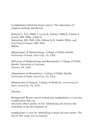
  • 17. participants were found for disease characteristics and breast cancer treatments, indicating that the study results may be generalized to breast cancer cases diagnosed in Iowa during 2004. Statistical analysis Univariate relative risk estimates (RRs) with 95% confidence intervals (95% CI) were calculated using unconditional logistic regression. Potential confounders were identified prior to analysis based on biologic plausibility. Estimates were adjusted for confounders that conferred a 10% or greater change from the crude RR. For factors of interest in which less than 20 subjects indicated they had the condition, confounders that presented a >20% change from the crude RR were adjusted for in the final model. RESULTS Cumulative incidence of lymphedema Arm lymphedema subsequent to breast cancer treatment was identified in 102 (19.5%) participants. The time between initial breast cancer treatment and onset of arm symptoms or
  • 18. physician-diagnosed lymphedema are graphed in Figure 1. The majority of lymphedema Tsai et al. Page 4 Front Womens Health. Author manuscript; available in PMC 2018 December 14. A u th o r M a n u scrip t A u th o r M a n u scrip t
  • 19. A u th o r M a n u scrip t A u th o r M a n u scrip t cases was persistent cases, and was diagnosed within two years after the initial breast cancer treatment.
  • 20. Participants’ characteristics At the time of interview, the average age of participants was 63 years and the mean BMI was 28.8 kg/m2. One-third (30.5%) of participants were college graduates and 70% were married. Neither education level nor marital status was associated with lymphedema (Table 2). Subjects who were under 50 at the time of interview were more likely to develop lymphedema than subjects aged 75+ years (RR=2.95, 95% CI: 1.25, 6.98). Participants with a BMI ≥30 (35.9%) were more likely to develop lymphedema (RR=2.15, 95% CI: 1.35, 3.42) than those with a BMI <30. An increasing trend in the RRs was observed as BMI increased over 30 (Table 2). Breast cancer disease and treatment For cancer characteristics, 87% of participants were classified as having stage I or II breast cancer and the mean tumor size was 19mm. In regards to breast cancer surgical treatments, 57% of women were treated with lumpectomy and 34% with sentinel node biopsy with an
  • 21. average of 8 nodes removed. Only 30.5% were detected with positive nodes and no trend was seen with increasing number of positive nodes (data not shown). Participants with > 10 lymph nodes removed were found to have an increased risk of developing lymphedema in the presence of radiation therapy. However this effect was reduced after adjustment for axillary dissection. Our results observed a trend of increasing risk as an increasing number of nodes was removed. Radiation therapy was received by 63% of women, and among those who received radiation, 30 stated that radiation was directed to the axillary area as well as the breast. Over half of the participants had chemotherapy and/or hormonal therapy as part of their breast cancer treatment. Lymphedema was associated with stage III cancer (RR=2.23, 95% CI: 1.09, 4.55), tumors ≥ 30mm (RR=2.76, 95% CI: 1.16, 6.58), and the presence of positive nodes (RR=1.88, 95% CI: 1.13, 3.13) (Table 3). Axillary dissection and radiation were found to interact (p=0.01). The combination of both axillary dissection and radiation
  • 22. therapy showed a slightly stronger association with lymphedema (RR=2.61, 95% CI: 1.27, 5.39) than either axillary dissection (RR=2.21, 95% CI: 1.32, 3.68) or radiation alone (RR=1.29, 95% CI: 0.81, 2.04). Radiation directed to the axillary area (RR=1.10, 95% CI: 0.62–1.93) and other treatment factors were not associated with lymphedema (Table 3). Chronic conditions Lymphedema was associated with chronic bronchitis (RR=3.45, 95% CI: 1.24, 9.63). A borderline increased risk for developing lymphedema was seen among participants who were diagnosed with osteoarthritis/ rheumatoid arthritis (RR=1.57, 95% CI: 0.93, 2.67) and/or kidney failure (RR=4.70, 95% CI: 0.89, 24.85). No association was found with high blood pressure, diabetes or other conditions reported after adjustment for age, BMI, and the interaction of axillary dissection and radiation. Tsai et al. Page 5 Front Womens Health. Author manuscript; available in PMC 2018 December 14.
  • 24. a n u scrip t A u th o r M a n u scrip t Arm activity and other personal factors No associations were found between lymphedema and specific arm activities including swimming, playing tennis, weightlifting or gardening. When analyses were restricted to participants who had the same level of arm activity before and after breast cancer diagnosis, no association between lymphedema and arm activity level either above or below the
  • 25. shoulders was found (Table 4). Surgery on dominant side (RR=1.49, 95% CI: 0.95–2.32), and air travel (RR=0.98, 95% CI: 0.63–1.52) were not associated with lymphedema in this study. An association was seen between infection and lymphedema (RR =8.51, 95% CI: 3.07, 23.61). However, all but one participant developed arm infection after lymphedema diagnosis. Discussion The prevalence of arm lymphedema in women diagnosed with breast cancer in Iowa in 2004 was 19.5% five-years after diagnosis, similar to results reported from previous studies [4, 12, 13]. This study, similar to other studies [14–19], found that BMI was associated with the development of lymphedema among these women. The association with increased BMI was evident both for the study definition of lymphedema and when defined only as physician- diagnosed cases. This suggests that the association seen was not an artifact of measurement error in lymphedema. Obesity, because of larger tissue volume
  • 26. and higher fat content, may have contributed to lymphedema development through increased difficulty of performing surgery or required alternative treatment techniques [20, 21]. In addition, obesity may increase lymphatic stress by exacerbating the inflammatory response or prolonging the surgical healing time [22]. Moreover, the increased amount of adipose tissue may act as a reservoir for lymphatic fluids [20]. Furthermore, one small study found that weight loss was correlated with a significant reduction in arm volume [23]. Obesity is also linked to chronic conditions such as high blood pressure and diabetes, which may further impair a lethargic lymphatic system by disrupting fluid balance. An increase in lymphedema risk was observed when both axillary dissection and radiation therapy were performed. A number of studies [24–29] have suggested that the addition of radiation therapy to axillary dissection increases the risk of lymphedema. Radiation after axillary dissection may have induced additional fibrosis that could compress or block
  • 27. lymphatic vessels. Participants in this study who had radiation and no axillary dissection were generally diagnosed with early stage breast cancer (stage I or II) and had less invasive treatments. Conversely, women who receive both axillary dissection and radiation therapy tended to be stage III and thus were treated more aggressively. This may further explain the interaction observed between axillary dissection and radiation therapy. Overall, published reports have both supported [16, 17, 29–32] and refuted [21, 33, 34] that increasing number of nodes excised is linked to arm lymphedema risk. This study did not find such an association after adjusting for axillary dissection. Axillary dissection was identified as a confounder because it was speculated that the association with the development of lymphedema may have been attributed to the intactness of the lymphatic network in the axilla rather than how many nodes were removed [30]. Axillary dissection is generally indicated in the presence of positive nodes and leads to an increased number of
  • 28. Tsai et al. Page 6 Front Womens Health. Author manuscript; available in PMC 2018 December 14. A u th o r M a n u scrip t A u th o r M a n u scrip t A u
  • 29. th o r M a n u scrip t A u th o r M a n u scrip t nodes excised. Axillary dissection, a procedure that disrupts the lymphatic network, remained associated with lymphedema even after adjusting for the number of lymph nodes removed.
  • 30. While breast cancer treatments are major contributors to lymphedema, the association between lymphedema and advance stages of cancer, positive nodes, or large tumors persisted even after adjusting for axillary dissection. It is possible that advanced disease or larger tumors may disrupt or damage regional lymphatics. The presence of most chronic conditions did not influence the subsequent development of lymphedema. While it was speculated that conditions such as high blood pressure and diabetes may exacerbate a damaged lymphatic system due to increased hydrostatic pressure [35], we did not find such an association in this study. Medications taken to control high blood pressure [14], may have negated the effect of increased hydrostatic pressure. Both chronic bronchitis and kidney conditions were linked to the development of lymphedema. Kidney failure may be associated with fluid retention that may cause edema [35], thus further complicating an already delicate lymphatic system. It is also possible that these subjects may have a surveillance bias for physician-diagnosed
  • 31. lymphedema due to visiting the doctor for these other conditions. This study’s findings are inconclusive since <20 subjects were diagnosed with these conditions. Arthritis and autoimmune diseases can contribute to lymphedema through inflammation to the joints, blood or lymph vessels, which may be reflected in the borderline association we saw. While most of the previously published studies did not find an association between age and lymphedema, we, similar to Geller et al.[14], found younger age was associated with developing lymphedema. It has been suggested that younger women may have advanced cancer which required more invasive treatments [36]. This study found that younger women under the age of 50 were more likely to have positive nodes (41% versus 19%) or be diagnosed at a higher stage after adjustment for confounders. They were also more likely to have axillary dissection. Moreover, younger women are more active outside of the home and may be more likely to notice the effects of mild lymphedema [37]. Also, older women tend
  • 32. to have extensive co-morbidity and might pay less attention to arm symptoms. Hence, arm symptoms related to lymphedema may have been under-reported by older women [37, 38]. While this study attempted to capture arm lymphedema in both an objective and subjective way (based on arm symptoms experienced within the last three months), under-reporting of arm symptoms (subjective method) is an issue because subclinical cases of lymphedema may be missed. Specific activities were not found to be associated with lymphedema. Among women who weight lifted, an increased amount of time spent weightlifting above the heart was not found to be associated with lymphedema. The results from this study were similar to another study in that none of the specific activities or overall arm activity level showed increased risk for lymphedema [39]. Arm lymphedema can lead to a reduction of arm activity level. In an attempt to avoid this bias, only subjects that reported no change in arm activity level both
  • 33. above and below the shoulders a year after resuming household activities (as compared to a year before breast cancer diagnosis) were included in the overall arm activity level analysis. Tsai et al. Page 7 Front Womens Health. Author manuscript; available in PMC 2018 December 14. A u th o r M a n u scrip t A u th o r M a n u scrip
  • 34. t A u th o r M a n u scrip t A u th o r M a n u scrip t With this restriction, arm activity level was not associated with lymphedema. However
  • 35. without this restriction, we found low level of arm activities below the shoulders was associated with lymphedema (RR=2.40, 95% CI: 1.38, 4.20) (data not shown). We believe that this may be a reflection of decreased level of arm activity due to the presence of lymphedema. Overall, our findings on arm activity do not support an association with post- operative arm exercise and arm lymphedema. Although air travel (41.6% of our subjects) has been speculated by both clinicians and breast cancer survivors to be a potential risk factor for lymphedema, such an association was not observed in our study or the study by Kilbreath et al [40]. It is probable that having lymphedema puts breast cancer survivors at risk for getting an arm infection due to decreased lymphatic circulation. Strengths This study was conducted using a population-based cohort of breast cancer survivors five to six years after breast cancer diagnosis, thereby avoiding erroneous inclusion of acute
  • 36. lymphedema cases. Participants reporting physician-diagnosed lymphedema were additionally asked if their condition has since resolved to decrease misclassification. Furthermore, objective and subjective assessments were applied to capture subclinical cases. Thirty-two percent of subjects reporting resolved lymphedema were later identified to have lymphedema through subclinical means. In addition, obtaining lymphedema status five or more years after breast cancer diagnosis allowed us to observe the long-term risk from treatments, as many studies have short follow-up times of 1–2 years after diagnosis or treatments. Limitations The biggest limitation was that all of our measures of lymphedema were self-reported. Due to caller identification and increased usage of cell phones, we were unable to reach as many subjects as we anticipated and 181 eligible women could not be traced from ICR information.
  • 37. Conclusion Among this cohort of breast cancer survivors, we found lymphedema to have a prevalence of 19.5% five years after diagnosis, with most developing lymphedema within the first 2 years after surgery. In particular, women with a high BMI were found to be at risk for developing lymphedema, suggesting that obesity may further promote inflammation which can lead to lymphatic impairment. Younger age was also associated with lymphedema development. The combination of axillary dissection and radiation therapy doubled the risk of developing lymphedema. Low level of arm activity was not found to be associated with lymphedema. Acknowledgments Funding This study was funded by the National Cancer Institute Grant Number: 5R03CA130031 (PI: Rebecca Tsai, PhD). Tsai et al. Page 8 Front Womens Health. Author manuscript; available in PMC 2018 December 14. A
  • 39. u scrip t A u th o r M a n u scrip t Abbreviations: BMI body mass index CATI Computer-assisted telephone interviewing ICR Iowa Cancer Registry RR Relative Risk CI Confidence Interval REFERENCES 1. Siegel R, Ma J, Zou Z, Jemal A (2014) Cancer statistics,
  • 40. 2014. CA: a cancer journal for clinicians 64:9–29. [PubMed: 24399786] 2. Surveillance Epidemiology and End Results, 2018 SEER Stat fact sheets: breast. Available at: http:// seer.cancer.gov/statfacts/html/breast.html [Accessed April 26 2018]. 3. Noone AM HN, Krapcho M, Miller D, Brest A, Yu M, Ruhl J, Tatalovich Z, Mariotto A, Lewis DR, Chen HS, Feuer EJ, Cronin KA (eds), SEER Cancer Statistics Review, 1975–2015, National Cancer Institute Bethesda, MD, https://seer.cancer.gov/csr/1975_2015/, based on November 2017 SEER data submission, posted to the SEER web site, 4 2018. 4. Velanovich V, Szymanski W (1999) Quality of life of breast cancer patients with lymphedema. Am J Surg 177:184–7; discussion 8. [PubMed: 10219851] 5. Kent H (1996) Breast-cancer survivors begin to challenge exercise taboos. CMAJ 155:969–71. [PubMed: 8837548] 6. Harris SR, Niesen-Vertommen SL (2000) Challenging the myth of exercise-induced lymphedema following breast cancer: a series of case reports. J Surg Oncol 74:95–8; discussion 8–9. [PubMed: 10914817] 7. McKenzie DC, Kalda AL (2003) Effect of upper extremity exercise on secondary lymphedema in breast cancer patients: a pilot study. J Clin Oncol 21:463–6. [PubMed: 12560436] 8. Lee TS, Kilbreath SL, Sullivan G, Refshauge KM, Beith JM,
  • 41. et al. (2009) Factors that affect intention to avoid strenuous arm activity after breast cancer surgery. Oncol Nurs Forum 36:454–62. [PubMed: 19581236] 9. Harris SR (2012) “We’re all in the same boat”: a review of the benefits of dragon boat racing for women living with breast cancer. Evidence-based complementary and alternative medicine : eCAM 2012:167651. [PubMed: 22811743] 10. Baumann FT, Reike A, Reimer V, Schumann M, Hallek M, et al. (2018) Effects of physical exercise on breast cancer-related secondary lymphedema: a systematic review. Breast cancer research and treatment. 11. Bloomquist K, Oturai P, Steele ML, Adamsen L, Moller T, et al. (2018) Heavy-Load Lifting: Acute Response in Breast Cancer Survivors at Risk for Lymphedema. Med Sci Sports Exerc 50:187–95. [PubMed: 28991039] 12. Petrek JA, Heelan MC (1998) Incidence of breast carcinoma-related lymphedema. Cancer 83:2776–81. [PubMed: 9874397] 13. Sakorafas GH, Peros G, Cataliotti L, Vlastos G (2006) Lymphedema following axillary lymph node dissection for breast cancer. Surg Oncol 15:153–65. [PubMed: 17187979] 14. Geller BM, Vacek PM, O’Brien P, Secker-Walker RH (2003) Factors associated with arm swelling after breast cancer surgery. J Women’s Health 12:921–30.
  • 42. 15. Helyer LK, Varnic M, Le LW, Leong W, McCready D (2010) Obesity is a risk factor for developing postoperative lymphedema in breast cancer patients. Breast J 16:48–54. [PubMed: 19889169] 16. Clark B, Sitzia J, Harlow W (2005) Incidence and risk of arm oedema following treatment for breast cancer: a three-year follow-up study. Q J Med 98:343–8. Tsai et al. Page 9 Front Womens Health. Author manuscript; available in PMC 2018 December 14. A u th o r M a n u scrip t A u th o r M
  • 44. http://seer.cancer.gov/statfacts/html/breast.html https://seer.cancer.gov/csr/1975_2015/ 17. Dominick SA, Madlensky L, Natarajan L, Pierce JP (2013) Risk factors associated with breast cancer-related lymphedema in the WHEL Study. Journal of cancer survivorship : research and practice 7:115–23. [PubMed: 23212606] 18. DiSipio T, Rye S, Newman B, Hayes S (2013) Incidence of unilateral arm lymphoedema after breast cancer: a systematic review and meta-analysis. Lancet Oncol 14:500–15. [PubMed: 23540561] 19. Ribeiro Pereira ACP, Koifman RJ, Bergmann A (2017) Incidence and risk factors of lymphedema after breast cancer treatment: 10 years of follow-up. Breast 36:67–73. [PubMed: 28992556] 20. Meric F, Buchholz TA, Mirza NQ, Vlastos G, Ames FC, et al. (2002) Long-term complications associated with breast-conservation surgery and radiotherapy. Ann Surg Oncol 9:543–9. [PubMed: 12095969] 21. Goffman TE, Laronga C, Wilson L, Elkins D (2004) Lymphedema of the arm and breast in irradiated breast cancer patients: risks in an era of dramatically changing axillary surgery. Breast J 10:405–11. [PubMed: 15327493] 22. Ahmed RL, Schmitz KH, Prizment AE, Folsom AR (2011) Risk factors for lymphedema in breast cancer survivors, the Iowa Women’s Health Study. Breast
  • 45. Cancer Res Treat 130:981–91. [PubMed: 21761159] 23. Shaw C, Mortimer P, Judd PA (2007) Randomized controlled trial comparing a low-fat diet with a weight-reduction diet in breast cancer-related lymphedema. Cancer 109:1949–56. [PubMed: 17393377] 24. Kissin MW, Querci della Rovere G, Easton D, Westbury G (1986) Risk of lymphoedema following the treatment of breast cancer. Br J Surg 73:580–4. [PubMed: 3730795] 25. Schunemann H, Willich N (1998) Lymphoedema of the arm after primary treatment of breast cancer. Anticancer Res 18:2235–6. [PubMed: 9703792] 26. Herd-Smith A, Russo A, Muraca MG, Del Turco MR, Cardona G (2001) Prognostic factors for lymphedema after primary treatment of breast carcinoma. Cancer 92:1783–7. [PubMed: 11745250] 27. Kwan W, Jackson J, Weir LM, Dingee C, McGregor G, et al. (2002) Chronic arm morbidity after curative breast cancer treatment: prevalence and impact on quality of life. J Clin Oncol 20:4242–8. [PubMed: 12377968] 28. Keramopoulos A, Tsionou C, Minaretzis D, Michalas S, Aravantinos D (1993) Arm morbidity following treatment of breast cancer with total axillary dissection: a multivariated approach. Oncology 50:445–9. [PubMed: 8233285]
  • 46. 29. Hinrichs CS, Watroba NL, Rezaishiraz H, Giese W, Hurd T, et al. (2004) Lymphedema secondary to postmastectomy radiation: incidence and risk factors. Ann Surg Oncol 11:573–80. [PubMed: 15172932] 30. Goldberg JI, Wiechmann LI, Riedel ER, Morrow M, Van Zee KJ (2010) Morbidity of sentinel node biopsy in breast cancer: the relationship between the number of excised lymph nodes and lymphedema. Ann Surg Oncol 17:3278–86. [PubMed: 20574774] 31. Petrek JA, Senie RT, Peters M, Rosen PP (2001) Lymphedema in a cohort of breast carcinoma survivors 20 years after diagnosis. Cancer 92:1368–77. [PubMed: 11745212] 32. Zou L, Liu FH, Shen PP, Hu Y, Liu XQ, et al. (2018) The incidence and risk factors of related lymphedema for breast cancer survivors post-operation: a 2-year follow-up prospective cohort study. Breast Cancer 25:309–14. [PubMed: 29397555] 33. Yen TW, Fan X, Sparapani R, Laud PW, Walker AP, et al. (2009) A contemporary, population- based study of lymphedema risk factors in older women with breast cancer. Ann Surg Oncol 16:979–88. [PubMed: 19194754] 34. Beaulac SM, McNair LA, Scott TE, LaMorte WW, Kavanah MT (2002) Lymphedema and quality of life in survivors of early-stage breast cancer. Arch Surg 137:1253–7. [PubMed: 12413312] 35. Paulus BM, Ali S, Zia AA, Munir A, Davis RC, Jr, et al.
  • 47. (2008) Causes and consequences of systemic venous hypertension. Am J Med Sci 336:489–97. [PubMed: 19092322] 36. Paskett ED, Naughton MJ, McCoy TP, Case LD, Abbott JM (2007) The epidemiology of arm and hand swelling in premenopausal breast cancer survivors. Cancer Epidemiol Biomarkers Prev 16:775–82. [PubMed: 17416770] Tsai et al. Page 10 Front Womens Health. Author manuscript; available in PMC 2018 December 14. A u th o r M a n u scrip t A u th o r M
  • 49. 37. Armer J, Fu MR (2005) Age differences in post-breast cancer lymphedema signs and symptoms. Cancer Nurs 28:200–7. [PubMed: 15915063] 38. Warmuth MA, Bowen G, Prosnitz LR, Chu L, Broadwater G, et al. (1998) Complications of axillary lymph node dissection for carcinoma of the breast: a report based on a patient survey. Cancer 83:1362–8. [PubMed: 9762937] 39. Ahmed RL, Thomas W, Yee D, Schmitz KH (2006) Randomized controlled trial of weight training and lymphedema in breast cancer survivors. J Clin Oncol 24:2765–72. [PubMed: 16702582] 40. Kilbreath SL, Ward LC, Lane K, McNeely M, Dylke ES, et al. (2010) Effect of air travel on lymphedema risk in women with history of breast cancer. Breast Cancer Res Treat 120:649–54. [PubMed: 20180016] Tsai et al. Page 11 Front Womens Health. Author manuscript; available in PMC 2018 December 14. A u th o r M a n
  • 51. o r M a n u scrip t Figure 1. Cumulative Incidence of Arm Lymphedema diagnosed by a physician or by having 4 + symptoms among breast cancer survivors in Iowa, 2004 Tsai et al. Page 12 Front Womens Health. Author manuscript; available in PMC 2018 December 14. A u th o r M a n u scrip
  • 54. t A u th o r M a n u scrip t A u th o r M a n u scrip t Tsai et al. Page 13 Table 1. Distribution of self-reported lymphedema assessments among study participants diagnosed with breast cancer
  • 55. in Iowa, 2004. Participants % N=522 Physician-diagnosed lymphedema Unresolved 44 8.4 Resolved 28 5.4 Not diagnosed 450 86.2 Arm measurement >2cm difference between arms 49 9.4 ≤ 2 cm difference between arms 351 67.2 Missing 122 23.4 Arm symptoms* Subjective indication of arm lymphedema 45 8.6 No subjective indication of arm lymphedema 477 91.4 Arm lymphedema based on any of the above Arm lymphedema 102 19.5 No arm lymphedema 420 80.5 *
  • 56. Subjective indication is present if subject has at least 2 major symptoms (shirt sleeve felt tight, arm felt swollen, heavy, tense or hard) and at least 4 total symptoms (major arm symptoms plus arm felt numb, stiff, or painful, rash on arm, other arm symptoms, cannot see knuckles or veins on hand, or rings felt tight) Front Womens Health. Author manuscript; available in PMC 2018 December 14. A u th o r M a n u scrip t A u th o r M a
  • 58. Table 2. Relative risk of demographic factors and lymphedema among subjects diagnosed with breast cancer in Iowa, 2004 Lymphedema Adjusted Yes No RR CI Age at interview 75–85 12 (11.8) 86 (20.5) Ref * 70–74 14 (13.7) 56 (13.3) 1.85 0.75, 4.55 65–69 16 (15.7) 51 (12.1) 1.77 0.72, 4.34 60–64 13 (12.8) 61 (14.5) 1.29 0.52, 3.18 55–59 13 (12.8) 60 (14.3) 1.40 0.56, 3.49 50–54 14 (13.7) 57 (13.6) 1.61 0.66, 3.94 25–49 20 (19.6) 49 (11.7) 2.95 1.25, 6.98 Trend RR comparing age 75–85 to 25–49 1.85 0.93, 3.70 Education ≤ High school 45 (44.5) 192 (46.0) Ref †
  • 59. Some college 25 (24.8) 98 (23.5) 0.90 0.50, 1.63 ≥ College 31 (30.7) 127 (30.5) 0.94 0.53, 1.65 Trend RR comparing ≥ College to ≤ High school 0.93 0.53, 1.64 Married No 33 (33.0) 124 (29.5) Ref ‡ Yes 67 (67.0) 296 (70.5) 0.76 0.46, 1.23 BMI (kg/m2) <18.5 0 4 (1.0) N/A 18.5–24.9 22 (21.8) 138 (33.1) Ref c 25–29.9 28 (27.7) 140 (33.6) 1.20 0.64, 2.23 30–34.9 20 (19.8) 83 (19.9) 1.50 0.76, 2.98 35–39.9 13 (12.9) 35 (8.4) 2.51 1.12, 5.65 40+ 18 (17.8) 17 (4.1) 5.58 2.45, 12.7 Trend RR comparing 40+ to 18.5–24.9 7.43 3.03, 18.2 RR= relative risk; CI = confidence interval; BMI= body mass index. Note: Total number of subjects may not add up to 522 due to missing data
  • 60. * Adjusted for BMI, axillary dissection, number of lymph nodes removed, any radiation, and any radiation x axillary dissection † Adjusted for BMI, age, axillary dissection, any radiation, and any radiation x axillary dissection ‡ Adjusted for age, axillary dissection, any radiation, and any radiation x axillary dissection Front Womens Health. Author manuscript; available in PMC 2018 December 14. A u th o r M a n u scrip t A u th
  • 62. t Tsai et al. Page 15 Table 3. Relative Risk of Lymphedema by Breast Cancer Treatment and Disease Factors Among Subjects Diagnosed with Breast Cancer in Iowa, 2004 Lymphedema Adjusted Yes No RR CI Stage Stage I 42 (42.0) 235 (58.2) Ref * Stage II 38 (38.0) 139 (34.4) 1.13 0.66, 1.92 Stage III 20 (20.0) 30 (7.4) 2.23 1.09, 4.55 Trend RR comparing Stage III to Stage I 1.99 0.99, 3.98 Primary tumor size (mm) 0–9 15 (15.8) 106 (26.6) Ref † 10–14 17 (17.9) 88 (22.1) 1.26 0.57, 2.75 15–29 39 (41.1) 158 (39.6) 1.60 0.78, 3.34
  • 63. 30+ 24 (25.3) 47 (11.8) 2.76 1.16, 6.58 Trend RR comparing 30+ to 0–9 2.65 1.14, 6.13 Surgery Lumpectomy 54 (52.9) 243 (58.1) Ref ‡ Mastectomy 13 (12.8) 56 (13.4) 1.09 0.39, 3.04 Modified radical mastectomy 35 (34.3) 119 (28.5) 1.53 0.75, 3.15 # of lymph node removed 0–2 17 (16.8) 126 (30.4) Ref * 3–6 21 (20.8) 108 (26.0) 1.10 0.50, 2.39 7–10 18 (17.8) 57 (13.73) 1.45 0.56, 3.78 >10 45 (44.6) 124 (29.9) 1.60 0.66, 3.84 Trend RR comparing >10 to 0–2 1.64 0.74, 3.61 Positive nodes No 54 (52.9) 300 (73.5) Ref * Yes 48 (47.1) 108(26.5) 1.88 1.13, 3.13 Radiation to the axilla §
  • 64. No 60 (65.2) 272 (61.8) Ref * Yes 32 (34.8) 107 (28.2) 1.10 0.62, 1.93 Chemotherapy No 40 (40.0) 208 (49.9) Ref * Yes 60 (60.0) 209 (50.1) 1.01 0.61, 1.67 Hormones therapy No 47 (47.0) 183 (44.6) Ref * Yes 53 (53.0) 227 (55.4) 0.92 0.58, 1.46 RR= relative risk; CI = confidence interval; BMI= body mass index Note: Total number of subjects may not add up to 522 due to missing data Front Womens Health. Author manuscript; available in PMC 2018 December 14. A u th o r M a
  • 66. th o r M a n u scrip t Tsai et al. Page 16 * Adjusted for age, axillary dissection, any radiation, and any radiation x axillary dissection † Adjusted for age, axillary dissection, chemotherapy, number of lymph nodes removed, any radiation, and any radiation x axillary dissection ‡ Adjusted for age, any radiation to axilla, axillary dissection, any radiation, and any radiation x axillary dissection § Among subjects who received radiation therapy Front Womens Health. Author manuscript; available in PMC 2018 December 14. A
  • 68. u scrip t A u th o r M a n u scrip t Tsai et al. Page 17 Table 4. Relative Risk of Arm Activities and Lymphedema Among Subjects Diagnosed with Breast Cancer in Iowa, 2004, Restricted to Women Who Reported no Change in Arm Activity Level Before and After Breast Cancer Diagnosis Lymphedema Crude Adjusted Arm activities Yes No RR CI RR CI Above the shoulders: *
  • 69. High 9 (20) 35 (13.3) Ref Ref † Medium 14 (31.1) 113 (43) 0.48 0.19, 1.21 0.51 0.19, 1.40 Low 22 (48.9) 115 (43.7) 0.74 0.31, 1.76 0.78 0.29, 2.07 Below the shoulders: * High 18 (40) 126 (47.9) Ref Ref † Medium 17 (37.8) 92 (35) 1.29 0.63, 2.65 1.38 0.63, 3.01 Low 10 (22.2) 45 (17.1) 1.56 0.67, 3.62 1.59 0.60, 4.24 RR= relative risk; CI = confidence interval. * Only included subjects that reported no change in arm activity level from before breast cancer diagnosis to after resuming household activities (N=308) † Adjusted for BMI, age, axillary dissection, any radiation, and any radiation x axillary dissection Front Womens Health. Author manuscript; available in PMC 2018 December 14.
  • 70. AbstractIntroductionMaterials and MethodsRecruitmentParticipant interviewLymphedema categorizationReliability and representativenessStatistical analysisRESULTSCumulative incidence of lymphedemaParticipants’ characteristicsBreast cancer disease and treatmentChronic conditionsArm activity and other personal factorsDiscussionStrengthsLimitationsConclusionReferencesFig ure 1.Table 1.Table 2.Table 3.Table 4. 1 Running head: ARTICLE CRITIQUE 8 EBP GRANT PROPOSAL Chapter 11: Lymphedema The purpose of this paper is to discuss lymphedema and critique an article on the topic. Lymphedema is when there is an obstruction in the lymphatic vessels that cause the tissues in the extremities to swell (Hubert & Vanmeter, 2018). This also allows for accumulation of lymph in the tissues as well. Commonly, this disorder is congenital and may involve the lymph nodes along with the vessels (Hubert & Vanmeter, 2018). It can also be caused by blockage of the lymph vessels due to parasitic worms (Hubert & Vanmeter, 2018). When the lymph states to build up in the body, the more the extremity swells (Huber & Vanmeter, 2018). As lymphedema continues to progress over time, the extremity becomes enlarged, firm and painful (Hubert & Vanmeter, 2018). Lymphedema can be chronic as well, which leads to frequent infection (Hubert & Vanmeter, 2018). According to Johns Hopkins Medicine (2020), lymphedema can occur after cancer surgery when lymph nodes are removed. Authors Rebecca J. Tsai, Leslie K. Dennis, Charles F. Lynch, Linda G. Snetselaar, Gideon K. D. Zamba, Carol Scott- Conner published an article on lymphedema after breast cancer
  • 71. entitled “Lymphedema following breast cancer: The importance of surgical methods and obesity” Published to Front Women’s Health in 2018. The purpose of the article is to discuss the research that the authors have conducted on the association between developing lymphedema after cancer surgery and personal risk factors (Tsai et al., 2018). This research will allow for them to better understand if there are certain factors that make a person more at risk for developing lymphedema after cancer surgery. The literature was drawn from a systemic approach. This is because it focuses on a specific question and critically appraises all relevant research. The review focuses on cause and effect meaning how does one issue effect the other. In this case, the authors are correlating developing lymphedema after surgery and certain risk factors that can affect this. Tsai et al. (2018) identify concern that the measures of the study are from subject self-reporting. The authors feel that there can be issues with self-reporting because the patients do not always participate until the end of the study. The aim of the study is to understand if there is a correlation between arm lymphedema and certain personal risk factors (Tasi et al., 2018). To achieve an accurate study, the authors used a population-based cohort design. This allows for better understanding of the research for this specific population of women. The sample was obtained in Iowa from 2004 to 2010. Tsai et al. (2018) states that the women who met the criteria, completed a short telephone interview about their lymphedema status, arm activities, demographics and chronic conditions that they currently have. The patients were confirmed to have lymphedema from physician reports and the presence of at least four major arm symptoms that can occur with the disease (Tsai et al., 2018). There were 522 participants in the study, which seems to be an adequate size for a population-based cohort design. The study showed that lymphedema was identified in 102 of the 522 patients (Tsai et al., 2018). The results showed that people who had radiation and some dissection if the axilla were
  • 72. more at risk for lymphedema after breast cancer surgery (Tsai et al., 2018). Tsai et al. (2018) states that the women who were the most likely to have lymphedema had a body mass index above 40. This shows that obesity, the characteristics of the cancer and the methods used in surgery were all major factors in the patient developing lymphedema (Tsai et al., 2018). The authors found that obesity promotes the inflammation of the body which leads to issues with lymphedema. The authors also found that women who were younger had a higher chance of developing lymphedema as well. The authors are satisfied with the results and do not mention a need for further testing (Tsai et al., 2018). The article is greatly recommended as a nurse and a future advanced practice nurse. This article is easy to comprehend and has pertinent information that can be used in practice. The information can be useful when assessing patients who have had breast cancer surgery. Knowing and understanding the risk factors for developing lymphedema after breast cancer surgery can allow for preventative care for the patients who are most at risk. It may not be completely curable, but there are certain steps that can be taken to reduce the symptoms or even keep it from starting in the first placed (Johns Hopkins Medicine, 2020). It is apparent that advanced practice nurses should understand the signs and symptoms of developing lymphedema so that it can be treated adequately.
  • 73. References Hubert, R. & VanMeter, K. C. (2018). Gould's pathophysiology for the health professions. St. Louis, MO: Elsevier Saunders. Johns Hopkins Medicine. (2020). Breast cancer: lymphedema after treatment. Retrieved from https://www.hopkinsmedicine.org/health/conditions-and- diseases/breast-cancer/breast-cancer-lymphedema-after- treatment Tsai R. J., Dennis, L. K., Lynch, C. F., Snetselaar, L. G., Zamba, G. K. D., Scott-Conner, C. (2018). Lymphedema following breast cancer: the importance of surgical methods and obesity. Front Women’s Health, 3(2), 1-17. References Lastname, C. (2008). Title of the source without caps except Proper Nouns or: First word after colon. The Journal or Publication Italicized and Capped, Vol#(Issue#), Page numbers. Lastname, O. (2010). Online journal using DOI or digital object identifier. Main Online Journal Name, Vol#(Issue#), 159- 192. doi: 10.1000/182 Lastname, W. (2009). If there is no DOI use the URL of the main website referenced. Article Without DOI Reference, Vol#(Issue#), 166-212. Retrieved from http://www.mainwebsite.org
  • 74. Journal of Clinical Medicine Article Optimizing Hydroxyurea Treatment for Sickle Cell Disease Patients: The Pharmacokinetic Approach Charlotte Nazon 1, Amelia-Naomi Sabo 2,3, Guillaume Becker 3,4, Jean-Marc Lessinger 2, Véronique Kemmel 2,3,* and Catherine Paillard 1,5,* 1 Hôpitaux Universitaires de Strasbourg, Centre de compétence pour les maladies constitutionnelles du globule rouge et de l’érythropoïèse, Service d’hématologie oncologie pédiatrique, Avenue Molière, 67200 Strasbourg, France; [email protected] 2 Laboratoire de Pharmacologie et Toxicologie Neurocardiovasculaire, Faculté de Médecine, 11 rue Humann, 67085 Strasbourg, France; [email protected] (A.-N.S.); [email protected] (J.-M.L.) 3 Hôpitaux Universitaires de Strasbourg, Hôpital de Hautepierre, Laboratoire de Biochimie et Biologie Moléculaire, Avenue Molière, 67200 Strasbourg, France; [email protected] 4 Hôpitaux Universitaires de Strasbourg, Service de la Pharmacie, Avenue Molière, 67200 Strasbourg, France 5 Laboratoire d’ImmunoRhumatologie Moléculaire, INSERM UMR_S 1109, LabEx Transplantex, Fédération de Médecine Translationnelle de Strasbourg, 4 rue Kirschleger,
  • 75. 67085 Strasbourg Cedex, France * Correspondence: [email protected] (V.K.); [email protected] (C.P.); Tel.: +33-(0)-3-88-12-75-33 (V.K.); +33-(0)-3-88-12-88-23 (C.P.) Received: 21 August 2019; Accepted: 11 October 2019; Published: 16 October 2019 ���������� ������� Abstract: Background: Hydroxyurea (HU) is a FDA- and EMA- approved drug that earned an important place in the treatment of patients with severe sickle cell anemia (SCA) by showing its efficacy in many studies. This medication is still underused due to fears of physicians and families and must be optimized. Methods: We analyzed our population and identified HU pharmacokinetic (PK) parameters in order to adapt treatment in the future. Working with a pediatric population, we searched for the most indicative sampling time to reduce the number of samples needed. Results: Nine children treated by HU for severe SCA were included for this PK study. HU quantification was made using a validated gas chromatography/mass spectrometry (GC/MS) method. Biological parameters (of effectiveness and compliance) and clinical data were collected. None of the nine children reached the therapeutic target defined by Dong et al. as an area under the curve (AUC) = 115 h.mg/L; four patients were suspected to be non- compliant. Only two patients had an HbF over 20%. The 2 h sample was predictive of the medication exposure (r2 = 0.887). Conclusions: It is
  • 76. urgent to be more efficient in the treatment of SCA, and pharmacokinetics can be an important asset in SCA patients. Keywords: sickle cell disease; sickle cell anemia; hydroxyurea; pharmacokinetics 1. Introduction Sickle cell anemia (SCA) is one of the most common inherited diseases in the world. It affects more than 300,000 infants born annually worldwide, and the epidemiologic projections show a growing tendency for the years to come (30% increase by 2050) [1]. It is an autosomal recessive disease affecting the red blood cells due to a point missense mutation on the hemoglobin beta chain. This mutation leads to a polymerization of the hemoglobin (Hb), which causes an increased density, dehydration, and deformation of red blood cells, forming the sickle cells [2]. Clinical manifestations of SCA include hemolytic anemia, vaso- occlusive crisis (VOC), and bacterial susceptibility and can have an impact on many organs [3–9]. J. Clin. Med. 2019, 8, 1701; doi:10.3390/jcm8101701 www.mdpi.com/journal/jcm http://www.mdpi.com/journal/jcm http://www.mdpi.com https://orcid.org/0000-0003-4680-4293 http://dx.doi.org/10.3390/jcm8101701 http://www.mdpi.com/journal/jcm https://www.mdpi.com/2077- 0383/8/10/1701?type=check_update&version=2
  • 77. J. Clin. Med. 2019, 8, 1701 2 of 10 Hydroxyurea (HU) is a FDA- and EMA-approved drug that earned an important place in the treatment of patients with severe SCA. It has shown its efficacy in multiple studies by reducing the morbi-mortality and frequency of VOC, transfusions, and hospitalizations for those patients [10–16]. Furthermore, HU treatment is associated with improvement in hemoglobin concentration illustrated by increasing mean corpuscular volume (MCV) and Hemoglobin F (HbF) levels [17]. One way to adjust HU dose, mostly used in American centers, is to introduce HU at 15–20 mg/kg/day and to make a dose escalation until a mild myelosuppression tolerated by the patient is obtained, which indicates that the maximum tolerated dose (MTD) has been reached [18]. The dose escalation of HU depends on three hematological parameters: the neutrophil count (1.5–3 G/L), the reticulocyte count (100–200 G/L), and the platelet count (>80 G/L) [17]. When the MTD is reached, the risk/benefit balance is optimal for the patient [19]. Despite the fact that the escalation to MTD has proven to be the best way to dose HU, many European centers use a fixed-dose strategy (20 mg/kg/day). The dose escalation method presents some drawbacks: it requires frequent outpatient visits and laboratory tests and usually takes 6 to 12 months; the treatment is usually suboptimal during that time [20–22]. The fact that there is a period during which the treatment is not providing any clinical improvement, and that frequent laboratory tests and medical consultations are needed, does not help
  • 78. patient adherence, which is already low in this population [23,24]. It has been highlighted by Brandow et al., who showed that the patients who refused HU gave the following reasons: fear of cancer and other side effects in the majority followed by not wanting to take medication, not wanting to have required laboratory monitoring, or not thinking the medication would work [23]. Regarding toxicities, even if we now know that the myelosuppression is transient [18,25], that the azoospermia caused by HU could be reversible [26], and that there is no evidence it has a genotoxic or a leukemogenic effect [15], the reticence among physicians and families is still strong, leading to an underuse of this treatment [23]. Studies showed that the escalation to MTD did not add any toxicities [27]. Organ damage begins early in life, worsens over time, and is irreversible. Accordingly, early optimal treatment in young patients who have not yet developed serious or irreversible organ damage is a necessity. Another difficulty remains in pharmacokinetic variations of HU. Indeed, for a same dose of HU, drug exposure may vary five times in adults and three times in children [25]. There are major inter-individual variations regarding absorption, profiles, distribution, and clearance of HU [28]. Logically, HU is not efficacious at the same dosage for everyone: In a study of Dong et al., posology at MTD ranges from 14.2 to 35.5 mg/kg/day [21]. It makes standard patient care impossible. For these reasons, a personalized dose optimization process that can rapidly identify MTD for individual SCA patients is highly desirable. The main goal of this study was to analyze our population
  • 79. and identify the pharmacokinetic parameters of HU to be able to adapt and optimize their treatment in the future. By doing so, we intend to have a model we can use to adapt the dose of HU and reach the MTD for an earlier clinical benefit for our patients more quickly. Working with a pediatric population, we wanted to find the most indicative sampling time to reduce the number of blood collections needed. 2. Experimental Section 2.1. Patients We included, prospectively, all the patients with SCA of <20 years of age, treated by HU attending our hospital (Hôpitaux Universitaires de Strasbourg, France), for a follow-up consultation between February and May 2018. More than 100 patients with SCA (SS or SC) are followed in our hospital, including 27 treated by HU. In our hospital, HU is introduced at 15 mg/kg/day and normally increased to reach MTD following hematological criteria: the neutrophil count (1.5–3 G/L), the reticulocyte count (100–200 G/L), and the platelet count (>80 G/L). Although, most of the time, if there is no clinical manifestation of SCA, no dose adjustment is done. J. Clin. Med. 2019, 8, 1701 3 of 10 All patients and/or parents/guardians provided written informed consent before enrollment in the study. This study received approval from the Institutional review board of Strasbourg University Hospital (DRCI 2018-project n◦6112) and the French data
  • 80. protection authority (CNIL-n◦2215437). 2.2. Study Design Plasma samples were collected at the following times: pre-dose and at 10 min, 20 min, 1 h, 2 h, 4 h, and 6 h after oral HU administration at the patient’s usual dose. Whole blood samples were transported and/or stored at 2–8 ◦C for a maximum of four h before centrifugation, and then aliquoted plasma was rapidly frozen at −20 ◦C. Demographic information and standard laboratory parameters were collected: Neutrophil, reticulocyte, and platelet counts were measured in turn to identify the individual tolerability of the treatment, hemoglobin, MCV, and the percentage of HbF to monitor the clinical efficacy of HU. We evaluated the adherence of our patients by asking them about the frequency of missed doses over the past six months. Genotypic data were collected, looking for associated alpha thalassemia and haplotype. 2.3. GC-MS The dosage of HU was made by our hospital’s Biochemistry and Molecular Biology Laboratory using a gas chromatography coupled with mass spectrometry (GC-MS) technique (version 2.0, CHU Strasbourg, Strasbourg, France), which can be used routinely. A Thermo Scientific™ ISQ Series (Thermo Fisher Scientific, Courtaboeuf, France) was used for the analysis. Chromatography was performed on a low polarity RTX-5-MS (30 m × 0.25 mm × 0.25
  • 81. µm) capillary column, and helium was the carrier gas. The total analysis time of a sample was 13 min. Data were obtained from the Thermo Xcalibur© software (© 2012 Thermo Fisher Scientific Inc., version 3.1, Courtaboeuf, France). It is a sensitive and specific method validated by our local laboratory that is feasible with a low quantity of plasma (50 µL). The measurement of this method ranges from 0.79 to 100 mg/L with a detection limit of 0.28 mg/L. 2.4. Modelling and Statistical Analysis The plasma concentration–time data was analyzed by a non- compartmental method to obtain a concentration–time curve. The pharmacokinetics’ parameters were defined for each patient: the maximum plasma concentration (Cmax), the time to reach the Cmax (Tmax), and the area under the curve (AUC). The Cmax were identified by a graphical analysis. The mean, standard deviation, and median were calculated using Microsoft Excel. The simulated AUC0–6h were calculated by the linear log trapezoidal rule (version 6, GraphPad Prism, San Diego, CA, USA). 2.5. Optimal Sampling Exploration of the relationship between HU concentration–time and exposition was made. The significant linear correlations were defined by the determination coefficient r2 > 0.5. AUC was tested by a non-compartmental method. Data analysis was made using the GraphPad Prism® program
  • 82. (version 6, GraphPad Prism, San Diego, CA, USA). 3. Results 3.1. Characteristics of Patients Nine patients were included, and their characteristics are shown in Table 1. These patients were on HU treatment for multiple VOC or acute chest syndrome (ACS). Most of them were treated by HU for more than four years, and their daily doses ranged from 12.9 to 24.6 mg/kg/day. J. Clin. Med. 2019, 8, 1701 4 of 10 Table 1. Patient characteristics. Demographic Characteristics Sex ratio M/F 0.8 (4/5) Age Mean ± standard deviation 14.4 (±3.7) Median 16.5 Weight Mean ± standard deviation 49.9 (±20.5) Median 49.1 Background (Number of Patients and Percentage) Cholecystectomy 3 (33%) Stroke 1 (11.1%)
  • 83. Abnormal Transcranial doppler episode 1 (11.1%) Osteonecrosis 2 (22.2%) Retinopathy 1 (11.1%) Splenic Sequestration 0 (0%) Pulmonary Hypertension 1 (11.1%) Cardiac Events 1 (11.1%) Kidney Failure 0 (0%) Events per Year: 2016–2018 Period Transfusion/Year Mean ± Standard Deviation 0.8 Median 0.3 (0–2) Hospitalization/Year Mean ± standard deviation 1.4 Median (range) 0.6 (0–5.0) VOC/Year Mean ± Standard Deviation 1.6 Median (range) 1 (0–5.6) ACS Number of Patients > 1 ACS 4 (45%) HU Dose (mg/kg/day) Mean ± Standard Deviation 19.0 (±4.0) Median (range) 20.4 (12.9–24.6) Time since Introduction of HU (Months)
  • 84. Mean ± Standard Deviation 63.5 (±44.6) Median (Range) 58.8 (11.2–138.8) Age at Introduction (Year) Mean ± Standard Deviation 8.5 (±4.4) Median (Range) 6.0 (4.0–16.0) HU: Hydroxyurea; VOC: vaso-occlusive crisis; ACS: acute chest syndrome. M: male; F: female. 3.2. Biological Parameters and Self-Reported Compliance Biological parameters are presented in Table 2. Seven patients had an HbF lower than 20%. Four children had a normal or low MCV. None of the patients reached the myelosuppression as defined earlier as a sign of MTD of HU. None of the patients showed a major hematological toxicity. However, Patient 8 had the lowest neutrophil count, middle MCV, and a low percentage of HbF. We evaluated the adherence of our patients by asking them about the frequency of missed doses over the past six months. We defined low compliance level as one missed-dose per week or more (n = 2), medium compliance level as one to three missed-doses per month (n = 4), and high compliance level as less than one dose-missed a month (Table 2) (n = 3). The genotypic profile was available only for Patient 8, who is not a carrier of alpha thalassemia and has a Benin/Benin (BEN/BEN) haplotype. J. Clin. Med. 2019, 8, 1701 5 of 10
  • 85. Table 2. Self-reported compliance, biological parameters, and HU intake characteristics of the nine children on HU. Patient Self-Reported Compliance Hb (g/dL) MCV (fL) Retic. (G/L) PNN (G/L) Platelets (G/L) HbF (%) Dose HU (mg/kg/day) Pre-HU Post-HU Pre-HU Post-HU 1 poor 8.6 81.2 87.5 284.0 6.0 427 3.3 7.0 17.1 2 good 9.0 86.3 111.9 161.2 4.9 282 8.8 20.5 20.9 3 poor 7.7 N/A 70.2 192.7 11.5 279 N/A 3.0 20.4 4 medium 7.1 83.0 95.8 242.2 14.2 81 4.8 7.9 21.4 5 medium 8.3 79.0 78.1 286.7 8.0 539 N/A 1.4 21.4 6 good 9.1 N/A 94.9 185.0 5.9 232 N/A 23.7 18.9 7 medium 7.8 88.0 80.4 325.2 9.2 589 5.7 7.4 24.6 8 medium 7.6 78.4 95.0 177.1 3.2 290 2.5 5.6 13.0 9 good 7.6 N/A 97.8 164.7 4.9 465 N/A 14.5 12.9 Mean ± SD 8.1 ± 0.7 90.2 ± 12.5 224.3 ± 61.5 7.5 ± 3.6 354 ± 163 10.1 ± 7.8 19.0 ± 4.0 Median 7.8 94.9 192.7 6.0 290.0 7.4 20.4 Pre-HU: parameters before HU initiation; Post-HU: parameters after HU initiation. 3.3. Pharmacokinetic Parameters
  • 86. Principal pharmacokinetic parameters are presented in Table 3 and mean HU concentration-time are represented in Figure 1. The AUC ranged from 43.3 to 113.5 h.mg/L with a median of 75.1 h.mg/L. None of the nine children reached 115 h.mg/L, which was the target-AUC in a study by Dong et al. [21]. Six of them had an AUC less than 100 h.mg/L. However, Patient 2 presented with an AUC of 113.5 h.mg/L and had one of the highest HbF percentages (20.5 %). Patient 6, who had the highest HbF (=23.7%), had an AUC of 74.0 h.mg/L, showing a non-optimal response. Every child except one had a time to reach the Cmax (Tmax) between 1 and 2.5 h. Table 3. Pharmacokinetics parameters of the nine children on HU. Patient Treatment Duration (Months) Dose (mg/kg/day) Cmax (mg/L) Tmax (hours) AUC (h.mg/L) 1 14.2 17.1 24.0 1.33 75.1 2 121.6 20.9 33.9 1.11 113.5 3 27.3 20.4 14.9 2.00 59.5 4 58.8 21.4 15.2 2.44 57.3 5 138.8 21.4 37.5 1.11 102.0 6 51.6 18.9 25.8 0.66 74.0 7 70.1 24.6 31.0 2.44 108.2 8 11.2 13.0 10.8 1.33 43.3 9 77.8 12.9 24.0 1.33 98.2 Mean ± Standard Deviation 63.5 (±44.6) 19.0 (±4.0) 24.1 (±9.1) 1.5 (±0.6) 81.3 (±25.2) Median 58.8 20.4 24.0 1.33 75.1
  • 87. J. Clin. Med. 2019, 8, x FOR PEER REVIEW 5 of 10 4 medium 7.1 8.3 9.1 7.8 7.6 7.6 83.0 95.8 242.2 14.2 81 4.8 7.9 21.4 5 medium 79.0 78.1 286.7 8.0 539 N/A 1.4 21.4 6 good N/A 94.9 185.0 5.9 232 N/A 23.7 18.9 7 medium 88.0 80.4 325.2 9.2 589 5.7 7.4 24.6 8 medium 78.4 95.0 177.1 3.2 290 2.5 5.6 13.0 9 good N/A 97.8 164.7 4.9 465 N/A 14.5 12.9 Mean ± SD 8.1 ± 0.7 90.2 ± 12.5 224.3 ± 61.5 7.5 ± 3.6 354 ± 163 10.1 ± 7.8 19.0 ± 4.0 Median 7.8 94.9 192.7 6.0 290.0 7.4 20.4 Pre-HU: parameters before HU initiation; Post-HU: parameters after HU initiation. 3.3. Pharmacokinetic Parameters Principal pharmacokinetic parameters are presented in Table 3 and mean HU concentration-time are represented in Figure 1. The AUC ranged from 43.3 to 113.5 h.mg/L with a median of 75.1 h.mg/L. None of the nine children reached 115 h.mg/L, which was the target-AUC in a study by Dong et al. [21]. Six of them had an AUC less than 100 h.mg/L. However, Patient 2 presented with an AUC of 113.5 h.mg/L and had one of the highest HbF percentages (20.5 %). Patient
  • 88. 6, who had the highest HbF (= 23.7%), had an AUC of 74.0 h.mg/L, showing a non-optimal response. Every child except one had a time to reach the Cmax (Tmax) between 1 and 2.5 hours. Table 3. Pharmacokinetics parameters of the nine children on HU. Patient Treatment Duration (Months) Dose (mg/kg/day) Cmax (mg/L) Tmax (hours) AUC (h.mg/L) 1 14.2 17.1 24.0 1.33 75.1 2 121.6 20.9 33.9 1.11 113.5 3 27.3 20.4 14.9 2.00 59.5 4 58.8 21.4 15.2 2.44 57.3 5 138.8 21.4 37.5 1.11 102.0 6 51.6 18.9 25.8 0.66 74.0 7 70.1 24.6 31.0 2.44 108.2 8 11.2 13.0 10.8 1.33 43.3 9 77.8 12.9 24.0 1.33 98.2 Mean ± Standard Deviation 63.5 (± 44.6) 19.0 (± 4.0) 24.1 (± 9.1) 1.5 (± 0.6) 81.3 (± 25.2) Median 58.8 20.4 24.0 1.33 75.1 Figure 1. Mean HU (± standard deviation) concentration–time plot (n = 9 patients).
  • 89. 3.4. Optimal Sampling Using a non-compartmental method, a more or less significant correlation appears between AUC and the concentrations measured at the same time of sampling. As shown in Figure 2a, the most significant correlation was obtained at the 2-hour sampling time (r2 = 0.8775). A less significant correlation (Figure 2b) was found for the 4-hour concentrations (r2 = 0.6058). The best correlation was found for the 2-hour samples, which could be sufficient to predict the patient AUC (Table 4). Figure 1. Mean HU (±standard deviation) concentration–time plot (n = 9 patients). 3.4. Optimal Sampling Using a non-compartmental method, a more or less significant correlation appears between AUC and the concentrations measured at the same time of sampling. As shown in Figure 2a, the most significant correlation was obtained at the 2-h sampling time (r2 = 0.8775). A less significant correlation (Figure 2b) was found for the 4-h concentrations (r2 = 0.6058). The best correlation was found for the 2-h samples, which could be sufficient to predict the patient AUC (Table 4). J. Clin. Med. 2019, 8, 1701 6 of 10 J. Clin. Med. 2019, 8, x FOR PEER REVIEW 6 of 10
  • 90. (a) (b) Figure 2. (a) Relation between area under the curve (AUC) and HU concentration-time for 2-hour samples; y = 3.5701x + 11.727. (b) Relation between AUC and HU concentration-time for 4-hour samples; y = 4.4637x + 28.402. Table 4. Determination coefficient r2 for the different sampling times. Sampling Time Determination Coefficient r2 0 0.0314 10 min 0.0146 20 min 0.0947 1 hour 0.3415 2 hours 0.8775 4 hours 0.6058 6 hours 0.4813 4. Discussion In this study, we analyzed the pharmacokinetic parameters of a population of nine children in order to appreciate the possibility of medical care improvement using PK analysis. This opportunity could allow us to adapt their treatment in a more efficient way. The GC/MS method had the specificity and sensitivity required for the therapeutic follow-up of HU. It was linear from 0.79 to 100 mg/L and had a detection limit of 0.28 mg/L. All these qualities are
  • 91. compatible with the plasma concentrations found in adults and infants [25,29]. Even if many HU dosage techniques have been published, only those using mass spectrometry as a detector are specific enough not to interfere with endogenous compounds and allow low concentration quantification [30–32]. Ware et al. described two phenotypic absorption profiles: “Fast” (defined as Cmax reached at 15 or 30 minutes) and “Slow” (Cmax reached at 60 or 120 minutes) [28]. In our study, we also observed a “fast” and “slow” absorption profile but with Cmax reached before two hours and after two hours, respectively. This indicates a slower absorption profile, but six out nine were with “fast” profiles and only three with a “slow” one. Ware et al. described the same proportion between slow and fast profiles despite the time-shift. We highlighted that our patient care is sub-optimal. None of the patients had reached the myelosuppression that was defined earlier as the MTD. Dong et al., using a Bayesian analysis approach, published that 115 h.mg/L could be chosen as the target AUC to reach at the HU initiation [21]. Regarding the pharmacokinetic parameters in our study, none of the patients reached the MTD. Our AUC results were in accordance with observations by Dong et al. before MTD was reached. Moreover, our low AUC results were in accordance with the first administration of HU found in Ref. [21,33]. The highest HbF was 23.7%, while McGann et al. showed that the average HbF of their population was 33.3 ± 9.1% after 12 months of treatment at MTD [33].
  • 92. r2 = 0.6058 r2 = 0.8775 Figure 2. (a) Relation between area under the curve (AUC) and HU concentration-time for 2-h samples; y = 3.5701x + 11.727. (b) Relation between AUC and HU concentration-time for 4-h samples; y = 4.4637x + 28.402. Table 4. Determination coefficient r2 for the different sampling times. Sampling Time Determination Coefficient r2 0 0.0314 10 min 0.0146 20 min 0.0947 1 h 0.3415 2 h 0.8775 4 h 0.6058 6 h 0.4813 4. Discussion In this study, we analyzed the pharmacokinetic parameters of a population of nine children in order to appreciate the possibility of medical care improvement using PK analysis. This opportunity could allow us to adapt their treatment in a more efficient way. The GC/MS method had the specificity and sensitivity required for the therapeutic follow-up of HU. It was linear from 0.79 to 100 mg/L and had a detection limit of 0.28 mg/L. All these qualities are compatible with the plasma concentrations found in adults and
  • 93. infants [25,29]. Even if many HU dosage techniques have been published, only those using mass spectrometry as a detector are specific enough not to interfere with endogenous compounds and allow low concentration quantification [30–32]. Ware et al. described two phenotypic absorption profiles: “Fast” (defined as Cmax reached at 15 or 30 min) and “Slow” (Cmax reached at 60 or 120 min) [28]. In our study, we also observed a “fast” and “slow” absorption profile but with Cmax reached before two hours and after two hours, respectively. This indicates a slower absorption profile, but six out nine were with “fast” profiles and only three with a “slow” one. Ware et al. described the same proportion between slow and fast profiles despite the time-shift. We highlighted that our patient care is sub-optimal. None of the patients had reached the myelosuppression that was defined earlier as the MTD. Dong et al., using a Bayesian analysis approach, published that 115 h.mg/L could be chosen as the target AUC to reach at the HU initiation [21]. Regarding the pharmacokinetic parameters in our study, none of the patients reached the MTD. Our AUC results were in accordance with observations by Dong et al. before MTD was reached. Moreover, our low AUC results were in accordance with the first administration of HU found in Ref. [21,33]. The highest HbF was 23.7%, while McGann et al. showed that the average HbF of their population was 33.3 ± 9.1% after 12 months of treatment at MTD [33].
  • 94. J. Clin. Med. 2019, 8, 1701 7 of 10 The first explanation for our patients not reaching MTD resides in the dose. The administrated doses in our population ranged from 12.9 to 24.6 mg/kg/day, while in the study by Dong et al., when the MTD was reached, doses were between 14.2 and 35.5 mg/kg/day. Despite their long treatment durations (between 11.2 and 138.8 months), we could see that the dose escalation to reach MTD was not done properly for these patients. It is clear that most of the patients did not have the appropriate dose. Secondly, four out of nine children were suspected as non- compliant due to their low or normal MCV; however, using laboratory parameters to assess HU adherence can be misleading since the increased MCV and HbF is not universal [23]. Moreover, the genotyping of our population was performed for only one patient, so the association with alpha thalassemia explaining a low MCV cannot be ruled out. MCV prior to HU initiation was not available for all the patients because the treatment was initiated in another country or medical center. Another limitation of our study is the use of patients in order to assess compliance. This method is often unreliable, but some patients admitted their non-adherence. The use of a combination of methods (laboratory parameters, pill counts, logbook) is necessary. Our results are yet consistent with the studies showing the physicians reticence to increase HU dosages and that the adherence of SCA patients to HU is average [23,24]. Another piece of information that has proven its importance is that of the beta haplotypes. Unlike
  • 95. the United States, which has a high predominance of BEN haplotypes, there is a larger proportion of patients with Central African Republic (CAR) haplotypes. It has been shown by Bernaudin et al. that BEN/BEN patients have a better response to HU than CAR/CAR patients [34]. Only one result of the haplotype was available, and the patient had a BEN/BEN haplotype. The rest of the population must be explored knowing the influence on the course of the disease it has. The one pitfall of this study is, of course, the sample size, which was mainly due to the difficulty of implementation. In fact, the most restrictive aspect of this method was the number of blood samples needed. Working with a pediatric population, we wanted to find an optimal sampling time that could predict the exposure to the medication. The best correlation between AUC and HU concentrations was found at two hours. This result is consistent with the literature [29,35] and would make PK analysis easier to apply routinely. Furthermore, a recent study (TREAT, ClinicalTrials.gov NCT02286154) demonstrated that the PK guided dose strategy, with a target set at 115 h.mg/L, was more efficient and without excess hematologic toxicities than classical dose escalation based on hematological parameters [33]. This pharmacokinetic approach, offering no additional toxicities and optimizing the dose of HU more efficiently and quickly, is an important asset in SCA patient care. Reaching MTD will help compliance by helping the patients see the quick benefits of taking this medication. This is all the more
  • 96. important given the necessity to be efficient before the occurrence of irreversible organ damage. 5. Conclusions It is urgent to be more efficient in the treatment of SCA patients. Specifically, risks of HU must be compared with the risks of untreated SCA; the natural history of clinically severe SCA is well known with a poor prognosis [9,36–38]. We need to stop underusing HU, which has proven its benefits for years. We have to embrace the concept of dose escalation, and we need to find ways to be optimal. The PK approach could be one such way. Author Contributions: Conceptualization, C.P. and V.K.; methodology, C.N., A.-N.S., V.K., and C.P.; software, A.-N.S. and G.B.; validation, A.-N.S., G.B., and V.K.; formal analysis, C.N., A.-N.S., V.K., and C.P.; investigation, C.N. and C.P.; resources, J.-M.L.; data curation, C.N., A.-N.S., V.K., and C.P..; writing—original draft preparation, C.N., and C.P.; writing—review and editing, C.N., A.-N.S., V.K., and C.P.; supervision, C.N., A.-N.S., V.K., and C.P.; project administration, J.-M.L. and C.P.; funding acquisition, J.-M.L., V.K., and C.P. Conflicts of Interest: The authors declare no conflict of interest. J. Clin. Med. 2019, 8, 1701 8 of 10 References 1. Piel, F.B.; Hay, S.I.; Gupta, S.; Weatherall, D.J.; Williams, T.N. Global burden of sickle cell anaemia in children
  • 97. under five, 2010–2050: Modelling based on demographics, excess mortality, and interventions. PLoS Med. 2013. [CrossRef] [PubMed] 2. Ware, R.E.; de Montalembert, M.; Tshilolo, L.; Abboud, M.R. Sickle cell disease. Lancet 2017, 390, 311–323. [CrossRef] 3. Ware, R.E.; Rees, R.C.; Sarnaik, S.A.; Iyer, R.V.; Alvarez, O.A.; Casella, J.F.; Shulkin, B.L.; Shalaby-Rana, E.; Strife, C.F.; Miller, J.H.; et al. Renal function in infants with sickle cell anemia: Baseline data from the BABY HUG trial. J. Pediatr. 2010, 156, 66–70. [CrossRef] [PubMed] 4. Niss, O.; Quinn, C.T.; Lane, A.; Daily, J.; Khoury, P.R.; Bakeer, N.; Kimball, T.R.; Towbin, J.A.; Malik, P.; Taylor, M.D. Cardiomyopathy with Restrictive Physiology in Sickle Cell Disease. JACC Cardiovasc. Imaging 2016, 9, 243–252. [CrossRef] 5. Niss, O.; Fleck, R.; Makue, F.; Alsaied, T.; Desai, P.; Towbin, J.A.; Malik, P.; Taylor, M.D.; Quinn, C.T. Association between diffuse myocardial fibrosis and diastolic dysfunction in sickle cell anemia. Blood 2017, 130, 205–213. [CrossRef] 6. Fitzhugh, C.D.; Lauder, N.; Jonassaint, J.C.; Telen, M.J.; Zhao, X.; Wright, E.C.; Gilliam, F.R.; De Castro, L.M. Cardiopulmonary complications leading to premature deaths in adult patients with sickle cell disease. Am. J. Hematol. 2010, 85, 36–40. [CrossRef] 7. Gladwin, M.T.; Sachdev, V.; Jison, M.L.; Shizukuda, Y.; Plehn, J.F.; Minter, K.; Brown, B.; Coles, W.A.; Nichols, J.S.; Ernst, I.; et al. Pulmonary hypertension as a risk factor for death in patients with sickle cell
  • 98. disease. N. Engl. J. Med. 2004, 350, 886–895. [CrossRef] 8. Kwiatkowski, J.L.; Zimmerman, R.A.; Pollock, A.N.; Seto, W.; Smith-Whitley, K.; Shults, J.; Blackwood-Chirchir, A.; Ohene-Frempong, K. Silent infarcts in young children with sickle cell disease. Br. J. Haematol. 2009, 146, 300–305. [CrossRef] 9. Ohene-Frempong, K.; Weiner, S.J.; Sleeper, L.A.; Miller, S.T.; Embury, S.; Moohr, J.W.; Wethers, D.L.; Pegelow, C.H.; Gill, F.M. Cerebrovascular accidents in sickle cell disease: Rates and risk factors. Blood 1998, 91, 288–294. 10. Thornburg, C.D.; Files, B.A.; Luo, Z.; Miller, S.T.; Kalpatthi, R.; Iyer, R.; Seaman, P.; Lebensburger, J.; Alvarez, O.; Thompson, B.; et al. Impact of hydroxyurea on clinical events in the BABY HUG trial. Blood 2012, 120, 4304–4310. [CrossRef] 11. Wang, W.C.; Ware, R.E.; Miller, S.T.; Iyer, R.V.; Casella, J.F.; Minniti, C.P.; Rana, S.; Thornburg, C.D.; Rogers, Z.R.; Kalpatthi, R.V.; et al. Hydroxycarbamide in very young children with sickle-cell anaemia: A multicentre, randomised, controlled trial (BABY HUG). Lancet 2011, 377, 1663–1672. [CrossRef] 12. Lopes de Castro Lobo, C.; Pinto, J.F.C.; Nascimento, E.M.; Moura, P.G.; Cardoso, G.P.; Hankins, J.S. The effect of hydroxcarbamide therapy on survival of children with sickle cell disease. Br. J. Haematol. 2013, 161, 852–860. [CrossRef] [PubMed] 13. Lê, P.Q.; Gulbis, B.; Dedeken, L.; Dupont, S.; Vanderfaeillie, A.; Heijmans, C.; Huybrechts, S.; Devalck, C.; Efira, A.; Dresse, M.-F.; et al. Survival among children and
  • 99. adults with sickle cell disease in Belgium: Benefit from hydroxyurea treatment. Pediatr. Blood Cancer 2015, 62, 1956–1961. [CrossRef] [PubMed] 14. Quarmyne, M.-O.; Dong, W.; Theodore, R.; Anand, S.; Barry, V.; Adisa, O.; Buchanan, I.D.; Bost, J.; Brown, R.C.; Joiner, C.H.; et al. Hydroxyurea effectiveness in children and adolescents with sickle cell anemia: A large retrospective, population-based cohort. Am. J. Hematol. 2017, 92, 77–81. [CrossRef] 15. Steinberg, M.H.; Barton, F.; Castro, O.; Pegelow, C.H.; Ballas, S.K.; Kutlar, A.; Orringer, E.; Bellevue, R.; Olivieri, N.; Eckman, J.; et al. Effect of hydroxyurea on mortality and morbidity in adult sickle cell anemia: Risks and benefits up to 9 years of treatment. JAMA 2003, 289, 1645–1651. [CrossRef] 16. Voskaridou, E.; Christoulas, D.; Bilalis, A.; Plata, E.; Varvagiannis, K.; Stamatopoulos, G.; Sinopoulou, K.; Balassopoulou, A.; Loukopoulos, D.; Terpos, E. The effect of prolonged administration of hydroxyurea on morbidity and mortality in adult patients with sickle cell syndromes: Results of a 17-year, single-center trial (LaSHS). Blood 2010, 115, 2354–2363. [CrossRef] 17. Ware, R.E. Optimizing hydroxyurea therapy for sickle cell anemia. Hematol. Am. Soc. Hematol. Educ. Program. 2015, 2015, 436–443. [CrossRef] http://dx.doi.org/10.1371/journal.pmed.1001484 http://www.ncbi.nlm.nih.gov/pubmed/23874164 http://dx.doi.org/10.1016/S0140-6736(17)30193-9 http://dx.doi.org/10.1016/j.jpeds.2009.06.060 http://www.ncbi.nlm.nih.gov/pubmed/19880138 http://dx.doi.org/10.1016/j.jcmg.2015.05.013
  • 100. http://dx.doi.org/10.1182/blood-2017-02-767624 http://dx.doi.org/10.1002/ajh.21569 http://dx.doi.org/10.1056/NEJMoa035477 http://dx.doi.org/10.1111/j.1365-2141.2009.07753.x http://dx.doi.org/10.1182/blood-2012-03-419879 http://dx.doi.org/10.1016/S0140-6736(11)60355-3 http://dx.doi.org/10.1111/bjh.12323 http://www.ncbi.nlm.nih.gov/pubmed/23590693 http://dx.doi.org/10.1002/pbc.25608 http://www.ncbi.nlm.nih.gov/pubmed/26173735 http://dx.doi.org/10.1002/ajh.24587 http://dx.doi.org/10.1001/jama.289.13.1645 http://dx.doi.org/10.1182/blood-2009-05-221333 http://dx.doi.org/10.1182/asheducation.V2015.1.436.3917688 J. Clin. Med. 2019, 8, 1701 9 of 10 18. Ware, R.E. How I use hydroxyurea to treat young patients with sickle cell anemia. Blood 2010, 115, 5300–5311. [CrossRef] 19. Zimmerman, S.A.; Schultz, W.H.; Davis, J.S.; Pickens, C.V.; Mortier, N.A.; Howard, T.A.; Ware, R.E. Sustained long-term hematologic efficacy of hydroxyurea at maximum tolerated dose in children with sickle cell disease. Blood 2004, 103, 2039–2045. [CrossRef] 20. Ware, R.E.; Davis, B.R.; Schultz, W.H.; Brown, R.C.; Aygun, B.; Sarnaik, S.; Odame, I.; Fuh, B.; George, A.; Owen, W.; et al. Hydroxycarbamide versus chronic transfusion for maintenance of transcranial doppler flow velocities in children with sickle cell anaemia-TCD With Transfusions Changing to Hydroxyurea (TWiTCH): A multicentre, open-label, phase 3, non-inferiority trial. Lancet 2016, 387, 661–670. [CrossRef]
  • 101. 21. Dong, M.; McGann, P.T.; Mizuno, T.; Ware, R.E.; Vinks, A.A. Development of a pharmacokinetic-guided dose individualization strategy for hydroxyurea treatment in children with sickle cell anaemia. Br. J. Clin. Pharmacol. 2016, 81, 742–752. [CrossRef] [PubMed] 22. Ware, R.E.; Helms, R.W. SWiTCH Investigators Stroke with Transfusions Changing to Hydroxyurea (SWiTCH). Blood 2012, 119, 3925–3932. [CrossRef] [PubMed] 23. Brandow, A.M.; Jirovec, D.L.; Panepinto, J.A. Hydroxyurea in children with sickle cell disease: Practice patterns and barriers to utilization. Am. J. Hematol. 2010, 85, 611–613. [CrossRef] [PubMed] 24. Thornburg, C.D.; Calatroni, A.; Telen, M.; Kemper, A.R. Adherence to hydroxyurea therapy in children with sickle cell anemia. J. Pediatr. 2010, 156, 415–419. [CrossRef] [PubMed] 25. Paule, I.; Sassi, H.; Habibi, A.; Pham, K.P.; Bachir, D.; Galactéros, F.; Girard, P.; Hulin, A.; Tod, M. Population pharmacokinetics and pharmacodynamics of hydroxyurea in sickle cell anemia patients, a basis for optimizing the dosing regimen. Orphanet J. Rare Dis. 2011. [CrossRef] 26. Hydroxycarbamide—An Overview. Available online: https://www.sciencedirect.com/topics/pharmacology- toxicology-and-pharmaceutical-science/hydroxycarbamide (accessed on 29 Jul 2019). 27. Hankins, J.S.; Ware, R.E.; Rogers, Z.R.; Wynn, L.W.; Lane, P.A.; Scott, J.P.; Wang, W.C. Long-term hydroxyurea therapy for infants with sickle cell anemia: The HUSOFT extension study. Blood 2005, 106, 2269–2275.
  • 102. [CrossRef] 28. Ware, R.E.; Despotovic, J.M.; Mortier, N.A.; Flanagan, J.M.; He, J.; Smeltzer, M.P.; Kimble, A.C.; Aygun, B.; Wu, S.; Howard, T.; et al. Pharmacokinetics, pharmacodynamics, and pharmacogenetics of hydroxyurea treatment for children with sickle cell anemia. Blood 2011, 118, 4985–4991. [CrossRef] 29. Wiczling, P.; Liem, R.I.; Panepinto, J.A.; Garg, U.; Abdel- Rahman, S.M.; Kearns, G.L.; Neville, K.A. Population pharmacokinetics of hydroxyurea for children and adolescents with sickle cell disease. J. Clin. Pharmacol. 2014, 54, 1016–1022. [CrossRef] 30. Scott, D.K.; Neville, K.; Garg, U. Determination of hydroxyurea in serum or plasma using gas chromatography-mass spectrometry (GC-MS). Methods Mol. Biol. 2010, 603, 279–287. 31. Garg, U.; Scott, D.; Frazee, C.; Kearns, G.; Neville, K. Isotope-dilution gas chromatography-mass spectrometry method for the analysis of hydroxyurea. Ther. Drug Monit. 2015, 37, 325–330. [CrossRef] 32. Marahatta, A.; Megaraj, V.; McGann, P.T.; Ware, R.E.; Setchell, K.D.R. Stable-Isotope Dilution HPLC-Electrospray Ionization Tandem Mass Spectrometry Method for Quantifying Hydroxyurea in Dried Blood Samples. Clin. Chem. 2016, 62, 1593–1601. [CrossRef] [PubMed] 33. McGann, P.T.; Niss, O.; Dong, M.; Marahatta, A.; Howard, T.A.; Mizuno, T.; Lane, A.; Kalfa, T.A.; Malik, P.; Quinn, C.T.; et al. Robust clinical and laboratory response to hydroxyurea using pharmacokinetically guided
  • 103. dosing for young children with sickle cell anemia. Am. J. Hematol. 2019, 94, 871–879. [CrossRef] [PubMed] 34. Bernaudin, F.; Arnaud, C.; Kamdem, A.; Hau, I.; Lelong, F.; Epaud, R.; Pondarré, C.; Pissard, S. Biological impact of α genes, β haplotypes, and G6PD activity in sickle cell anemia at baseline and with hydroxyurea. Blood Adv. 2018, 2, 626–637. [CrossRef] [PubMed] 35. Estepp, J.H.; Wiczling, P.; Moen, J.; Kang, G.; Mack, J.M.; Liem, R.; Panepinto, J.A.; Garg, U.; Kearns, G.; Neville, K.A. Hydroxycarbamide in children with sickle cell anaemia after first-dose vs. chronic therapy: Pharmacokinetics and predictive models for drug exposure: Pharmacokinetics of first vs. chronic hydroxycarbamide. Br. J. Clin. Pharmacol. 2018, 84, 1478– 1485. [CrossRef] 36. Leikin, S.L.; Gallagher, D.; Kinney, T.R.; Sloane, D.; Klug, P.; Rida, W. Mortality in children and adolescents with sickle cell disease. Cooperative Study of Sickle Cell Disease. Pediatrics 1989, 84, 500–508. http://dx.doi.org/10.1182/blood-2009-04-146852 http://dx.doi.org/10.1182/blood-2003-07-2475 http://dx.doi.org/10.1016/S0140-6736(15)01041-7 http://dx.doi.org/10.1111/bcp.12851 http://www.ncbi.nlm.nih.gov/pubmed/26615061 http://dx.doi.org/10.1182/blood-2011-11-392340 http://www.ncbi.nlm.nih.gov/pubmed/22318199 http://dx.doi.org/10.1002/ajh.21749 http://www.ncbi.nlm.nih.gov/pubmed/20658592 http://dx.doi.org/10.1016/j.jpeds.2009.09.044 http://www.ncbi.nlm.nih.gov/pubmed/19880135 http://dx.doi.org/10.1186/1750-1172-6-30 https://www.sciencedirect.com/topics/pharmacology-toxicology-
  • 104. and-pharmaceutical-science/hydroxycarbamide https://www.sciencedirect.com/topics/pharmacology-toxicology- and-pharmaceutical-science/hydroxycarbamide http://dx.doi.org/10.1182/blood-2004-12-4973 http://dx.doi.org/10.1182/blood-2011-07-364190 http://dx.doi.org/10.1002/jcph.303 http://dx.doi.org/10.1097/FTD.0000000000000145 http://dx.doi.org/10.1373/clinchem.2016.263715 http://www.ncbi.nlm.nih.gov/pubmed/27694393 http://dx.doi.org/10.1002/ajh.25510 http://www.ncbi.nlm.nih.gov/pubmed/31106898 http://dx.doi.org/10.1182/bloodadvances.2017014555 http://www.ncbi.nlm.nih.gov/pubmed/29555644 http://dx.doi.org/10.1111/bcp.13426 J. Clin. Med. 2019, 8, 1701 10 of 10 37. Platt, O.S.; Brambilla, D.J.; Rosse, W.F.; Milner, P.F.; Castro, O.; Steinberg, M.H.; Klug, P.P. Mortality in sickle cell disease. Life expectancy and risk factors for early death. N. Engl. J. Med. 1994, 330, 1639–1644. [CrossRef] 38. Miller, S.T.; Sleeper, L.A.; Pegelow, C.H.; Enos, L.E.; Wang, W.C.; Weiner, S.J.; Wethers, D.L.; Smith, J.; Kinney, T.R. Prediction of adverse outcomes in children with sickle cell disease. N. Engl. J. Med. 2000, 342, 83–89. [CrossRef] © 2019 by the authors. Licensee MDPI, Basel, Switzerland. This article is an open access article distributed under the terms and conditions of the Creative Commons Attribution (CC BY) license (http://creativecommons.org/licenses/by/4.0/). http://dx.doi.org/10.1056/NEJM199406093302303
  • 105. http://dx.doi.org/10.1056/NEJM200001133420203 http://creativecommons.org/ http://creativecommons.org/licenses/by/4.0/. Copyright of Journal of Clinical Medicine is the property of MDPI Publishing and its content may not be copied or emailed to multiple sites or posted to a listserv without the copyright holder's express written permission. However, users may print, download, or email articles for individual use. Introduction Experimental Section Patients Study Design GC- MS Modelling and Statistical Analysis Optimal Sampling Results Characteristics of Patients Biological Parameters and Self-Reported Compliance Pharmacokinetic Parameters Optimal Sampling Discussion Conclusions References 1 RUNNING HEAD: SICKLE CELL ANEMIA 2 SICKLE CELL ANEMIA Discussion Post – Week 4 Sickle Cell Anemia Chapter 10 Regis College
  • 106. Jennifer Pike The purpose of this discussion post is to critique a research article focused on the treatment of Sickle Cell Anemia (SCA) with Hydroxyurea. SCA is attributed to an inherited characteristic that leads to the formation of abnormal hemoglobin, referred to as hemoglobin S (HbS) (Hubert & VanMeter, 2018). There are over 300,000 infants born each year worldwide with sickle cell anemia, it is an autosomal recessive disease. The clinical manifestations of SCA can include hemolytic anemia, vaso-occlusive crisis, and bacterial susceptibility that can have a lasting impact on many of the organs (Becker et al., 2019). Many of the signs and symptoms of SCA do not appear until a child is around 12 months of age when their fetal hemoglobin has been replaced by the HbS, the amount of hemoglobin that is replaced by HbS determines how severe the illness will be (Hubert & VanMeter 2018). One of the medications used for the treatment of SCA is Hydroxyurea which when effective for an individual is able to reduce the frequency of vaso-occlusive episodes and prolong the lifespan. The article titled, “Optimizing Hydroxyurea Treatment for Sickle Cell Disease Patients: The Pharmacokinetic Approach,” the article was published in the Journal of Clinical Medicine in October 2019. The authors include Charlotte Nazon, Amelia- Naomi Sabo, Guillaume Becker, Jean-Marc Lessinger, Veronique Kemmel, and Catherine Paillard. The purpose of the article presented is to optimize and encourage the use of Hydroxyurea in SCA patients by showing the efficacy in reducing the frequency of vaso-occlusive episodes, hospitalizations, need for blood transfusions, and overall improved quality of life. The authors used a method of dose increasing in order to reach optimal level of treatment. The participants of the study were patients with SCA that were under 20 years of age and being treated with Hydroxyurea currently by the authors hospital of affiliation between February and May 2018. The sample size consisted of 9 patients that
  • 107. were on Hydroxyurea for multiple vaso-occlusive events or acute chest syndrome. According to the researchers most of the participants in the study had already been on Hydroxyurea for more than four years with a daily dose of 12.9 to 24.6 mg/kg/day. Plasma samples of the participant was collected at pre-determined times including, pre-dose, at 10 minutes, 20 minutes, 1 hour, 2 hour, 4 hour, and the 6 hour mark after dosing of the patients usual dose. The levels of neutrophil count, reticulocyte count and platelet count were monitored in order to ensure maximum dose tolerance could be reached without the risk of toxicity. None of the patients in the study were able to reach maximum dose tolerance with myelosuppression. The inability of the participants to reach maximum dose tolerance was attributed to two factors. The dose used on the patients in this study was smaller than the doses used in previous studies with better results. Another factor that was brought up was patients’ compliance to taking the medication. Four out of the 9 patients were suspected as being non-compliant due their low MCV levels. The authors described another gap and limitation of this research study is using the patients as reporting compliance as this can often be unreliable method of knowing whether or not they were truly compliant. Another large limitation of this study is the sample size of only 9 patients, this doesn’t allow for the most accurate or widely accepted number of evidence having such a small sample size. What is made apparent in this research study is the importance of proper hydroxyurea dosing to improve the lives of patients with SCA. This is important for any APRN who is treating a patient with SCA at the level of preventive care to optimize and pro-long their lives. The use of Hydroxyurea is widely known to help reduce vaso-occlusive episodes is not as widely used as it could or should be. SCA being prominent starting in early childhood can bring some fears in use of such a strong medication. Parents are often fearful of the medication and the side effects that can happen without having significant
  • 108. knowledge of the benefits of the medication (Bogen et al., 2015). As APRNs taking care of children or anyone with SCD it is important to be up to date of the proper dosing and usage of Hydroxyurea because it is one of the only medications that is approved to help. Bogen et al., 2015, says that providers should offer hydroxyurea to all pediatric patients with severe SCD to encourage that the parents have shared decision making when it comes to the treatment of their child. This article is important to highlight the usage of the medication and I would recommend this to any provider or colleague who was treating children with SCD who needed guidance or any education on the dosage and use of the medication. References: Becker, G., Kemmel, V., Lessinger, J.-M., Paillard, C., Nazon, C., & Sabo, A.-N. (2019). Optimizing Hydroxyurea Treatment for Sickle Cell Disease Patients: The Pharmacokinetic Approach. Journal of Clinical Medicine, 8(10), 1701–1701. https://doi.org/10.3390/jcm8101701 Bogen, D. L., Creary, S., Krishnamurti, L., Ross, D., & Zickmund, S. (2015). Hydroxyurea therapy for children with sickle cell disease: Describing how caregivers make this decision. BMC Research Notes, 1. https://doi.org/10.1186/s13104-015-1344-0 Hubert, R., & VanMeter, K. (2018). Gould’s Pathophysiology for the Health Professions (Sixth). Elsevier.