3. • The abdomen is the part of the trunk inferior
to the thorax.
• Its musculomembranous walls surround a
large cavity, which is bounded
superiorly by the diaphragm &
inferiorly by the pelvic inlet.
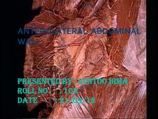
4. Abdominal wall
Bounded superiorly by : xiphoid process &
costal margins.
Posteriorly by : vertebral column.
Inferiorly by : upper parts of pelvic bones.
5. Anterolateral Muscles
o There are 5 muscles in the anterolateral
group of abdominal wall muscles.
o 3 flat muscles whose fibres begin
posterolaterally, pass anteriorly, & are
replaced by an aponeurosis as the muscle
continues towards the midline.
1. External oblique
2. Internal oblique
3. Transversus abdominis
6. o 2 vertical muscles, near the mildline which
are enclosed within a tendinous sheath
formed by aponeuroses of flat muscles.
1. Rectus abdominis
2. Pyramidalis.
7. 1. External oblique muscle:
Most superficial out of the three flat muscles.
Immediately superficial to superficial
fascia.
Its laterally placed muscle fibres pass in an
inferomedial direction,while its large
aponeurotic component covers the anterior part
to the midline.
At the midline, aponeuroses are entwined
forming the linea alba, which extends from
xiphoid process to pubic symphysis.
8. 2. Internal oblique muscle
Deep to external oblique muscle.
2nd of the 3 flat muscles.
Most of its muscle fibres pass in a
superolateral direction.
Its lat muscular components end anteriorly as
an aponeurosis that blends into linea alba at
the midline.
9. 3. Transversus abdominis
Deep to internal oblique.
Ends in an anterior aponeurosis, which blends
with the linea alba at the midline.
Transversalis fascia
Each of the three flat muscles is covered on
the ant & post surfaces by a layer of deep
fascia. Only the layer deep to the
transversus abdominis is remarkable.
10. Vertical muscles
1. Rectus abdominis
long, flat muscle and extends the length of the
anterior abd wall.
It is a paired muscle, separated in the midline by
linea alba.
It widens & thins as it ascends from the pubic
symphysis to costal margin.
11. 2. Pyramidalis muscle
2nd vertical muscle.
This small, triangular muscle which may be
absent, is anterior to the rectus abdominis,
has its base on the pubis, and apex is
attached superiorly & medially to the linea
alba.
12. Rectus Sheath
Aponeurotic sheath covering the rectus
abdominis.
Completely encloses the upper ¾ of rectus
abdominis & covers the anterior surface of
lower ¼ of the muscle
The posterior surface of lower quarter of
rectus abdominis is in direct contact with the
transversalis fascia.
13. Innervation
The skin, muscles and parietal peritoneum of
the anterolateral abdominal wall are supplied
byT7 toT12 and L1 spinal nerves.
NervesT7 toT9 supply skin from xiphoid
process to just above umbilicus.
T10 supplies skin around umbilicus.
T11,T12 &L1 supply skin from just below the
umbilicus to, and including the pubic region.
14. Arterial supply & venous
drainage.
Superficially :
1) Musculophrenic artery
2) Superficial epigastric artery
& Superficial circumflex iliac.
At a deeper level :
1) Sup. Epigastric artery
2)10th & 11th intercostal
arteries & subcostal artery
3) Inf. Epigastric artery
Veins of similar names are reponsible for
venous drainage.
15. Lymphatic drainage
Superficial lymphatics :
1) Axillary nodes
2) Superficial inguinal nodes
Deep lymphatic drainage follows the deep
arteries back to parasternal nodes along with
internal thoracic artery.
18. 3) Paraumbilical hernia
Loop of intestine protrude through the linea
alba around the region of umbilicus.
19. 4) Femoral hernia
Occurs more in females due to larger pelvis,
smaller blood vessels and larger femoral
canal.
Surgery is essential for its treatment.
22. 6) Incisional hernia
Occurs through the anterolateral abdominal
wall when incisions are made for the surgery,
involving cutting of spinal nerves.
23. •Internal Hernia
Remnants of the vitellointestinal duct may
form a tumour at the umbilicus (raspberry red
tumour)
Persistance of a patent vitellointestine duct
results in a faecal fistula at the umbilicus.
24. Ventral hernia
Supraumbilical median incisions through
the linea alba have various advantages
as being
1) Bloodless
2) Safety to muscles & nerves
However, It tends to leave a
postoperative weakness through which
a ventral hernia may develop.
25. Infraumbilical Median Incision
Safer, because the close approx. of recti
prevents formation of any ventral hernia.
Paramedian incisions are more sound than
median incisions.
The rectus muscle is retracted laterally to
protect the nerves supplying it from any
injury.