Cephalometrics
•
12 likes•3,097 views

The Indian Dental Academy is the Leader in continuing dental education , training dentists in all aspects of dentistry and offering a wide range of dental certified courses in different formats.for more details please visit www.indiandentalacademy.com

Recommended
More Related Content
What's hot
What's hot (20)
Viewers also liked
Viewers also liked (9)
Similar to Cephalometrics
Similar to Cephalometrics (20)
More from Indian dental academy
More from Indian dental academy (20)
Recently uploaded
Recently uploaded (20)
Cephalometrics
- 2. CEPHALOMETRICS Cephalometric radiography:- is the production of skull radiographs ,which are useful in making measurements of the cranium and oro-facial complex. PACINI- IN 1922 DEMONSTRATED THE BASIC PROCEDURE OF CEPHALOMETRICS. It was in 1931, HOFRATH in GERMANY and BROADBENT in UNITED STATES published articles in which they had refined the technique and applied these principles to orthodontics.www.indiandentalacademy.com
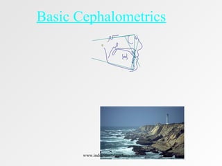
- 3. 15"15"60"60" Source PlaneSource Plane X-ray SourceX-ray Source Patient in Head Positioning Device Patient in Head Positioning Device Mid-saggital Plane Mid-saggital Plane Film PlaneFilm Plane X-ray Film in Cassette X-ray Film in Cassette CephalostatCephalostat www.indiandentalacademy.com
- 4. Cephalometrics is a technique employing oriented radiographs for the purpose of making head measurements. Purpose of CephalometricsPurpose of Cephalometrics •Study craniofacial growthStudy craniofacial growth •DiagnosisDiagnosis •Planning orthodontic treatmentPlanning orthodontic treatment •Evaluation of treated casesEvaluation of treated cases www.indiandentalacademy.com
- 5. Cephalometrics is a technique employing oriented radiographs for the purpose of making head measurements. Purpose of Cephalometrics • Study craniofacial growth • Diagnosis • Planning orthodontic treatment • Evaluation of treated cases www.indiandentalacademy.com
- 6. 15"15"60"60" Source PlaneSource Plane X-ray SourceX-ray Source Patient in Head Positioning Device Patient in Head Positioning Device Mid-saggital PlaneMid-saggital Plane Film PlaneFilm Plane X-ray Film in Cassette X-ray Film in Cassette Cephalostat www.indiandentalacademy.com
- 9. White Black Israeli Chinese Japanese SNA 82 85 82 82 81 SNB 80 81 78 79 77 ANB 2 4 4 3 4 U1-NA 4 mm, 22 7 mm, 23 5 mm, 24 5 mm, 24 6 mm, 24 L1-NB 4 mm, 25 10 mm, 34 6 mm, 29 6 mm, 27 8 mm, 31 U1-L1 131 119 124 126 120 GoGn-SN 32 32 35 32 34 L1-MnPl 93 100 93 93 96 L1-FH 62 51 57 57 57 Y axis 61 63 61 61 62 from Proffit,Contemporary Orthodontics, 1992 Cephalometric Values for Selected Groups www.indiandentalacademy.com
- 10. What Are We Trying to Accomplish? • Find out skeletal classification – anteroposterior – vertical • Find out angulation of incisors • Consider soft tissue – facial profile – airway considerations www.indiandentalacademy.com
- 11. What Are We Trying to Accomplish? (In other words) • Is the patient Class I, II, III skeletal? • Does the patient have a skeletal open bite growth pattern, or a deep bite growth pattern, or a normal growth pattern? • Are the maxillary/mandibular incisors proclined, retroclined or normal? • Is the facial profile protrusive, retrusive, or straight; can the patient breathe normally? www.indiandentalacademy.com
- 14. USES OF CEPHALOMETRICS 1. semi-longitudinal growth studies of population. Study of growth & development : - Cephalograms are used for longitudinal,cross-sectional and – --- By superimposing two ceph.radiographs taken over a period of time will help us in knowing the amount and direction of growth. 2..Diagnosis of a case 3.Treatment planning. 4.Prognosis 5.It serves as a record. www.indiandentalacademy.com
- 15. 6.To differentiate the facial types. 7.Study cranio-facial abnormalities. 8.Growth prediction. 9.To study facial asymmetry. 10.For studying the soft tissue morphology. -using >RICKETS STEINER’S HOLDAWAY’S BURSTONE etc., www.indiandentalacademy.com
- 16. Limitations of cephalometrics 1. It gives a 2 dimensional view of a three dimensional object. 2. Reliability of cephalometrics is not always accurate, as there can be errors in identifying the landmarks or tracing etc. www.indiandentalacademy.com
- 19. CEPHALOMETRICS • DEFINITION • Scientific study of the measurement of the head. • CEPHALOMETRIC RADIOGRAPHY is a standardized method of production of skull radiographs,which are useful in making measurements of the cranium and the orofacial complex.The radiograph thus obtained is called a cephalogram. www.indiandentalacademy.com
- 20. DISCOVERED BY- • IN 1931,HOFRATH IN GERMANY AND BOARDBENT IN U.S.A • Provided a standardized cephalometric technique using a high power x-ray machine and a head holder called a CEPHALOSTAT. www.indiandentalacademy.com
- 21. TYPES OF CEPHALOGRAMS • LATERAL FRONTAL www.indiandentalacademy.com
- 22. WHY CEPHALOMETRICS? • Aids in orthodontic diagnosis by enabling the study of skeletal,dental and soft tissue structures of the craniofacial region. • Aids in establishing the facial type. • Helps in the classification of skeletal and dental abnormalities. • Helps in treatment planning. www.indiandentalacademy.com
- 23. • Aids in evaluating the treatment results and recognizing changes brought about by treatment. • Aids in predicting growth changes and changes associated with surgical treatment. • Study of relapse in orthodontics. • Valuable aid in research work. www.indiandentalacademy.com
- 24. OBTAINING A CEPHALOGRAM • CEPHALOMETRIC EQUIPMENT • CEPHALOSTAT,X-RAY SOURCE & A CASETTE HOLDER. • Cephalostat-2 ear rods-prevent movement of the head in the horizontal plane. • Vertical stabilization of the head-orbital pointer that contacts the lower border of the left orbit. www.indiandentalacademy.com
- 25. • The upper part of the face is supported with the help of a forehead clamp which is positioned above the region of the nasal bridge. • The distance between the x-ray source and the midsagittal plane of the patient is 5 feet. www.indiandentalacademy.com
- 26. CEPHALOMETRIC LANDMARKS • LANDMARK-Is a point serving as a guide for measurement.An ideal landmark is located reliably on the skull and behaves consistently during growth. • It should not be assumed that all the landmarks are equally reliable and valid. • The reliability,reproducibility and dependability of a landmark is affected by-www.indiandentalacademy.com
- 27. #The quality of the cephalogram. #The experience of the tracer. #Confusion with other landmarks. The cephalometric landmarks should have the following attributes- A) Should be easily seen on the radiograph. B)Uniform in outline. www.indiandentalacademy.com
- 28. • C)Should be easily reproducible. • D)Landmarks should permit valid quantitative and qualitative measurements of lines and angles projected from them. • E)Measurements should be amenable to statistical analysis. www.indiandentalacademy.com
- 30. CLASSIFICATION OF LANDMARKS • 1)ANATOMIC LANDMARKS • 2)DERIVED LANDMARKS • 3)UNILATERAL LANDMARKS • 4)BILATERAL LANDMARKS • 5)IMPLANTS www.indiandentalacademy.com
- 31. • ANATOMIC LANDMARKS • They represent actual anatomic structures on the skull e.g. ANS,Na • DERIVED LANDMARKS • These are obtained secondarily from anatomic structures in a cephalogram.e.g.Ar(Articulare),Ptm(Pterygom axillary fissure) www.indiandentalacademy.com
- 32. • IMPLANTS • They are artificially inserted radio opaque markers, usually made of inert metal. • They are ‘PRIVATE POINTS’ and their position can vary from subject to subject. • They are ideal for longitudinal studies on the same subject. www.indiandentalacademy.com
- 33. UNILATERAL LANDMARKS • NASION-The most anterior point midway between the frontal and the nasal bones in the frontonasal suture. www.indiandentalacademy.com
- 34. ANTERIOR NASAL SPINE/ANS • It is the tip of the sharp bony process of the maxilla in the midline. www.indiandentalacademy.com
- 35. PROSTHION • The lowest and the most anterior point on the alveolar process in the median plane between the central incisors. www.indiandentalacademy.com
- 36. SUBSPINALE/POINT A • It is the deepest point in the midline between the ANS and the alveolar crest, between the two central incisors. It is also called as subspinale. Pink dot-pt.Awww.indiandentalacademy.com
- 37. INFRADENTALE/(Id) • The highest and the most anterior point in the alveolar bone in the midline between the lower central incisors. Blue dot-(Id) www.indiandentalacademy.com
- 38. SUPRAMENTALE/Pt.B • It is the deepest point in the midline between the alveolar crest and the mental process. Pink dot-pt.Bwww.indiandentalacademy.com
- 39. POGONION(Pog) • It is the most anterior point of the bony chin in the median plane. Red dot-(Pog) www.indiandentalacademy.com
- 40. MENTON(Me) • It is the most inferior midline point on the mandibular symphysis. Yellow dot-Me www.indiandentalacademy.com
- 41. GNATHION(Gn) • It is the most antero -inferior point on the symphysis of the chin. It is constructed by intersecting a line drawn perpendicular to the line connecting menton and pogonion. Orange dot-(Gn) www.indiandentalacademy.com
- 42. BASION(Ba) • It is the median point on the anterior margin of foramen magnum. www.indiandentalacademy.com
- 43. POSTERIOR NASAL SPINE(PNS) • The most posterior point in the bony hard palate in the sagittal plane. • Marks the distal limit of the maxilla. www.indiandentalacademy.com
- 44. SELLA(S) • The point representing the midpoint of sella tursica. www.indiandentalacademy.com
- 45. • GLABELLA:It is the most prominent point of the forehead in the mid-saggital plane. • SUBNASALE:The point where the lowest border of the nose meets the outer contour of the upper lip. ●G ●Sn www.indiandentalacademy.com
- 46. BILATERAL LANDMARKS • 1)ORBITALE(Or) • The lowest point on the inferior bony margin of the orbit. www.indiandentalacademy.com
- 47. G0NION • It is a constructed point at the junction of ramal plane and mandibular plane. www.indiandentalacademy.com
- 48. CONDYLION/(Co) • The most superior point on the head of condyle. www.indiandentalacademy.com
- 49. ARTICULARE/(Ar) • It is a point at the junction of the posterior border of the ramus and inferior border of the basal part of the occipital bone. Blue dot-(Ar) www.indiandentalacademy.com
- 50. PTERYGOMAXILLARY POINT/Ptm • It is the intersection of the inferior border of foramen rotundum with the posterior wall of pterygomaxillary fissure. • It is a bilateral tear drop shaped area of radiolucency. www.indiandentalacademy.com
- 51. PORION/(Po) • The highest bony point on the upper margin of the external auditory meatus. www.indiandentalacademy.com
- 52. BOLTON POINT • The highest point at the posterior condylar notch of the occipital bone. Bo www.indiandentalacademy.com
- 53. • THE KEY RIDGE-The lowest most point on the contour of the anterior wall of the infratemporal fossa. • CHELION:It is the lateral terminus of the oral slit on the outer corner of the mouth. www.indiandentalacademy.com
- 54. LINES AND PLANES IN CEPHALOMETRY • Cephalometrics makes use of certain lines or planes. These lines are obtained from connecting two landmarks. • Based on their orientation the lines or planes are classified into: • Horizontal and vertical planes. www.indiandentalacademy.com
- 55. HORIZONTAL PLANES • 1)S.N.PLANE- It is the cranial line between the center of sella tursica and the anterior point of the fronto nasal suture(nasion). • It represents the anterior cranial base. www.indiandentalacademy.com
- 56. FRANKFORT HORIZONTAL PLANE • This plane connects the lowest point of the orbit(orbitale)and the superior point of the external auditary meatus(porion). www.indiandentalacademy.com
- 57. OCCLUSAL PLANE • It is a denture plane bisecting the posterior occlusion of the permanent molars and premolars(or deciduous molars in mixed dentition)and extends anteriorly. www.indiandentalacademy.com
- 58. MANDIBULAR PLANE • Several mandbiular planes are used in cephalometrics,based on the analysis being done. The most commonly used ones are- • TWEEDS-Tangent to the lower border of the mandible. • STEINERS-A line connecting gonion and gnathion. • DOWNS-A line connecting gonion and menton. www.indiandentalacademy.com
- 59. PALATAL PLANE • It is a line linking the anterior nasal spine of the maxilla and the posterior nasal spine of the palatine bone. www.indiandentalacademy.com
- 60. BASION-NASION PLANE • Line connecting the basion and nasion. • Represents the cranial base. www.indiandentalacademy.com
- 61. VERTICAL PLANE • A-POG LINE: It is a line from point A on the maxilla to pogonion on the mandible. www.indiandentalacademy.com
- 62. FACIAL PLANE • It is a line from the anterior point of the frontonasal suture (nasion)to the most anterior point of the mandible(pogonion). www.indiandentalacademy.com
- 63. BOLTON’S PLANE • This is a plane that connects the Bolton's points posterior to the occipital condyles and nasion. Bo www.indiandentalacademy.com
- 64. FACIAL AXIS • It is point from Ptm point to cephalometric gnathion. www.indiandentalacademy.com
- 65. ESTHETIC PLANE/E PLANE • Is a line between the most anterior point of the soft tissue nose and the soft tissue chin. www.indiandentalacademy.com
- 67. FRONTAL CEPHALOGRAM (P-A VIEW) This provides an antero-posterior view of the skull. www.indiandentalacademy.com
- 69. The goal of lateral ceph Analysis is to establish the Antero-posterior and Vertical relationships of the Five major cranio-facial units. 1.The cranium and cranial base 2.The naso-maxillary complex 3.The Mandible 4.The Maxillary dentition 5.The Mandibular dentition. www.indiandentalacademy.com
- 70. STEINERS ANALYSIS By Cecil C. STEINER------1960 – Selected what he considered to be the most meaningful parameters and evolved a composite analysis,which he believed would provide the maximum clinical information with the least number of measurements. Certain measurements were then selected and the means (or ) Averages were determined on a series of individuals with normal Occlusions. www.indiandentalacademy.com
- 71. Steiner’s analysis is divided into 3 parts:- 1. Skeletal Analysis 2. Dental Analysis 3. Soft tissue analysis www.indiandentalacademy.com
- 79. Soft Tissue Analysis ‘S-LINE’ : According to Steiner the lips in a well balanced faces Should touch a line extending from the soft tissue contour of the Chin to the middle of the ‘S’ formed by the lower border of the Nose. THIS LINE IS REFERRED TO AS THE ‘S’ LINE. www.indiandentalacademy.com
- 80. THE ‘S’ LINE STEINER’S ‘S’ LINE www.indiandentalacademy.com
- 84. ‘’WE CALL OUR LANGUAGE THE MOTHER TONGUE BECAUSE THE FATHER SELDOM GETS TO SPEAK.’’ www.indiandentalacademy.com
- 88. DOWN’S ANALYSIS DOWN’S :-Study based on 20 Caucasian subjects ,ranged In age from 12 to 17 years of age and were equally divided As to sex. All the individuals possessed clinically excellent occlusions. -1952 www.indiandentalacademy.com
- 89. DOWN –divided his analysis into 2 components---- 1. The skeletal component helped in defining the underlying facial type . 2. The dental component is used to establish if the dentition is placed normally in relation to the underlying bony structures. www.indiandentalacademy.com
- 93. Y-AXIS (GROWTH AXIS) Angle formed by joining Sella-gnathion line with The F-H plane. Mean value- - 59degrees www.indiandentalacademy.com
- 96. DOWN’S POLYGON The graph divided into:- 1.Skeletal pattern on the top half of the graph 2.Denture polygon on the lower half of the graph. Wigglegram:- helps us in Visualizing the type of malocclusion I.e, Skeletal or dental.. www.indiandentalacademy.com
- 99. A functional occlusal plane is drawn through the overlapping Cusps of first pre-molars and first molars. Perpendiculars are drawn to the occlusal plane from points A and B . The points of contact of these perpendiculars on the occlusal Plane are termed AO and BO. The distance between points AO and BO gives the antero- Posterior relation between the two jaws,. www.indiandentalacademy.com
- 102. TWEED’S TRIANGLE TWEED’S DIAGNOSTIC FACIAL TRIANGLEwww.indiandentalacademy.com
- 103. TWEED- developed this analysis as an aid to: •Anchorage preparation •To treatment planning •And determining the prognosis of orthodontic cases Tweed’s analysis is based primarily on the deflection of the mandible as measured by the Frankfurt Mandibular plane Angle(FMA) and the posture of the lower incisor. It is done to determine the final position the lower incisor Should occupy at the end of the treatment. Dr.Tweed established that prognosis could be predicted relatively accurately based on the configuration of the triangle www.indiandentalacademy.com
- 104. Tweeds triangle is formed by: 1. Frankfurt horizontal plane 2. The mandibular plane 3. The long axis of lower incisor.s The three angle’s formed are: • Frankfort-Mandibular Plane(FMA) Angle---- • FH Plane with mandibular plane. • Lower Incisor to Mandibular plane(IMPA)Angle----- • Long axis of lower incisor with mandibular plane. • Frankfort Mandibular Incisor Angle (FMIA)-------- • Long axis of lower incisor with FH plane. FMA=25degrees IMPA=90degrees FMIA=65degrees.www.indiandentalacademy.com
- 105. Based on the FMA angle the prognosis can be predicted:- 1. FMA 16 to 28 degrees- prognosis good Apprx. 60% of malocclusions have FMA between 16 to 28 2.FMA from 28 to 35 degrees prognosis fair. Extractions necessary in majority of cases 3.FMA above 35 degrees , prognosis bad ,,extractions frequently complicate the problem. www.indiandentalacademy.com
- 106. OTHER IMPORTANT CEPHALOMETRIC ANALYSIS McNamara's ANALYSIS RAKOSI’S ANALYSIS GRUMMON’S –FRONTAL ASYMMETRY ANLYSIS SCHWARZ ANALYSIS NHP –ANALYSIS BURSTONE’S-HARD TISSUE ANALYSIS(COGS) BURSTONE’S –SOFTY TISSUE ANALYSIS(COGS) HOLDAWAY’S SOFT TISSUE ANALYSIS ARNETT-SOFT TISSUE CEPH.ANLYSIS(STCA) www.indiandentalacademy.com