
Recommended
More Related Content
What's hot
What's hot (20)
Similar to Coarctation - Wetzel
Similar to Coarctation - Wetzel (20)
Recently uploaded
Recently uploaded (20)
Coarctation - Wetzel
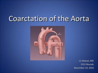
- 1. Coarctation of the AortaCoarctation of the Aorta Liz Wetzel, MDLiz Wetzel, MD CICU RoundsCICU Rounds November 19, 2010November 19, 2010
- 2. ObjectivesObjectives • Review Anatomy/Lesion DevelopmentReview Anatomy/Lesion Development • Discuss DR Presentation and ManagementDiscuss DR Presentation and Management • Review Post-natal Evaluation and TransportReview Post-natal Evaluation and Transport • Describe possible presentation in non-prenatallyDescribe possible presentation in non-prenatally diagnosed cases and timing of duct closurediagnosed cases and timing of duct closure • Discuss potential CICU Course and outcomesDiscuss potential CICU Course and outcomes
- 3. Development of the aortic archDevelopment of the aortic arch The left fourth arch vessel becomes the arch of the aorta. The left 6The left fourth arch vessel becomes the arch of the aorta. The left 6thth becomes part of the left pulmonary artery and the ductus arteriosis.becomes part of the left pulmonary artery and the ductus arteriosis. Sadler, TW. Langman’s Medical Embryology 8th edition. Philadelphia: Lippincott Williams& Wilkins,2000: 239-243.
- 4. Development continuedDevelopment continued Gittenberger-De Groot, A.C. Bartelings, M.M. Deruiter, M.C. Poelmann, R.E. Basics of Cardiac Development for the Understanding of Congenital Heart Malformations. Pediatric Research. 2005; 57 (2): 169-176. Molin, D. DeRuiter, M.C, Wisse, L.J, Azhar, M., Doetschman, T., Poelmann, R. E., Gittenberger-de Groot, A. C. Altered apoptosis pattern during pharyngeal arch artery remodeling is associated with aortic arch malformations in Tgfβ2 knock-out mice. Cardiovascular Research. 2002; 56: 312-322.
- 5. Development of CoarctationDevelopment of Coarctation • Abnormal development ofAbnormal development of left 4left 4thth and 6and 6thth aortic archesaortic arches • Represents 5-10% of allRepresents 5-10% of all congenital cardiac lesionscongenital cardiac lesions • More common in boys thanMore common in boys than girlsgirls • No real impact prior to birthNo real impact prior to birth due to presence of PDAdue to presence of PDA unless there is fetal closureunless there is fetal closure
- 6. Ductus Tissue TheoryDuctus Tissue Theory • Due to a migration ofDue to a migration of ductus smooth muscle cellsductus smooth muscle cells into the periductal aortainto the periductal aorta with subsequentwith subsequent constriction and narrowingconstriction and narrowing of the aortic lumenof the aortic lumen • Evident when ductus closesEvident when ductus closes Hemodynamic TheoryHemodynamic Theory • Reduced intrauterineReduced intrauterine blood flow causesblood flow causes underdevelopment ofunderdevelopment of aortic archaortic arch • Results from reducedResults from reduced volume of blood flowvolume of blood flow through the fetal aorticthrough the fetal aortic arch and isthmusarch and isthmus
- 7. Other TheoriesOther Theories • May be due to a defect in the vascular wall of theMay be due to a defect in the vascular wall of the ascending aortaascending aorta • Vascular apoptosis may have a roleVascular apoptosis may have a role (Molin et al 2002)(Molin et al 2002) • Recessive genetic mutation found in zebrafishRecessive genetic mutation found in zebrafish (Weinstein et(Weinstein et al 1995)al 1995) • Autosomal dominant inheritance of non-syndromic leftAutosomal dominant inheritance of non-syndromic left ventricular outflow tract obstructionventricular outflow tract obstruction (Wessels et al 2005)(Wessels et al 2005)
- 8. DR Presentation & ManagementDR Presentation & Management • Follow normal NRP guidelines for resuscitation and beFollow normal NRP guidelines for resuscitation and be sure to have a stable airwaysure to have a stable airway • This is not a lesion where you would expect acuteThis is not a lesion where you would expect acute delivery room decompensationdelivery room decompensation • Should not be a blue baby due to the heart defectShould not be a blue baby due to the heart defect • Admit to the NICUAdmit to the NICU
- 9. Management in the NICUManagement in the NICU • Support the airway (intubation if necessary)Support the airway (intubation if necessary) • Echocardiogram and CXREchocardiogram and CXR • Ideally establish umbilical accessIdeally establish umbilical access • PGEPGE11 infusioninfusion (0.03 to 0.05 mcg/kg/min)(0.03 to 0.05 mcg/kg/min) • Correct acidosis and electrolyte abnormalitiesCorrect acidosis and electrolyte abnormalities • Blood pressure support as indicatedBlood pressure support as indicated • Main goal is to stabilize the patient and get themMain goal is to stabilize the patient and get them transferred to a cardiac intensive care unittransferred to a cardiac intensive care unit
- 10. Transport Issues and Hand-off to cardsTransport Issues and Hand-off to cards • Be confident you have a stable airwayBe confident you have a stable airway – Risk of apnea with PGERisk of apnea with PGE11 infusion, consider caffeine?infusion, consider caffeine? • Have secure IV access with fluids runningHave secure IV access with fluids running – PGEPGE11 infusion can cause vasodilatation and can result ininfusion can cause vasodilatation and can result in relative hypovolemia in neonatesrelative hypovolemia in neonates • Report recent blood gas with electrolytes including ionizedReport recent blood gas with electrolytes including ionized calcium and potentially a lactatecalcium and potentially a lactate • Full set of vitals including 4 extremity blood pressuresFull set of vitals including 4 extremity blood pressures
- 11. Undiagnosed Coarctation PresentationUndiagnosed Coarctation Presentation • Decreased or absent femoral pulses,Decreased or absent femoral pulses, tachypnea, grunting, poor feeding,tachypnea, grunting, poor feeding, signs of CHF, abnormal 4 extremitysigns of CHF, abnormal 4 extremity blood pressuresblood pressures • If coming from home can present to ED in shock with multi-If coming from home can present to ED in shock with multi- organ dysfunction and severe metabolic acidosisorgan dysfunction and severe metabolic acidosis • CXR with cardiomegaly, pulmonary congestionCXR with cardiomegaly, pulmonary congestion Sharland, G.K, Chan, KY, Allen, LD. Coarctation of the aorta: difficulties in prenatal diagnosis. British Heart Journal. 1994; 71: 70-75. http://www.heartonline.org/congenital.htm
- 12. Potential CXR ProgressionPotential CXR Progression DAY FIVE OF LIFEDAY FIVE OF LIFEDAY THREE OF LIFEDAY THREE OF LIFE
- 13. Closure of the ductClosure of the duct • Functional and Anatomic Closure of DuctFunctional and Anatomic Closure of Duct – Closure occurs in three steps:Closure occurs in three steps: 1.1.constriction of ductal smooth muscle;constriction of ductal smooth muscle; 2.2.hypoxia/ischemia of medial smooth muscle;hypoxia/ischemia of medial smooth muscle; 3.3.remodeling resulting in permanent closure (Koch et. alremodeling resulting in permanent closure (Koch et. al 2006)2006) •In term infantsIn term infants functional closurefunctional closure can occur as early ascan occur as early as 12-1512-15 hours of age,hours of age, if greater than 72 hours it isif greater than 72 hours it is considered persistent,considered persistent, truetrue anatomicanatomic closureclosure can takecan take weeksweeks Neoreviews Controversies in the Management of PDA (Gien 2008)
- 14. Timing of Ductal ClosureTiming of Ductal Closure • In >95% of neonates >1500g closure usually begins within 96 hours (Koch et. al 2006) • Spontaneous closure occurs in >34% of ELBW neonates (Koch et. al 2006)
- 15. Potential surgical interventionPotential surgical intervention • First surgery was done experimentally in animals in 1944First surgery was done experimentally in animals in 1944 – Blalock and Park– Blalock and Park • 1. Resection with end-to-end anastomosis1. Resection with end-to-end anastomosis • 2. Patch aortoplasty2. Patch aortoplasty • 3. Left subclavian patch aortoplasty3. Left subclavian patch aortoplasty • 4. Bypass grafts between ascending and descending4. Bypass grafts between ascending and descending aortaaorta Rothman, Abraham. Coarctation of the Aorta: An Update. Current Problems in Pediatrics. 1998; 37-60.
- 16. Cincinnati Children’s ExperienceCincinnati Children’s Experience • Preferred approach here is end-to-end anastomosisPreferred approach here is end-to-end anastomosis • Most important determinant of outcome is how fast it isMost important determinant of outcome is how fast it is detected and how soon they head to the ORdetected and how soon they head to the OR • Typically in the OR within 12 hours of admission to CICUTypically in the OR within 12 hours of admission to CICU • Usual length of stay is 2 days in CICU and a total of 5Usual length of stay is 2 days in CICU and a total of 5 days in the hospitaldays in the hospital (unless very sick prior to OR)(unless very sick prior to OR) – Less than 5% need re-interventionLess than 5% need re-intervention Courtesy of Dr. Angela Lorts; Cardiac Critical Care Staff; Cincinnati Children’s Heart Institute
- 17. Surgical outcomesSurgical outcomes • Acute mortality ranged from 3% to 32%, strongly correlated withAcute mortality ranged from 3% to 32%, strongly correlated with complexity of associated cardiovascular lesionscomplexity of associated cardiovascular lesions • Lowest in those with isolated coarcation (<2%)Lowest in those with isolated coarcation (<2%) • Restenosis rate was 3-41%Restenosis rate was 3-41% Rothman, Abraham. Coarctation of the Aorta: An Update. Current Problems in Pediatrics. 1998; 37-60.
- 18. Survival DataSurvival Data • Quaegegbeur et alQuaegegbeur et al. reported on a multi-institutional study that looked. reported on a multi-institutional study that looked at 326 severely symptomatic neonates with coarctation and with orat 326 severely symptomatic neonates with coarctation and with or without VSD.without VSD. • The 1 month survival was 93% and the 24 month survival was 84%.The 1 month survival was 93% and the 24 month survival was 84%. Quaegebeur, J.M, Jonas, R.A, Weinberg, A.D, Blackstone, E.H, Kirklin, J.W. Outcomes in seriously ill neonates with coarctation of the aorta, A multiinstitutional study. The Journal of Thoracic and Cardiovascular Surgery. 1994; 108: 841-854.
- 19. Post-operative complicationsPost-operative complications • HoarsenessHoarseness • Ipsilateral diaphragm paralysisIpsilateral diaphragm paralysis • ChylothoraxChylothorax • Vessel injury/bleedingVessel injury/bleeding • Rebound HTNRebound HTN • Post-coartectomy syndromePost-coartectomy syndrome • Paralysis due to spinal cord ischemiaParalysis due to spinal cord ischemia
- 20. Long term complicationsLong term complications • Re-stenosis: influenced by presence of residualRe-stenosis: influenced by presence of residual ductal tissue within the aortaductal tissue within the aorta • Hypertension: more likely in repair at a later ageHypertension: more likely in repair at a later age • Neurologic abnormalitiesNeurologic abnormalities – Ultrasound abnormalitiesUltrasound abnormalities – microcephalymicrocephaly
- 21. Neurologic abnormalitiesNeurologic abnormalities Preoperative neurobehavioral abnormalities: abnormalPreoperative neurobehavioral abnormalities: abnormal tone, posturing, weak cry, poor suck, poor auditory andtone, posturing, weak cry, poor suck, poor auditory and visual orientingvisual orienting Abnormal ultrasound findings: ventriculomegaly, IVH, basalAbnormal ultrasound findings: ventriculomegaly, IVH, basal ganglia calcification, widened subarachnoid spaces foundganglia calcification, widened subarachnoid spaces found preoperativelypreoperatively Limperopoulos C, Majnemer A, Shevell M, Rosenblatt, Rohlicek C, Tchervenkov C. Neurologic Status of Newborns With Congenital Heart Defects Before Open Heart Surgery.Pediatrics. 1999; 103(2): 402-408.
- 22. THANK YOU TO MY ADVISOR DR. KRAWCZESKI ANY QUESTIONS?????
- 23. References • Brouwer, R.M, Erasmsus, M.E, Ebels, T, Eijgelaar, A. Influence of age on sruvival, late hypertension, and recoarctation in elective aortic coarctationBrouwer, R.M, Erasmsus, M.E, Ebels, T, Eijgelaar, A. Influence of age on sruvival, late hypertension, and recoarctation in elective aortic coarctation repair: Including long-term results after elective aortic coarctation repair with a follow-up from 25 to 44 years.repair: Including long-term results after elective aortic coarctation repair with a follow-up from 25 to 44 years. The Journal of Thoracic andThe Journal of Thoracic and Cardiovascular SurgeryCardiovascular Surgery. 1994; 108: 525-531.. 1994; 108: 525-531. • Chang RK, Gurvitz M, rodriguez S. Missed Diagnosis of Critical Congenital Heart Disease.Chang RK, Gurvitz M, rodriguez S. Missed Diagnosis of Critical Congenital Heart Disease. Arch Pediatr Adolesc Med.Arch Pediatr Adolesc Med. 2008; 162(10): 969-974..2008; 162(10): 969-974.. • De-Wahl Granelli, A, Mellander M, Sunnegardh J, Sandberg K, Ostman-Smith I. Screening for duct-dependent congential heart diseaswe with pulseDe-Wahl Granelli, A, Mellander M, Sunnegardh J, Sandberg K, Ostman-Smith I. Screening for duct-dependent congential heart diseaswe with pulse oximetry: A critical evaluation of strategies to maximize sensitivity.oximetry: A critical evaluation of strategies to maximize sensitivity. Acta PediatricaActa Pediatrica. 2005;94: 1590-1596. 2005;94: 1590-1596 • Gittenberger-De Groot, A.C. Bartelings, M.M. Deruiter, M.C. Poelmann, R.E. Basics of Cardiac Development for the Understanding of Congenital HeartGittenberger-De Groot, A.C. Bartelings, M.M. Deruiter, M.C. Poelmann, R.E. Basics of Cardiac Development for the Understanding of Congenital Heart Malformations.Malformations. Pediatric Research.Pediatric Research. 2005; 57 (2): 169-176.2005; 57 (2): 169-176. • Johnson BA and Ades A. Delivery Room and Early Postnatal Management of Neonates Who Have Prenatally Diagnosed Congenital Heart DiseaseJohnson BA and Ades A. Delivery Room and Early Postnatal Management of Neonates Who Have Prenatally Diagnosed Congenital Heart Disease. Clinics. Clinics in Perinatologin Perinatology. 2005; 32: 921-946.y. 2005; 32: 921-946. • Limperopoulos C, Majnemer A, Shevell M, Rosenblatt, Rohlicek C, Tchervenkov C. Neurologic Status of Newborns With Congential Heart Defects BeforeLimperopoulos C, Majnemer A, Shevell M, Rosenblatt, Rohlicek C, Tchervenkov C. Neurologic Status of Newborns With Congential Heart Defects Before Open Heart Surgery.Open Heart Surgery.Pediatrics.Pediatrics. 1999; 103(2): 402-408.1999; 103(2): 402-408. • Molin, D. DeRuiter, M.C, Wisse, L.J, Azhar, M., Doetschman, T., Poelmann, R. E., Gittenberger-de Groot, A. C. Altered apoptosis pattern during pharyngeal arch artery remodelling is associated with aortic arch malformations in Tgfβ2 knock-out mice. Cardiovascular Research. 2002; 56: 312-322. • Polin,Fox,Abman. Fetal and Neonatal Physiology 3rd edition. Mechanisms Regulating Closure of the Ductus Arteriosis. Saunders. Pennsylvania 2004: 743-747. • Quaegebeur, J.M, Jonas, RShultz A, Localio A, Clark B, Ravishankar C, Videon N, Kimmel S. Epidemiadn ologic Features of the Presentation of Critical Congenital Heart Disease: Implications for Screening. Pediatrics. 2008;121(4) 751-757. • .A, Weinberg, A.D, Blackstone, E.H, Kirklin, J.W. Outcomes in seriously ill neonates with coarctation of the aorta, A multiinstitutional study. The Journal of Thoracic and Cardiovascular Surgery. 1994; 108: 841-854. • Rothman, Abraham. Coarctation of the Aorta: An Update. Current Problems in Pediatrics. 1998; 37-60.
- 24. References • Sadler, TW. Langman’s Medical Embryology 8th edition. Philedelphia: Lippincott Williams& Wilkins,2000: 239-243. • Sharland, G.K, Chan, KY, Allen, LD. Coarctation of the aorta: difficulties in prenatal diagnosis.Sharland, G.K, Chan, KY, Allen, LD. Coarctation of the aorta: difficulties in prenatal diagnosis. British Heart JournalBritish Heart Journal. 1994; 71: 70-75.. 1994; 71: 70-75. • Weinstein, BM, Stemple, DL, Dreiver W, Fishman, MC. Gridlock, a localized heritable vascular patterning defect in the zebrafish.Weinstein, BM, Stemple, DL, Dreiver W, Fishman, MC. Gridlock, a localized heritable vascular patterning defect in the zebrafish. Nat Med.Nat Med. Nov 1995;Nov 1995; 1(11): 1143-1147.1(11): 1143-1147. • Wessels, M.W, Berger, R, Frohn-Mulder, I, Roos-Hesselink, J.W, Hoogeboom, J, Mancini, G.S, Bartelings, M.M, De Krijger, R, Wladimiroff, J.W,Wessels, M.W, Berger, R, Frohn-Mulder, I, Roos-Hesselink, J.W, Hoogeboom, J, Mancini, G.S, Bartelings, M.M, De Krijger, R, Wladimiroff, J.W, Niermeijer, M.F, Grossfeld, P, Willems, P.J. Autosomal Dominant Inheritance of Left Ventricular Outflow Tract Obstruction.Niermeijer, M.F, Grossfeld, P, Willems, P.J. Autosomal Dominant Inheritance of Left Ventricular Outflow Tract Obstruction. American Journal ofAmerican Journal of Medical GeneticsMedical Genetics. 2005; 134A: 171-179.. 2005; 134A: 171-179. • Zehr K, Gillinov M, Redmond M, Greene PS, Kan J, Gardner TJ, Reitz B, Cameron D. Repair of Coarctation of the Aorta in Neonates and Infants: AZehr K, Gillinov M, Redmond M, Greene PS, Kan J, Gardner TJ, Reitz B, Cameron D. Repair of Coarctation of the Aorta in Neonates and Infants: A Thirty-Year Experience.Thirty-Year Experience. Annals of ThoracicAnnals of Thoracic Surgery. 1995; 59: 33-41.Surgery. 1995; 59: 33-41. • http://www.heartonline.org/congenital.htm • Title page image: www.odlarmad.com
- 25. Survival Data based on AgeSurvival Data based on Age Brouwer, R.M, Erasmsus, M.E, Ebels, T, Eijgelaar, A. Influence of age on sruvival, late hypertension, and recoarctation in elective aortic coarctation repair: Including long-term results after elective aortic coarctation repair with a follow-up from 25 to 44 years. The Journal of Thoracic and Cardiovascular Surgery. 1994; 108: 525-531.
Editor's Notes
- The pharyngeal arches form during the 4 th and 5 th weeks of development. Each arch gets its own cranial nerve and artery-the arteries are the aortic arches. The A.A arise from the aortic sac and are embedded in the mesenchyme of the pharyngeal arches. The 5 th arch either never forms or forms incompletely. The first and second aortic arch vessels form the arteries of the head neck and trunk. The third becomes the common carotid. The right fourth becomes the proximal part of the right subclavian. The left fourth becomes the arch of the aorta . The right 6 th becomes part of the right pulmonary artery. The left 6 th becomes part of the left pulmonary artery and the ductus arteriosis. During the 6 th to 8 th week the primitive pattern is transformed into the adult pattern. In human infants, aortic arch anomalies are distinguished in those related to the B-segment (located between the left carotid and the left subclavian artery) and the A-segment or isthmus (located between the left subclavian artery and the ductus arteriosus). B-segment abnormalities are often seen in relation to the 22q11 deletion syndrome and are thought to relate to neural crest defects. The latter is under discussion since the finding of Tbx1 as a candidate gene (45, 50, 64a). In the human infant, hypoplasia or coarctation of the aorta at the isthmus site is not specifically linked to neural crest–related syndrome or a specific gene.
- The heart tube contacts the dorsal aorta through the first bilateral set of pharyngeal arch arteries. These are followed in time by a 2 nd , 3 rd , 4 th and 6 th set. This system is remodeled into the aortic arch in mammals. Figure 7. Schematic representation of the development of the bilateral pharyngeal arch artery system into a left-sided aortic arch ( a–d ). Special attention is paid to the 4th and 6th arch arteries. The right 6th arch artery disappears during early development only followed by left 6th arch artery(ductus arteriosus) closure after birth. The left and right 4th arch segments normally remain persistent, the left one as part of the aortic arch and the right one as part of the right subclavian artery. The 4th arch artery has morphogenetic characteristics that make it specifically vulnerable for the development of aortic arch abnormalities as, e.g. in the 22q11 deletion syndrome. Molin et. al has reported in Cardiovascular Research using mouse model to look at vascular apoptosis during pharyngeal arch artery remodeling. Their data showed that apoptosis accompanies normal PAA remodeling and that alterations in the process coincide with PAA malformations.
- THERE ARE TWO MAJOR THEORIES ABOUT ETIOLOGY OF COARCTATION: Ductal tissue theory and hemodynamic theory
- Aberrant ductal tissue exists partially or circumferentially in the aortic isthmus and neonatal constriction of this tissue leads to obstruction Problems with this theory: Some coarctation occurs in the presence of a patent ductus and distant from the insertion of the ductus arteriosus Isthmus is the section of aorta between the left subclavian and the aortic end of the ductus arteriosus. Does not explain isolated coarctation without associated intracardiac lesions
- Impaired elastic properties….. TO LOOK AT VASCULAR APOPTOSIS: Molin et al used a knockout mice model that develops aortic arch malformations to show that aberrant apoptosis was demonstrated in both fourth arch arteries. TO LOOK AT THE GENETIC ROLE : Weinstein et al found a recessive mutation gridlock , that causes a focal malformation resembling coarctation in humans. 2. Wessel et al described four new families with presumed AD inheritance of LVOTO (which included some cases of coarctation)
- Rush University Medical Center www.rush.edu/rumc/page-1098987357987.html Graham et. Al showed improvement in systemic blood pressure after starting PGE infusion, paper from 1978. (Direct effect on the vasculature and smooth muscle of ductus arteriosis)
- PGE 1: quickly metabolized, 60 to 80% on first pass through the lungs Other side effects include fever and rash Rarely: gastric outlet obstruction, cortical hyperostosis, leukocytosis and seizures (Johnson and Ades et al. 2005)
- Detection in utero is difficult: Retrospective 10 year study at a tertiary referral center in London. 8000 cases referred for echo. 615 had heart malformations. 54 correctly diagnosed with coarc, 24 suspected but not confirmed by echo, 9 missed that had normal fetal echocardiograms. Some features that suggest the presence of coarctation: enlargement of right ventricle, isthmus and transverse aortic arch diameters less than 3% for gestational age, hypoplasia of left-sided structures, decreased or reversal of flow in the PFO
- Term male, born to mom with pre-eclampsia, was transferred to NICU on day of life 3 with presumed sepsis, respiratory distress
- In many cases it may be when the duct is closing or after the duct has closed that these patients are identified Functional Closure of the lumen : By smooth muscle constriction Anatomic Occlusion of the lumen : Over the next several days due to extensive neointimal thickening and loss of smooth muscle cells from the inner muscle media. Increased intimal thickening and fragmentation of the internal elastic lamina after delivery. Constriction produces ischemic hypoxia of the vessel wall, inhibits local PGE2 and NO production and produces smooth muscle apoptosis and further remodeling. Increase in arterial partial pressure of oxygen (oxygen constricts in ductus) Decrease in circulating PGE2 (inc in PGE removal from lung and loss of production from placenta) Decrease in blood pressure within duct lumen due to decrease in pulmonary vascular resistance Decrease in number of PGE2 receptors in the ductus wall
- 50% of ELBW neonates were closed by about 3 days and 100% of those by 8 post-natal days (of the ones that would close) It is delayed in premature infants due to the decrease intrinsic tone of the extremely immature ductus, elevated circulating concentrations of PGE2 due to the decreased ability of the premature lung to clear PGE2, and increased sensitivity to PGE2 and NO not due to increased receptors but rather due to enhanced coupling b/w receptors and downstream pathways.
- Left subclavian is turned down and used to enlarge the narrowing. They don’t typically develop respiratory problems until they are really sick. During surgery patients have a period of cross clamping the aorta. Bypass is usually unnecessary as patients tend to tolerate the cross clamping in part due to upper to lower body collaterals. Usually done by a left thoracotomy approach but may be anterior sternotomy if indicated.
- The preferred approach here is resection with end-to-end anastomosis. The most important determinant of their outcomes is how fast it is detected and how soon they are operated on. They develop end organ dysfunction quickly after their duct closes. The typical course is a trip to the OR within 12 hours of admission to the CICU.
- Surgical coarctation series published over the prior decade, each with >100 patients. The highest mortality rate affected patients with very complex lesions, intermediate (5-15%) for those with coexisting VSD, and was lowest (<2%) in those with isolated coarctation. Overall neonates do well as long as there are not other associated cardiac lesions.
- Cincinnati Children’s was one of the 27 participating centers for this study. 435 neonates entered the 27 institutions, 171 had isolated coarctation, 155 had associated VSD and 109 had coarctation with other major congenital cardiac anomalies. Time period was 1990-1992. Most infants were on PGE’s, intubated and some required blood pressure support. Mean BW was 2.97kg (1.61-4.04), median age was 6 days (0-23 days), and median time before first procedure was 3 days (0-17 days).
- Hoarseness: damage to recurrent laryngeal nerve as it loops around PDA Phrenic nerve damage Thoracic duct damage as it crosses behind the aorta HTN: baroreceptor mediated increase in sympathetic activity and reflex vasospasm distal to coarc Post-syndrome: early post op period. Increase in blood flow and pressure in the mesenteric arteries = abdominal distention/pain/vomiting/decreased bowel sounds. Delay feeds, control blood pressure. Paralysis due to spinal cord ischemia
- Study of 56 infants (>36 weeks, BW appropriate for gestational age, known CHD requiring surgery, no syndromes, not HLHS, no indication of perinatal asphyxia not directly attributed to the heart). 5 patients had coarctation with aortic arch hypoplasia. Patients were examined by a neurologist and an occupational therapist. 56% demonstrated one or more abnormal neurologic finding. Used the ENNAS: formal neurologic assessment. One study has shown that ENNAS has a sensitivity from 83-100% for cognitive and motor performance at school entry.
- www.odlarmed.com
- Development of the Heart Ventricular and Outflow Tract Septation Thomas A. Marino, Ph.D. Department of Anatomy & Cell Biology Temple University School of Medicine
- You can see from this data that patients having their repair when they are less than 10 years of age have the best probability of survival.