SlideShare a Scribd company logo
1 of 165
ADVANVE DIPLOMA IN
NEUROREHABILITATION
APPLIED NEUROSCIENCE (NR4043)APPLIED NEUROSCIENCE (NR4043)
OVERVIEW THE ORGANIZATION OFOVERVIEW THE ORGANIZATION OF
NERVOUS SYSTEM (Part 1)NERVOUS SYSTEM (Part 1)
Learning Outcome
• Describe the organization/functions of
CNS
• Describe the structure/function of PNS
• Describe the organization/function of
lymbic system
• Explain the brain protection
(meninges/CSF/BBB) and blood supply.
Introduction
• Classification
i. Central Nervous System
- Brain, Spinal Cord
ii. Peripheral Nervous System
- peripheral nerves (spinal & cranial),
ganglion, receptor
Develoment of Central
Nervous System
• Consist of 4 major parts:
– Brain stemBrain stem
– DiencephalonDiencephalon
– CerebrumCerebrum
– CerebellumCerebellum
BRAIN
CEREBRUM:
Main functions:Main functions:
• Seat of intelligence
– It provides the ability to
• read, write and speak;
• Make calculations
• Compose music
• To remember the past
• Plan for the future
• Imagine things that have never existed before
• Centre of sensory perception
• Initiate and coordinate skeletal muscle
contraction
Cerebral cortex and it’s structures:Cerebral cortex and it’s structures:
• Consists of cerebral cortex (gray matter),
internal region of cerebral white matter and
gray matter nuclei deep within the white
matter.
• Thick : 2 – 4 mm, contains billions of
neurons.
• This gray matter is made of cell bodies of
neurons.
• During embryonic development, when brain
size increases rapidly, the gray matter of the
cortex enlarges much faster than the deeper
white matter.
• As a result, cerebral cortex rolls and folds
upon itself so that it could fit into the cranial
cavity.
• The folds called gyri / gyrus or
convolutions.
• The deeper grooves between folds are
known as fissures.
• The shallow grooves between folds are
termed as sulci / sulcus.
• The most prominent fissure, is the
longitudinal fissure which separates
the cerebrum into right and left halves
called cerebral hemisphere.cerebral hemisphere.
• Within the longitudinal fissure between the
cerebral hemispheres is the falx cerebri.
• The hemispheres are connected internally by
the corpus callosum. (broad band of white
matter containing axons that extend between
the hemispheres)
• Each hemisphere is further subdivided
into five lobes.
• The lobes are named after the bones
that cover them:
–Frontal lobes.
–Parietal lobes.
–Temporal lobes.
–Occipital lobes.
–Insula
• A fifth part of the cerebrum, the insula, cannot be seen at
the surface of the brain because it lies within the lateral
cerebral sulcus, deep to the parietal, frontal, and
temporal lobes.
Main boundaries in cerebrum:Main boundaries in cerebrum:
Sulcus and fissures
• Central sulcus
– Separates the frontal and parietal lobes.
• Longitudinal fissure
– Separates the cerebrum into right and left
hemispheres.
• Lateral cerebral sulcus
– Separates the frontal and temporal lobes.
• Parieto-occipital sulcus
– Separates parietal and occipital lobes.
Major Gyrus of
cerebrum:
• Precentral gyrus
– Located immediately
anterior to the
central sulcus.
– It contain primary
motor area.
• Postcentral gyrus
– is located
immediately
posterior to the
central sulcus,
contains the primary
somatosensory area
of the cerebral
cortex.
• The cerebral white matter consists of
myelinated and unmyelinated axons that
transmit impulses.
• There are 3 types of tracts:-
– Association tracts.
– Commissural tracts
– Projection tracts.
CEREBRAL WHITE MATTERCEREBRAL WHITE MATTER
• association
tracts;
contain
axons that
conduct
nerve
impulses
between gyri
in the same
hemisphere.
• Commissural
tracts; contain axon
that conduct nerve
impulses from gyri in
one cerebral
hemisphere to
corresponding gyri in
the other cerebral
hemisphere. 3
important commissural
tracts are the:
• Corpus callosum
(largest fiber bundle
in the brain – 300
million fibers)
• Anterior commissure
• Posterior
commissure
• Projection tracts;
contain axons that
conduct nerve impulses
from the cerebrum to
lower parts of the CNS
(thalamus, brainstem, or
spinal cord) or lower
parts of the cerebrum to
the cerebellum. An
example; is the internal
capsule, which is the
thick band of white
matter that contains
both ascending and
descending axons.
• projection tracts
• From brain to spinal cord & vice verca,
forms internal capsule
• Commissural tracts
• Cross to opposite hemisphere:
»Corpus callosum
»Anterior and posterior
commissures
• Association tracts
• Connect lobes and gyri within a
hemisphere.
Summary of TractsSummary of Tracts
• The frontal lobe forms the
anterior portion of each
cerebral hemisphere.
• Extends from precentral
sulcus up to the end of brain
anteriorly.
Cerebral lobes: FrontalCerebral lobes: Frontal
• It is bordered posteriorly by a central sulcus
(fissure of Rolando), which passes out from the
longitudinal fissure at a right angle and inferioly
by a lateral sulcus (fissure of Sylvius) which exits
the undersurface of the brain along its sides.
• Extend from central sulcus to parietooccipital
sulcus posteriorly.
• The parietal lobe is posterior to the frontal lobe and
is separated from it by the central sulcus.
Cerebral lobes: ParietalCerebral lobes: Parietal
• The temporal
lobe lies
inferior to the
frontal and
parietal lobes
and is
separated
from them by
the lateral
sulcus.
Cerebral lobes: TemporalCerebral lobes: Temporal
• The occipital lobe forms
the posterior portion of
each cerebral hemisphere
and is separated from the
cerebellum by a shelflike
extension of duramater
called tentorium cereblli.
• Extends from
parietooccipital sulcus to
the inferior end of brain
posteriorly.
• The occipital,parietal and
temporal lobes have no
distinct boundary.
Cerebral lobes: OccipitalCerebral lobes: Occipital
• A fifth part of the
cerebrum, the insula –
cannot be seen at the
surface of the brain
because it lies within the
lateral cerebral sulcus,
deep to the parietal,
frontal and temporal
lobes.
• Is on the lateral surface
(medially) of the cerebrum
Cerebral lobes: Insula
Cerebral lobes and it’s functionsCerebral lobes and it’s functions
• Frontal
– Voluntary motor functions
– Voluntary scanning movements of the eyes.
– Planning, mood, smell and social judgement
– Intellect
– Personality
– Complex learning ability
– Recall of information
– Initiative
– Reasoning
– Articulation of speech
Cerebral lobes and it’s functionsCerebral lobes and it’s functions
• Parietal
– Receives and integrates sensory
information.
– Examples: touch, proprioception, pain,
itching, tickle, temperature.
– Interprets the meaning of a speech.
Cerebral lobes and it’s functionsCerebral lobes and it’s functions
• Temporal
– Areas for hearing (auditory perception) and
smell (olfactory perception).
• Occipital
– Visual center of brain
– Involved in visual perception
– Relates present and past visual experiences
and is essential for recognizing and
evaluating what is seen.
Cerebral lobes and it’s functionsCerebral lobes and it’s functions
Functional Area of
Cerebral Cortex
Functional Organization of the
Cerebral Cortex
Functional Organization of the
Cerebral Cortex
• Specific types of sensory, motor and
integrative signals are processed in certain
regions of the cerebral cortex.
• Sensory areas receive sensory information and
are involved in perception, the conscious
awareness of a sensation.
• Motor areas control the execution of voluntary
movements.
• Association areas deal with more complex
integrative functions (memory, emotions,
reasoning, will, judgments, personality traits
and intelligence)
Brodmann’s AreaBrodmann’s Area
• Functional area of cerebral cortex
• Define by German Anatomist, Dr Korbinian
Brodmann
• Base on cytoarchitecture of the neuron in
cerebral cortex.
• Split the cortex into 52 areas
• Consist of
i. Motor areas (primary & association)
ii.Sensory areas (primary & association)
MOTOR AREASMOTOR AREAS
• Motor output from the cerebral cortex flows
mainly from the anterior part of each
hemisphere.
MOTOR AREASMOTOR AREAS
IMPORTANT MOTOR AREASIMPORTANT MOTOR AREAS
• Primary motor area
• Broca`s speech area
Primary motor area (4)Primary motor area (4)
• Located in the precentral gyrus of the frontal lobe.
• Each region in the primary motor area controls
voluntary contractions of specific muscles or groups of
muscles.
• Electrical stimulation of any point in the primary motor
area causes contraction of specific skeletal muscle
fibers on the opposite side of the body.
• More cortical area is devoted to those muscles involved
in skilled, complex or delicate movement.
• For instance, the cortical region devoted to muscles
that move the fingers is much larger than the region for
muscles that move the toes.
• Form corticospinal and corticobulbar/nuclear tracts
Broca’s speech area
(44, 45)
Broca’s speech area
(44, 45)
• Located in the frontal lobe close to the lateral cerebral
sulcus. (dominant hemisphere)
• Is involved in the articulation of speech.
• In most people, Broca’s speech area is localized in the
left cerebral hemisphere.
• Neural circuits between Broca’s speech area, premotor
area and primary motor area activate muscles of the
larynx, pharynx, and mouth and breathing muscles.
• The coordinated contractions of your speech and
breathing muscles enable you to speak your thoughts.
• In stroke patient when this areas is affected they still
have clear thoughts, but are unable to form words.(...)
MOTOR ASSOCIATION AREASMOTOR ASSOCIATION AREAS
Premotor area (6,8)Premotor area (6,8)
• Is a motor association area that is immediately anterior
to the primary motor area.
• Neurons in this area communicate with the primary
motor cortex, the sensory association areas in the
parietal lobe, the basal ganglia and the thalamus.
• The premotor area deals with learned motor activities
of complex and sequential nature
• It generates nerve impulses that cause specific groups
of muscles to contract in a specific sequence, as when
you write your name.
• The premotor area also serves as a memory bank for
such movements.
Frontal eye field area (8)Frontal eye field area (8)
• Is in the frontal cortex
• It is sometime included in the premotor area.
• It controls voluntary scanning movements of the eyes
– like those you just used in reading this sentence.
• Eyes move to the opposite site simultaneously –
conjugate movements.
SENSORYSENSORY
AREASAREAS
SENSORYSENSORY
AREASAREAS
Primary Sensory AreasPrimary Sensory AreasPrimary Sensory AreasPrimary Sensory Areas
• Sensory information arrives mainly in the
posterior half of both cerebral hemisphere in
regions behind the central sulci
• The cortex, primary sensory areas have most
direct connections with peripheral sensory
receptors.
• Sensory areas are:-
– Primary somatosensory area- postcentral gyrus.Primary somatosensory area- postcentral gyrus.
– Primary visual area- occipital lobe.Primary visual area- occipital lobe.
– Primary auditory area- temporal lobe.Primary auditory area- temporal lobe.
– Primary gustatory area- base of the postcentralPrimary gustatory area- base of the postcentral
gyrus.gyrus.
– Primary olfactory area- temporal lobePrimary olfactory area- temporal lobe
Secondary Sensory AreasSecondary Sensory AreasSecondary Sensory AreasSecondary Sensory Areas
• Secondary sensory areas and sensory association
areas often are adjacent to the primary areas and
from other brain regions.
• Secondary sensory areas and sensory association
areas integrate sensory experiences to generate
meaningful patterns of recognition and awareness.
• A person with damage in the primary visual area
would be blind in at least part of his visual field, but
a person with damage to a visual association area
might see normally yet be unable to recognize her
best friend.
IMPORTANTIMPORTANT
SENSORY AREASSENSORY AREAS
IMPORTANTIMPORTANT
SENSORY AREASSENSORY AREAS
1. Primary somatosensory area1. Primary somatosensory area
(1,2,3)(1,2,3)
1. Primary somatosensory area1. Primary somatosensory area
(1,2,3)(1,2,3)
• Located posterior to the
central sulcus of each
cerebral hemisphere in the
postcentral gyrus of each
parietal lobe.
• It extends from the lateral
cerebral sulcus, along the
lateral surface of the parietal
lobe to the longitudinal
fissure and then along the
medial surface of the parietal
lobe within the longitudinal
fissure.
• Receives nerve impulse for touch,
pressure, vibration, itch, tickle,
temperature, pain, proprioception and is
involved in the perception of these
somatic sensations.
• The side of cortical area receiving
impulses from a particular part of the
body depends on the number of
receptors present there rather than on
the size of the body part.
• A larger region of the somatosensory
area receives impulses from the lips and
fingertips than from the thorax or hip.
(sensory homunculus)
2. Primary visual area (17)2. Primary visual area (17)2. Primary visual area (17)2. Primary visual area (17)
• Located at the
posterior tip of
tip of the
occipital lobe
mainly on the
medial surface.
• Receives visual
information
and is involved
in visual
perception.
3. Primary auditory area (41,42)3. Primary auditory area (41,42)3. Primary auditory area (41,42)3. Primary auditory area (41,42)
• Located in the
superior part of
the temporal
lobe near the
lateral cerebral
sulcus
• Receives
information for
sound and is
involved in
auditory
perception.
4. Primary gustatory area (43)4. Primary gustatory area (43)4. Primary gustatory area (43)4. Primary gustatory area (43)
• Located at the
base of the
postcentral
gyrus superior
to the lateral
cerebral
sulcus in the
parietal cortex.
• Receives
impulses for
taste and is
involved in
gustatory
perception.
5. Primary olfactory area (34)5. Primary olfactory area (34)5. Primary olfactory area (34)5. Primary olfactory area (34)
• Located in
the temporal
lobe on the
medial
aspect (and
thus not
visible)
• Receives
impulses for
smell and is
involved in
olfactory
perception.
ASSOCIATION SENSORY AREASASSOCIATION SENSORY AREASASSOCIATION SENSORY AREASASSOCIATION SENSORY AREAS
• Secondary sensory areas /sensory
association areas often are adjacent to the
primary areas and from other brain regions.
• Secondary sensory areas / sensory
association areas integrate sensory
experiences to generate meaningful patterns
of recognition and awareness.
• A person with damage in the primary visual
area would be blind in at least part of his
visual field, but a person with damage to a
visual association area might see normally
yet be unable to recognize her best friend.
ASSOCIATION AREASASSOCIATION AREASASSOCIATION AREASASSOCIATION AREAS
• Consist of larger
areas of the
occipital, parietal
and temporal
lobes and
anterior surface
frontal lobe.
• Association
areas are
connected with
one another by
association
tracts.
1. Somatosensory association area1. Somatosensory association area
(5,7)(5,7)
1. Somatosensory association area1. Somatosensory association area
(5,7)(5,7)
• Is just posterior to and receives input from the
primary somatosensory area, as well as from
the thalamus and other parts of the brain.
• It allows to determine the expect shape and
texture of an object without looking at it, to
determine the orientation of one object with
respect to another as they are felt, and to sense
the relationship of one body part to another.
• Another role of the somatosensory association
area is the storage of memories of past somatic
sensory experiences, enabling you to compare
current sensations with previous experiences.
• For example, the somatosensory association
area allows you to recognize objects such as a
pencil and a paperclip simply by touching
them.
2. Visual association area2. Visual association area
(18, 19)(18, 19)
2. Visual association area2. Visual association area
(18, 19)(18, 19)• Located in the occipital lobe, receives sensory impulses
from the primary visual area and the thalamus.
• It relates present and past visual experiences and is
essential for recognizing and evaluating what is seen.
• For example, the visual association area allows you to
recognize an object such as a spoon simply by looking
at it.
• Corresponding roughly to areas 20, 21 and 37 in the
inferior temporal lobe, receives nerve impulses from
the visual associations area.
• This area stores information about faces, and it allows
you to recognize people by their faces.
(prosopagnosia)
• The facial recognition area in the right hemisphere is
usually more dominant than the corresponding region
in the left hemisphere.
3. Facial recognition area (20,21)3. Facial recognition area (20,21)3. Facial recognition area (20,21)3. Facial recognition area (20,21)
• Located inferior and posterior to the primary auditory
area in the temporal cortex
• Allows you to recognize a particular sound as speech,
music or noise.
4. Auditory association area (22)4. Auditory association area (22)4. Auditory association area (22)4. Auditory association area (22)
• Corresponding roughly to area 11 along the lateral part
of the frontal lobe
• Receives sensory impulses from the primary olfactory
area.
5.Secondary olfactory area (11)5.Secondary olfactory area (11)5.Secondary olfactory area (11)5.Secondary olfactory area (11)
• This area allows you to identify odors and to
discriminate among different odors.
• During olfactory processing, the orbital frontal cortex
of the right hemisphere exhibits greater activity than
the corresponding region in the left hemisphere.
6. Wernicke’s area (39,40)6. Wernicke’s area (39,40)6. Wernicke’s area (39,40)6. Wernicke’s area (39,40)
• A broad region in the left temporal and parietal
lobes (dominant hemisphere), interprets the
meaning of speech by recognizing spoken
words.
• It is active as you translate words into thoughts.
7. Common integrative area7. Common integrative area7. Common integrative area7. Common integrative area
• Is bordered by somatosensory, visual and auditory
association areas.
• It receives nerve impulses from these areas and from
the primary gustatory area, primary olfactory area, the
thalamus and parts of the brain stem.
• This area integrates sensory interpretations from the
association areas and impulses from other areas,
allowing the formation of thoughts based on a variety of
sensory inputs.
• It then transmits signals to other parts of the brain for
the appropriate response to the sensory signals it has
interpreted.
• is an extensive area
in the anterior
portion of the
frontal lobe.
• This area has
numerous
connections with
other areas of the
cerebral cortex,
thalamus,
hypothalamus,
limbic system and
cerebellum.
8. Prefrontal cortex (frontalPrefrontal cortex (frontal
association area) (9,10,11)association area) (9,10,11)
8. Prefrontal cortex (frontalPrefrontal cortex (frontal
association area) (9,10,11)association area) (9,10,11)
Prefrontal cortex (frontal associationPrefrontal cortex (frontal association
area)area)
The prefrontal cortex is concerned with make up of a
person`s:
• Personality
• Intellect
• Complex learning abilities
• Recall of information
• Initiative
• Judgement
• Foresight
• Reasoning
• Conscience
• Intuition
• Mood
• Planning for future
• Development of abstract ideas.
Prefrontal cortex (cont…)
• Damage to bilateral prefrontal cortices make a
person become:
– rude,
– inconsiderate
– Incapable of accepting advice
– Moody
– Inattentive
– Less creative
– Unable to plan for the future
• Frontal
– Voluntary motor functions
– Planning, mood, smell and social judgement
• Parietal
– Receives and integrates sensory information
• Occipital
– Visual center of brain
• Temporal
– Areas for hearing, smell, learning, memory,
emotional behaviour.
SUMMARY FOR CEREBRAL LOBESUMMARY FOR CEREBRAL LOBE
FUNCTIONSFUNCTIONS
SUMMARY FOR CEREBRAL LOBESUMMARY FOR CEREBRAL LOBE
FUNCTIONSFUNCTIONS
• Includes reading, writing, speaking and
understanding words.
• Wernicke`s area
– Permit recognition of spoken and written language
and creates plan of speech.
• Broca`s area
– Generates motor signals for larynx, tongue, cheeks
and lips.
– Transmits to primary motor cortex for action
SUMMARY: LANGUAGESUMMARY: LANGUAGE
• Slight anatomical differences between the
right and left hemispheres exist.
• 2/3rd
of the population, the Wernicke's area is
50% larger on the left side than on the right
side.
• Physiological differences also exists.
• This functional asymmetry is termed as
hemispheric lateralization.
HEMISPHERIC LATERALIZATIONHEMISPHERIC LATERALIZATIONHEMISPHERIC LATERALIZATIONHEMISPHERIC LATERALIZATION
Most obvious examples are:
• Left hemisphere receives somatic sensory
signals from and controls muscles on the
right side of the body, whereas the right
hemisphere receives sensory signals from
and controls the left side of the body.
• In most people the left hemisphere is more
important for reasoning, numerical and
scientific skills, spoken and written language,
and the ability to use and understand sign
language.
• The right hemisphere is more specialized for:
– musical and artistic awareness;
– spatial and pattern perception;
– recognition of faces and emotional content
of language;
– Discrimination of different smells
– Generating mental images of sight, sound,
touch, taste and smell to compare
relationships among them.
HEMISPHERIC LATERALIZATIONHEMISPHERIC LATERALIZATIONHEMISPHERIC LATERALIZATIONHEMISPHERIC LATERALIZATION
Neuroanatomy & Physiology
Basal Ganglia and its Clinical
Correlation
By Hermizan Halihanafiah
Anatomical Classification
Neurologic structure Basal nuclei
Corpus striatum Caudate nuclei and lentiform nuclei
Lentiform nuclei Putamen & globus pallidus
Neostriatum / striatum Caudate nuclei & putamen
Amygdaloid body Amygdaloid nuclei
Claustrum Claustrum
Functional Classification
Nuclei Discription
Corpus striatum Caudate nuclei and lentiform
nuclei
Subthalamic nuclei Diencephalic origin
Subtantia nigra Masses of grey matter located at
midbrain
Basal Ganglia Disorder
• Hypokinesia
• Hyperkinesia
Hypokinesia
• Parkinsonism
- bradykinesia / akinesia
- resting tremors / pill rolling
- lead pipe regidity/cogwheel rigidity
Hyperkinesia
• Chorea – Huntington
- Sydenham’s / St Vitus Dance
• Hemiballism
• Athetosis
• Dystonia
• Tardive dyskinesia
• Wilson disease – tremor, dyskinesia,
rigidity
Internal Capsule
Internal capsule
• Projection tracts (ascending and
descending tracts)
• V shaped of white matter
• Located in between lenticular nuclei
(laterally) and thalamic / caudate nuclei
(medially)
Internal capsule
• It divide into:
1.anterior limb
2.Genu
3.Posterior limb
4.Retrolenticular portion
5.Sublenticular portion
Genu
• Refers to the flexure of internal capsule
• Consist of corticobulbar/corticonuclear
tracts
• Which originate from primary motor area
(head and facial areas)
• Terminate at the cranial somatic motor
nuclei (III, IV, V, VI, VII, IX, X, XI, XII)
ANTERIOR LIMB
 Located in between lenticular nuclei and
caudate nuclei
 Huge connection between thalamus and
frontal lobe
 Three major tracts formed ALIC
1.thalamocortical/thalamofrontal
2.Frontothalamic and frontopontine
Posterior Limb
• The largest fibers of IC
• Consist most of the ascending and
descending tracts
• Located inbetween lentiform nuclei and
thalamus
• Contains fibers of:
1.Corticospinal tracts
2.Somaticsensory tracts (PCML, ASTT,
LSTT and TTT)
3.Optic radiation from thalamus to visual
cortex (occipital lobe)
4.Acoustic fibers to temporal lobes
Meninges
Ventricle System and CSF
Blood Brain Barrier
Lymbic System
1. MENINGES
• Is a protective covering of the brain and
spinal cord.
• The cranial bones and cranial meninges
surround and protect the brain.
• The cranial meninges are continuous with
the spinal meninges.
• Consists of 3 layers:
– Dura mater (outer most)
– Arachnoid mater (middle)
– Pia mater (inner most)
1.1. Dura mater
• cranial dura mater has 2 layers and spinal
dura mater has only 1 layer.
• 2 dural layers; periosteal and meningeal
layers around the brain are fused together
except where they separate to enclose the
dural venous sinuses that drain venous blood
from the brain and deliver it into the internal
jugular veins.(p498)
• There is no epidural space in the brain.
• Blood vessels enter brain tissue pass along
the surface of the brain, and as they penetrate
inward, they are sheathed by a loose – fitting
sleeve of pia mater.
Extensions of the Dura Mater
Copyright 2009, John Wiley & Sons,
Inc.
Extensions of the dura mater
3 extensions of the dura mater (Dural Folds)
separate parts of the brain (p498):
• Falx cerebri – the largest dural folds,
separates the 2 hemispheres of the
cerebrum, lies in the longitudinal fissure and
anchored anteriorly by crista galli.
• Falx cerebelli – separates the 2 hemispheres
of the cerebellum.
• Tentorium cerebelli – horizontally separates
the cerebrum and the cerebellum.
Falx CerebriDura mater
Falx Cerebri
Tentorium Cerebelli
1.1. Dura mater (cont…)
• spinal dura mater forms a loose sheath round the
spinal cord, extending from the foramen magnum to
the second sacral vertebra.
• It encloses the filum terminale and fuses with the
periosteum of the coccyx.
• It is an extension of the inner layer of cerebral dura
mater and is separated from the periosteum of the
vertebrae and ligaments within the neural canal
(vertebral canal) by the epidural or extradural space,
containing blood vessels and areolar tissue.
• Nerves enter and leave the spinal cord, pass through
the epidural space.
1.2. Arachnoid mater
• Is a serous membrane lies between the dura and pia
mater.
• Is separated from the dura mater by the subdural
space, and from pia mater by subarachnoid space
which contains cerebrospinal fluid.
• It passes over the convolutions of the brain and
accompanies the inner layer of dura mater in the
formation of the falx cerebri, tentorium cerebelli and
falx cerebelli.
• It continues downwards to envelop the spinal cord
and ends by merging with the dura mater at the level
of the 2nd
sacral vertebra.
1.3. Pia mater
• Is a fine connective tissue containing many minute
blood vessels.
• It adheres to the brain, completely covering the
convolutions and dipping into each fissure.
• It continues downwards surrounding the spinal cord.
• Beyond the end of the cord it continues as the filum
terminale, pierces the arachnoid tube and goes on,
with the dura mater, to fuse with the periosteum of
the coccyx.
Spinal Meninges
Cranial Meninges
2. VENTRICLES
Within the brain there are 4 irregular shaped
cavities or ventricles containing CSF (p499):
• 2 lateral ventricles (right and left)
• 3rd
ventricle
• 4th
ventricle
2.1. Lateral ventricles
• lie within the cerebral hemisphere, one on each side of
the median plane just below the corpus callosum
(p502).
• Anteriorly the lateral ventricles are separated from each
other by a thin membrane called septum pellucidum,
and they are lined with ciliated epithelium (p502). .
• Leteral ventricles communicate with 3rd
ventricle by
interventricular foramina (foramina of Monro).
• Consists network of blood capillaries called choroid
plexus.
2.2. Third ventricles
• Is a cavity situated below the lateral ventricles
between the right and left halves of the thalamus.
• Is a narrow cavity along the midline superior to the
hypothalamus.
• Communicates with the 4th
ventricle by a canal, the
cerebral aqueduct or aqueduct of the midbrain
(Aqueduct of Sylvius).
• Consists network of blood capillaries called choroid
plexus.
2.3. Fourth ventricles
• Is a diamond shaped cavity situated below and behind
the 3rd
ventricle, between the cerebellum and pons.
• Is continuous below with the central canal of the spinal
cord and communicates with the subarachnoid space by
two lateral aperture (Foramina of Luschka) and one
median aperture (foramina of Magendie).
• CSF enters the subarachnoid space through these
openings and through the open distal end of the central
canal of the spinal cord.
• Consists network of blood capillaries called choroid
plexus.
3. CEREBROSPINAL FLUID
• Is a colorless liquid that protects the brain
and spinal cord from chemical and physical
injuries.
• It carries oxygen, glucose and other needed
chemicals from the blood to neurons and
neuroglia (brain tissues).
• CSF continuously circulates through cavities
in the brain and spinal cord and around the
brain and spinal cord in the subarachnoid
space (between arachnoid and pia mater)
3. CEREBROSPINAL FLUID
• The total volume of CSF is 80 to 150 ml in an adult.
• CSF contains glucose, proteins, lactic acid , urea,
cations (Nat
, Kt
,Cat
, Mg2t
) and anions (Cl-
and HCO3-
);
it also contains some white blood cells.
• The CSF contributes to homeostasis in 3 main
ways:-
– Mechanical protection
– Chemical protection
– Circulation
3. CEREBROSPINAL FLUID
CSF contributes to Homeostasis in three main
ways:
Mechanical protection
• Serves as shock-absorbing medium that
protects the delicate tissues of the brain and
spinal cord from jolts that would otherwise
cause them to hit the bony walls of the
cranial and vertebral cavities.
• The fluid also buoys the brain so that it
‘floats’ in the cranial cavity.
3. CEREBROSPINAL FLUID
Chemical protection
• It provides an optimal chemical environment
for accurate neuronal signaling.
• Even slight changes in the ionic composition
of CSF within the brain can seriously disrupt
production of action potential and
postsynaptic potentials.
Circulation
• CSF allows exchange of nutrients and waste
products between the blood and nervous
tissue.
3. CEREBROSPINAL FLUID
3.1. Functions of CSF
• provide support and protection for brain and
spinal cord.
• Maintain a constant pressure around the
brain and spinal cord.
• Acts as cushion of fluid that absorb shock.
(shock absorber)
• Carries nutrient, oxygen and needed
chemicals from blood to neuron and
neuroglia.
• Carries out waste product from neuron and
neuroglia to blood.
3. CEREBROSPINAL FLUID
3.2. Formation of CSF
• Produced at the choroid plexus, network of
capillaries in the walls of the ventricles.
• The capillaries are covered by ependymal
cells that form CSF from blood plasma by
filtration and secretion.
• The blood-CSF fluid barrier permits certain
substances to enter the CSF but excludes
others, protecting the brain and spinal cord
from potentially harmful blood borne
substance.
Figure 8.37c
3. CEREBROSPINAL FLUID
3.3. Circulation of CSF
• Formed in the choroid plexuses of each
lateral ventricles - flows into the 3rd
ventricle
through 2 narrow, oval openings, the
interventricular foramina (of Monro).
• More CSF is added by the choroid plexus in
the roof of the 3rd
ventricle.
• The fluid then flows through the aqueduct of
the midbrain (cerebral aqueduct @ of
Sylvius) which passes through the midbrain,
into the 4th
ventricle.
3. CEREBROSPINAL FLUID
3.3. Circulation of CSF
• The choroid plexus of the 4th
ventricle
contributes more fluid.
• CSF enters the subarachnoid space through
3 openings in the roof of the 4th
ventricles; a
median aperture (of foramen of Magendie)
and the paired lateral apertures (foramen of
Luschka), one on each side.
• CSF then circulates in the central canal of
the spinal cord and in the subarachnoid
space around the surface of the brain and
spinal cord.
Figure 7.14Figure 7.14 Arrows showing the flow of cerebrospinal fluid.Arrows showing the flow of cerebrospinal fluid.
Copyright © Elsevier Ltd 2005. All rights reserved.Copyright © Elsevier Ltd 2005. All rights reserved.
3. CEREBROSPINAL FLUID
3.4. Reabsorption of CSF
• CSF is gradually reabsorbed into the blood through
arachnoid villi, fingerlike extensions of the
arachnoid that project into the dural venous sinuses,
especially the superior sagittal sinus (a cluster of
arachnoid villi is called an arachnoid granulation)
• Normally CSF is reabsorbed as rapidly as it is
formed by the choroid plexuses, at a rate of about
20mL/hr (480mL/day).
• Because the rates of formation and reabsorption are
the same, the pressure of CSF normally is constant.
Summary of the formation, circulation, and
reasorption of CSF
Lateral ventriclesLateral ventricles
3rd
ventricles3rd
ventricles
4th
ventricles4th
ventricles
Subarachnoid spaceSubarachnoid space
Arachnoid villi of dural
venous sinuses
Arachnoid villi of dural
venous sinuses
Heart and lungsHeart and lungs
Lateral ventricles
choroid plexuses
Lateral ventricles
choroid plexuses
3rd ventricles
choroid plexuses
3rd ventricles
choroid plexuses
4th
ventricles
choroid plexuses
4th
ventricles
choroid plexuses
CSF
CSF
CSF
Through interventricular
foramina
Through cerebral
aqueduct
Through lateral and
Median apertures
Venous blood
Blood Brain Barrier (BBB)
• The existence of BBB protect the brain cells from
harmful substances and pathogens by preventing
passage of many substances from blood into brain cells.
• Endothelial cells of the blood vessels in the choroid
plexus which are joined by tight junction form the BBB.
• The processes of many astrocytes press up against the
capillaries and secrete chemical that maintain the
permeability characteristic of tight junction.
• A few water soluble substances such as glucoses cross
the BBB by active transport.
• Other substances such as creatinine, urea, and most
ions, cross the BBB very slowly.
• Other substances such as
protein and antibiotic drugs do
not pass at all through BBB.
• However, lipid-soluble
substances, such as oxygen,
carbon dioxide, alcohol, and
most anesthetic agents easily
cross the BBB.
• Trauma, certain toxins, and
inflammation can cause a
breakdown of the BBB.
Blood Brain Barrier (BBB)
• Part of the cerebrum
and diencephalon are
grouped together as a
lymbic system.
• Lymbic refers to deep
portion of the cerebrum
that form a ring around
the diencephalon.
• Sometimes called “the
emotional brain”.
THE LIMBIC SYSTEM
The Limbic System
THE LIMBIC SYSTEM
The LimbicThe Limbic
SystemSystem
The Limbic SystemThe Limbic System
• The limbic system includes many structures in
the cerebral cortex and sub-cortex of the brain.
• Limbic lobe – a rim of cerebral cortex on the
medial surface of each hemisphere.
• It includes the cingulate gyrus which lies
above the corpus callosum
• Located in the frontal lobe of cerebrum.
• Cingulate gyrus : Autonomic functions
regulating heart rate, blood pressure and
cognitive and attentional processing
COMPONENTS OF THE
LIMBIC SYSTEM (p 517)
The Limbic SystemThe Limbic System
• parahippocampal gyrus which is in the
temporal lobe below.
• Parahippocampal gyrus : Plays a role in the
formation of spatial memory.
• The hippocampus is a portion of the
parahippocampal gyrus that extends into the
floor of the lateral ventricle.
• Hippocampus : Required for the formation of
long-term memories
COMPONENTS OF THE LIMBIC SYSTEM
• Dendate gyrus lies between the hippocampus
and parahippocampal gyrus.
• Dentate gyrus : thought to contribute to new
memories and to regulate happiness.
• The amygdala is composed of several groups
of neurons located close to the tail of the
caudate nucleus (basal ganglia).
• Amygdala : Involved in signaling the cortex of
motivationally significant stimuli such as those
fear
COMPONENTS OF THE LIMBIC SYSTEM
• The septal nuclei are located within the septal area
formed by the regions under the corpus callosum and
the paraterminal gyrus (a cerebral gyrus).
• The mammillary bodies of the hypothalamus are two
round masses close to the midline near the cerebral
peduncles.
• Mammillary body : Important for the formation of
memory
• Two nuclei of thalamus, the anterior nucleus and the
medial nucleus, participate in limbic circuits.
• Thalamus: The "relay station" to the cerebral cortex
COMPONENTS OF THE LIMBIC SYSTEM
The Limbic SystemThe Limbic System
• The olfactory bulbs are flattened bodies of the
olfactory pathway that rest on the cribriform
plate.
• Olfactory bulb: Olfactory sensory input
• The fornix, stria terminals, stria medullaris,
medial forebrain bundle, and mammillothalamic
tract are linked by bundles of interconnectimg
myelinated axons.
COMPONENTS OF THE LIMBIC SYSTEM
The Limbic System
• Limbic lobe; includes the cingulate gyrus and
parahippocampal gyrus and hippocampus gyrus.
• Dentate gyrus
• Amygdala
• Septal nuclei
• Mammillary bodies of the hypothalamus
• Anterior and medial nucleus
• Olfactory bulbs
• Fornix, stria terminals, stria medullaris, medial
forebrain bundle, mammillothalamic tract.
Summary:components Of The Limbic
System
The Limbic SystemThe Limbic System
• The limbic system is sometimes called the
emotional brain because it plays a primary
role in a range of emotions, including pain,
pleasure, docility, affection, and anger.
• It also involved in olfaction (smell) and
memory.
• The amygdala involves fear and aggression.
• The hippocampus, together with other parts
of the cerebrum function in memory. People
with damage to limbic system structures
forget recent events and cannot commit
anything to memory.
FUNCTIONS OF THE LIMBIC SYSTEM

More Related Content

What's hot

Anatomy and Physiology: Brain Stem
Anatomy and Physiology: Brain StemAnatomy and Physiology: Brain Stem
Anatomy and Physiology: Brain Stem
Vijay Kevlani
 
Anatomy of cerebellum
Anatomy of cerebellumAnatomy of cerebellum
Anatomy of cerebellum
MBBS IMS MSU
 
Anatomy of meninges, ventricles, cerebrospinal fluid
Anatomy of meninges, ventricles, cerebrospinal fluidAnatomy of meninges, ventricles, cerebrospinal fluid
Anatomy of meninges, ventricles, cerebrospinal fluid
MBBS IMS MSU
 

What's hot (20)

Cerebral cortex
Cerebral cortexCerebral cortex
Cerebral cortex
 
Anatomy of midbrain
Anatomy of midbrainAnatomy of midbrain
Anatomy of midbrain
 
The brainstem
The brainstemThe brainstem
The brainstem
 
Cerebellum-Connections and Functions
Cerebellum-Connections and FunctionsCerebellum-Connections and Functions
Cerebellum-Connections and Functions
 
Anatomy of limbic system
Anatomy of  limbic  systemAnatomy of  limbic  system
Anatomy of limbic system
 
Anatomy and Physiology: Brain Stem
Anatomy and Physiology: Brain StemAnatomy and Physiology: Brain Stem
Anatomy and Physiology: Brain Stem
 
Cerebellum
CerebellumCerebellum
Cerebellum
 
Cranial meninges
Cranial meningesCranial meninges
Cranial meninges
 
Third ventricle
Third ventricleThird ventricle
Third ventricle
 
The cerebellum
The cerebellumThe cerebellum
The cerebellum
 
Basal ganglia
Basal gangliaBasal ganglia
Basal ganglia
 
Anatomy of cerebellum
Anatomy of cerebellumAnatomy of cerebellum
Anatomy of cerebellum
 
Medial longitudinal bundle
Medial longitudinal bundle Medial longitudinal bundle
Medial longitudinal bundle
 
Anatomy of cerebellum
Anatomy of cerebellumAnatomy of cerebellum
Anatomy of cerebellum
 
Thalamus ppt
Thalamus pptThalamus ppt
Thalamus ppt
 
Anatomy of meninges, ventricles, cerebrospinal fluid
Anatomy of meninges, ventricles, cerebrospinal fluidAnatomy of meninges, ventricles, cerebrospinal fluid
Anatomy of meninges, ventricles, cerebrospinal fluid
 
Cerebellum
CerebellumCerebellum
Cerebellum
 
Commissural fibres
Commissural fibresCommissural fibres
Commissural fibres
 
MIDBRAIN basic anatomy and applied aspects.
MIDBRAIN basic anatomy and applied aspects.MIDBRAIN basic anatomy and applied aspects.
MIDBRAIN basic anatomy and applied aspects.
 
NEUROANATOMY - I
NEUROANATOMY - INEUROANATOMY - I
NEUROANATOMY - I
 

Similar to Neuroscience : Neuroanatomy of Cerebrum

Functional neuroanatomy of brain
Functional neuroanatomy    of brainFunctional neuroanatomy    of brain
Functional neuroanatomy of brain
Karrar Husain
 

Similar to Neuroscience : Neuroanatomy of Cerebrum (20)

anatomy cerebrum.pptx
anatomy cerebrum.pptxanatomy cerebrum.pptx
anatomy cerebrum.pptx
 
cerebrum.pptx
cerebrum.pptxcerebrum.pptx
cerebrum.pptx
 
The Brain
The BrainThe Brain
The Brain
 
The Human Brain lectures Brain structures parts functions
The Human Brain lectures Brain structures  parts functionsThe Human Brain lectures Brain structures  parts functions
The Human Brain lectures Brain structures parts functions
 
Functional neuroanatomy of brain
Functional neuroanatomy    of brainFunctional neuroanatomy    of brain
Functional neuroanatomy of brain
 
Central Nervous System.pdf
Central Nervous System.pdfCentral Nervous System.pdf
Central Nervous System.pdf
 
Anatomy of the brain
Anatomy of the brainAnatomy of the brain
Anatomy of the brain
 
Human brain
Human brainHuman brain
Human brain
 
Central Nervous system: Brain
Central Nervous system: BrainCentral Nervous system: Brain
Central Nervous system: Brain
 
8. Central Nervous System (Human Anatomy and Physiology)
8. Central Nervous System (Human Anatomy and Physiology)8. Central Nervous System (Human Anatomy and Physiology)
8. Central Nervous System (Human Anatomy and Physiology)
 
Central Nervous system review
Central Nervous system reviewCentral Nervous system review
Central Nervous system review
 
Nervous system final
Nervous system finalNervous system final
Nervous system final
 
General anatomy 19 (b)
General anatomy 19 (b)General anatomy 19 (b)
General anatomy 19 (b)
 
Anatomy & Physiology of Nervous System.pptx
Anatomy & Physiology of Nervous System.pptxAnatomy & Physiology of Nervous System.pptx
Anatomy & Physiology of Nervous System.pptx
 
Cerebrum
CerebrumCerebrum
Cerebrum
 
Nervous System.pdf
Nervous System.pdfNervous System.pdf
Nervous System.pdf
 
Nerve physiology in brain
Nerve physiology in brainNerve physiology in brain
Nerve physiology in brain
 
Anatomy and physiology of brain
Anatomy and physiology of brainAnatomy and physiology of brain
Anatomy and physiology of brain
 
Binder1.pdf
Binder1.pdfBinder1.pdf
Binder1.pdf
 
ORGANIZATION OF THE HUMAN NERVOUS SYSTEM.pptx
ORGANIZATION OF THE HUMAN NERVOUS SYSTEM.pptxORGANIZATION OF THE HUMAN NERVOUS SYSTEM.pptx
ORGANIZATION OF THE HUMAN NERVOUS SYSTEM.pptx
 

More from Sado Anatomist

More from Sado Anatomist (20)

Neuroscience : Neuroanatomy of Ascending & Descending Tracts
Neuroscience : Neuroanatomy of Ascending & Descending TractsNeuroscience : Neuroanatomy of Ascending & Descending Tracts
Neuroscience : Neuroanatomy of Ascending & Descending Tracts
 
Blood Supply, Venous Drainage and Lymphatic Drainage of Lower Limb
Blood Supply, Venous Drainage and Lymphatic Drainage of Lower LimbBlood Supply, Venous Drainage and Lymphatic Drainage of Lower Limb
Blood Supply, Venous Drainage and Lymphatic Drainage of Lower Limb
 
Peripheral Nerves of the Lower Limb
Peripheral Nerves of the Lower LimbPeripheral Nerves of the Lower Limb
Peripheral Nerves of the Lower Limb
 
Lumbar and Sacral Plexus
Lumbar and Sacral PlexusLumbar and Sacral Plexus
Lumbar and Sacral Plexus
 
Tarsal, Metatarsal and Phalanges of the Foot
Tarsal, Metatarsal and Phalanges of the FootTarsal, Metatarsal and Phalanges of the Foot
Tarsal, Metatarsal and Phalanges of the Foot
 
Tibia and Fibula
Tibia and FibulaTibia and Fibula
Tibia and Fibula
 
Femoral Triangle and Compartments of the Thigh
Femoral Triangle and Compartments of the ThighFemoral Triangle and Compartments of the Thigh
Femoral Triangle and Compartments of the Thigh
 
Pelvic girdle, Femur, Sacroiliac joint and Hip Joint
Pelvic girdle, Femur, Sacroiliac joint and Hip JointPelvic girdle, Femur, Sacroiliac joint and Hip Joint
Pelvic girdle, Femur, Sacroiliac joint and Hip Joint
 
Receptor
ReceptorReceptor
Receptor
 
Nerve Conduction and Synapse
Nerve Conduction and SynapseNerve Conduction and Synapse
Nerve Conduction and Synapse
 
ANATOMY MUSKULOSKELETAL - MUSCULAR SYSTEM 2
ANATOMY MUSKULOSKELETAL - MUSCULAR SYSTEM 2ANATOMY MUSKULOSKELETAL - MUSCULAR SYSTEM 2
ANATOMY MUSKULOSKELETAL - MUSCULAR SYSTEM 2
 
Anatomy of vertebral column
Anatomy of vertebral columnAnatomy of vertebral column
Anatomy of vertebral column
 
Macroscopic & Microscopic Structure of Skeletal System
Macroscopic & Microscopic Structure of Skeletal SystemMacroscopic & Microscopic Structure of Skeletal System
Macroscopic & Microscopic Structure of Skeletal System
 
Macroscopic & Microscopic Structure of Muscular System
Macroscopic & Microscopic Structure of Muscular SystemMacroscopic & Microscopic Structure of Muscular System
Macroscopic & Microscopic Structure of Muscular System
 
AUTONOMIC NERVOUS SYSTEM
AUTONOMIC NERVOUS SYSTEMAUTONOMIC NERVOUS SYSTEM
AUTONOMIC NERVOUS SYSTEM
 
Spinal cord and peripheral nerve
Spinal cord and peripheral nerveSpinal cord and peripheral nerve
Spinal cord and peripheral nerve
 
Organization and nerve cells
Organization and nerve cellsOrganization and nerve cells
Organization and nerve cells
 
Anatomy Muskuloskeletal Pectoral Girdle & Upper Limb
Anatomy Muskuloskeletal Pectoral Girdle & Upper LimbAnatomy Muskuloskeletal Pectoral Girdle & Upper Limb
Anatomy Muskuloskeletal Pectoral Girdle & Upper Limb
 
Benefits of Ramadan : Health and Fitness Perspective
Benefits of Ramadan : Health and Fitness PerspectiveBenefits of Ramadan : Health and Fitness Perspective
Benefits of Ramadan : Health and Fitness Perspective
 
Thoracic, lumbar , sacrum & coccyx vertebrae
Thoracic, lumbar , sacrum & coccyx vertebraeThoracic, lumbar , sacrum & coccyx vertebrae
Thoracic, lumbar , sacrum & coccyx vertebrae
 

Recently uploaded

Recently uploaded (20)

Call Girls Rishikesh Just Call 8250077686 Top Class Call Girl Service Available
Call Girls Rishikesh Just Call 8250077686 Top Class Call Girl Service AvailableCall Girls Rishikesh Just Call 8250077686 Top Class Call Girl Service Available
Call Girls Rishikesh Just Call 8250077686 Top Class Call Girl Service Available
 
💕SONAM KUMAR💕Premium Call Girls Jaipur ↘️9257276172 ↙️One Night Stand With Lo...
💕SONAM KUMAR💕Premium Call Girls Jaipur ↘️9257276172 ↙️One Night Stand With Lo...💕SONAM KUMAR💕Premium Call Girls Jaipur ↘️9257276172 ↙️One Night Stand With Lo...
💕SONAM KUMAR💕Premium Call Girls Jaipur ↘️9257276172 ↙️One Night Stand With Lo...
 
Best Rate (Guwahati ) Call Girls Guwahati ⟟ 8617370543 ⟟ High Class Call Girl...
Best Rate (Guwahati ) Call Girls Guwahati ⟟ 8617370543 ⟟ High Class Call Girl...Best Rate (Guwahati ) Call Girls Guwahati ⟟ 8617370543 ⟟ High Class Call Girl...
Best Rate (Guwahati ) Call Girls Guwahati ⟟ 8617370543 ⟟ High Class Call Girl...
 
Call Girls Service Jaipur {8445551418} ❤️VVIP BHAWNA Call Girl in Jaipur Raja...
Call Girls Service Jaipur {8445551418} ❤️VVIP BHAWNA Call Girl in Jaipur Raja...Call Girls Service Jaipur {8445551418} ❤️VVIP BHAWNA Call Girl in Jaipur Raja...
Call Girls Service Jaipur {8445551418} ❤️VVIP BHAWNA Call Girl in Jaipur Raja...
 
Night 7k to 12k Chennai City Center Call Girls 👉👉 7427069034⭐⭐ 100% Genuine E...
Night 7k to 12k Chennai City Center Call Girls 👉👉 7427069034⭐⭐ 100% Genuine E...Night 7k to 12k Chennai City Center Call Girls 👉👉 7427069034⭐⭐ 100% Genuine E...
Night 7k to 12k Chennai City Center Call Girls 👉👉 7427069034⭐⭐ 100% Genuine E...
 
Call Girls Vasai Virar Just Call 9630942363 Top Class Call Girl Service Avail...
Call Girls Vasai Virar Just Call 9630942363 Top Class Call Girl Service Avail...Call Girls Vasai Virar Just Call 9630942363 Top Class Call Girl Service Avail...
Call Girls Vasai Virar Just Call 9630942363 Top Class Call Girl Service Avail...
 
Call Girls Madurai Just Call 9630942363 Top Class Call Girl Service Available
Call Girls Madurai Just Call 9630942363 Top Class Call Girl Service AvailableCall Girls Madurai Just Call 9630942363 Top Class Call Girl Service Available
Call Girls Madurai Just Call 9630942363 Top Class Call Girl Service Available
 
Most Beautiful Call Girl in Bangalore Contact on Whatsapp
Most Beautiful Call Girl in Bangalore Contact on WhatsappMost Beautiful Call Girl in Bangalore Contact on Whatsapp
Most Beautiful Call Girl in Bangalore Contact on Whatsapp
 
Call Girls Service Jaipur {9521753030} ❤️VVIP RIDDHI Call Girl in Jaipur Raja...
Call Girls Service Jaipur {9521753030} ❤️VVIP RIDDHI Call Girl in Jaipur Raja...Call Girls Service Jaipur {9521753030} ❤️VVIP RIDDHI Call Girl in Jaipur Raja...
Call Girls Service Jaipur {9521753030} ❤️VVIP RIDDHI Call Girl in Jaipur Raja...
 
Night 7k to 12k Navi Mumbai Call Girl Photo 👉 BOOK NOW 9833363713 👈 ♀️ night ...
Night 7k to 12k Navi Mumbai Call Girl Photo 👉 BOOK NOW 9833363713 👈 ♀️ night ...Night 7k to 12k Navi Mumbai Call Girl Photo 👉 BOOK NOW 9833363713 👈 ♀️ night ...
Night 7k to 12k Navi Mumbai Call Girl Photo 👉 BOOK NOW 9833363713 👈 ♀️ night ...
 
Premium Bangalore Call Girls Jigani Dail 6378878445 Escort Service For Hot Ma...
Premium Bangalore Call Girls Jigani Dail 6378878445 Escort Service For Hot Ma...Premium Bangalore Call Girls Jigani Dail 6378878445 Escort Service For Hot Ma...
Premium Bangalore Call Girls Jigani Dail 6378878445 Escort Service For Hot Ma...
 
Russian Call Girls Service Jaipur {8445551418} ❤️PALLAVI VIP Jaipur Call Gir...
Russian Call Girls Service  Jaipur {8445551418} ❤️PALLAVI VIP Jaipur Call Gir...Russian Call Girls Service  Jaipur {8445551418} ❤️PALLAVI VIP Jaipur Call Gir...
Russian Call Girls Service Jaipur {8445551418} ❤️PALLAVI VIP Jaipur Call Gir...
 
Manyata Tech Park ( Call Girls ) Bangalore ✔ 6297143586 ✔ Hot Model With Sexy...
Manyata Tech Park ( Call Girls ) Bangalore ✔ 6297143586 ✔ Hot Model With Sexy...Manyata Tech Park ( Call Girls ) Bangalore ✔ 6297143586 ✔ Hot Model With Sexy...
Manyata Tech Park ( Call Girls ) Bangalore ✔ 6297143586 ✔ Hot Model With Sexy...
 
Pondicherry Call Girls Book Now 9630942363 Top Class Pondicherry Escort Servi...
Pondicherry Call Girls Book Now 9630942363 Top Class Pondicherry Escort Servi...Pondicherry Call Girls Book Now 9630942363 Top Class Pondicherry Escort Servi...
Pondicherry Call Girls Book Now 9630942363 Top Class Pondicherry Escort Servi...
 
Independent Call Girls Service Mohali Sector 116 | 6367187148 | Call Girl Ser...
Independent Call Girls Service Mohali Sector 116 | 6367187148 | Call Girl Ser...Independent Call Girls Service Mohali Sector 116 | 6367187148 | Call Girl Ser...
Independent Call Girls Service Mohali Sector 116 | 6367187148 | Call Girl Ser...
 
VIP Hyderabad Call Girls Bahadurpally 7877925207 ₹5000 To 25K With AC Room 💚😋
VIP Hyderabad Call Girls Bahadurpally 7877925207 ₹5000 To 25K With AC Room 💚😋VIP Hyderabad Call Girls Bahadurpally 7877925207 ₹5000 To 25K With AC Room 💚😋
VIP Hyderabad Call Girls Bahadurpally 7877925207 ₹5000 To 25K With AC Room 💚😋
 
Jogeshwari ! Call Girls Service Mumbai - 450+ Call Girl Cash Payment 90042684...
Jogeshwari ! Call Girls Service Mumbai - 450+ Call Girl Cash Payment 90042684...Jogeshwari ! Call Girls Service Mumbai - 450+ Call Girl Cash Payment 90042684...
Jogeshwari ! Call Girls Service Mumbai - 450+ Call Girl Cash Payment 90042684...
 
The Most Attractive Hyderabad Call Girls Kothapet 𖠋 9332606886 𖠋 Will You Mis...
The Most Attractive Hyderabad Call Girls Kothapet 𖠋 9332606886 𖠋 Will You Mis...The Most Attractive Hyderabad Call Girls Kothapet 𖠋 9332606886 𖠋 Will You Mis...
The Most Attractive Hyderabad Call Girls Kothapet 𖠋 9332606886 𖠋 Will You Mis...
 
Independent Call Girls In Jaipur { 8445551418 } ✔ ANIKA MEHTA ✔ Get High Prof...
Independent Call Girls In Jaipur { 8445551418 } ✔ ANIKA MEHTA ✔ Get High Prof...Independent Call Girls In Jaipur { 8445551418 } ✔ ANIKA MEHTA ✔ Get High Prof...
Independent Call Girls In Jaipur { 8445551418 } ✔ ANIKA MEHTA ✔ Get High Prof...
 
Low Rate Call Girls Bangalore {7304373326} ❤️VVIP NISHA Call Girls in Bangalo...
Low Rate Call Girls Bangalore {7304373326} ❤️VVIP NISHA Call Girls in Bangalo...Low Rate Call Girls Bangalore {7304373326} ❤️VVIP NISHA Call Girls in Bangalo...
Low Rate Call Girls Bangalore {7304373326} ❤️VVIP NISHA Call Girls in Bangalo...
 

Neuroscience : Neuroanatomy of Cerebrum

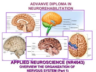
  • 1. ADVANVE DIPLOMA IN NEUROREHABILITATION APPLIED NEUROSCIENCE (NR4043)APPLIED NEUROSCIENCE (NR4043) OVERVIEW THE ORGANIZATION OFOVERVIEW THE ORGANIZATION OF NERVOUS SYSTEM (Part 1)NERVOUS SYSTEM (Part 1)
  • 2. Learning Outcome • Describe the organization/functions of CNS • Describe the structure/function of PNS • Describe the organization/function of lymbic system • Explain the brain protection (meninges/CSF/BBB) and blood supply.
  • 3. Introduction • Classification i. Central Nervous System - Brain, Spinal Cord ii. Peripheral Nervous System - peripheral nerves (spinal & cranial), ganglion, receptor
  • 5.
  • 6. • Consist of 4 major parts: – Brain stemBrain stem – DiencephalonDiencephalon – CerebrumCerebrum – CerebellumCerebellum BRAIN
  • 7.
  • 8. CEREBRUM: Main functions:Main functions: • Seat of intelligence – It provides the ability to • read, write and speak; • Make calculations • Compose music • To remember the past • Plan for the future • Imagine things that have never existed before • Centre of sensory perception • Initiate and coordinate skeletal muscle contraction
  • 9. Cerebral cortex and it’s structures:Cerebral cortex and it’s structures: • Consists of cerebral cortex (gray matter), internal region of cerebral white matter and gray matter nuclei deep within the white matter. • Thick : 2 – 4 mm, contains billions of neurons. • This gray matter is made of cell bodies of neurons. • During embryonic development, when brain size increases rapidly, the gray matter of the cortex enlarges much faster than the deeper white matter. • As a result, cerebral cortex rolls and folds upon itself so that it could fit into the cranial cavity.
  • 10.
  • 11.
  • 12. • The folds called gyri / gyrus or convolutions. • The deeper grooves between folds are known as fissures. • The shallow grooves between folds are termed as sulci / sulcus. • The most prominent fissure, is the longitudinal fissure which separates the cerebrum into right and left halves called cerebral hemisphere.cerebral hemisphere.
  • 13.
  • 14. • Within the longitudinal fissure between the cerebral hemispheres is the falx cerebri. • The hemispheres are connected internally by the corpus callosum. (broad band of white matter containing axons that extend between the hemispheres)
  • 15. • Each hemisphere is further subdivided into five lobes. • The lobes are named after the bones that cover them: –Frontal lobes. –Parietal lobes. –Temporal lobes. –Occipital lobes. –Insula • A fifth part of the cerebrum, the insula, cannot be seen at the surface of the brain because it lies within the lateral cerebral sulcus, deep to the parietal, frontal, and temporal lobes.
  • 16.
  • 17. Main boundaries in cerebrum:Main boundaries in cerebrum: Sulcus and fissures • Central sulcus – Separates the frontal and parietal lobes. • Longitudinal fissure – Separates the cerebrum into right and left hemispheres. • Lateral cerebral sulcus – Separates the frontal and temporal lobes. • Parieto-occipital sulcus – Separates parietal and occipital lobes.
  • 18.
  • 19.
  • 20. Major Gyrus of cerebrum: • Precentral gyrus – Located immediately anterior to the central sulcus. – It contain primary motor area. • Postcentral gyrus – is located immediately posterior to the central sulcus, contains the primary somatosensory area of the cerebral cortex.
  • 21.
  • 22. • The cerebral white matter consists of myelinated and unmyelinated axons that transmit impulses. • There are 3 types of tracts:- – Association tracts. – Commissural tracts – Projection tracts. CEREBRAL WHITE MATTERCEREBRAL WHITE MATTER
  • 24. • Commissural tracts; contain axon that conduct nerve impulses from gyri in one cerebral hemisphere to corresponding gyri in the other cerebral hemisphere. 3 important commissural tracts are the: • Corpus callosum (largest fiber bundle in the brain – 300 million fibers) • Anterior commissure • Posterior commissure
  • 25.
  • 26. • Projection tracts; contain axons that conduct nerve impulses from the cerebrum to lower parts of the CNS (thalamus, brainstem, or spinal cord) or lower parts of the cerebrum to the cerebellum. An example; is the internal capsule, which is the thick band of white matter that contains both ascending and descending axons.
  • 27. • projection tracts • From brain to spinal cord & vice verca, forms internal capsule • Commissural tracts • Cross to opposite hemisphere: »Corpus callosum »Anterior and posterior commissures • Association tracts • Connect lobes and gyri within a hemisphere. Summary of TractsSummary of Tracts
  • 28. • The frontal lobe forms the anterior portion of each cerebral hemisphere. • Extends from precentral sulcus up to the end of brain anteriorly. Cerebral lobes: FrontalCerebral lobes: Frontal
  • 29. • It is bordered posteriorly by a central sulcus (fissure of Rolando), which passes out from the longitudinal fissure at a right angle and inferioly by a lateral sulcus (fissure of Sylvius) which exits the undersurface of the brain along its sides.
  • 30. • Extend from central sulcus to parietooccipital sulcus posteriorly. • The parietal lobe is posterior to the frontal lobe and is separated from it by the central sulcus. Cerebral lobes: ParietalCerebral lobes: Parietal
  • 31. • The temporal lobe lies inferior to the frontal and parietal lobes and is separated from them by the lateral sulcus. Cerebral lobes: TemporalCerebral lobes: Temporal
  • 32. • The occipital lobe forms the posterior portion of each cerebral hemisphere and is separated from the cerebellum by a shelflike extension of duramater called tentorium cereblli. • Extends from parietooccipital sulcus to the inferior end of brain posteriorly. • The occipital,parietal and temporal lobes have no distinct boundary. Cerebral lobes: OccipitalCerebral lobes: Occipital
  • 33. • A fifth part of the cerebrum, the insula – cannot be seen at the surface of the brain because it lies within the lateral cerebral sulcus, deep to the parietal, frontal and temporal lobes. • Is on the lateral surface (medially) of the cerebrum Cerebral lobes: Insula
  • 34.
  • 35. Cerebral lobes and it’s functionsCerebral lobes and it’s functions
  • 36. • Frontal – Voluntary motor functions – Voluntary scanning movements of the eyes. – Planning, mood, smell and social judgement – Intellect – Personality – Complex learning ability – Recall of information – Initiative – Reasoning – Articulation of speech Cerebral lobes and it’s functionsCerebral lobes and it’s functions
  • 37. • Parietal – Receives and integrates sensory information. – Examples: touch, proprioception, pain, itching, tickle, temperature. – Interprets the meaning of a speech. Cerebral lobes and it’s functionsCerebral lobes and it’s functions • Temporal – Areas for hearing (auditory perception) and smell (olfactory perception).
  • 38. • Occipital – Visual center of brain – Involved in visual perception – Relates present and past visual experiences and is essential for recognizing and evaluating what is seen. Cerebral lobes and it’s functionsCerebral lobes and it’s functions
  • 40. Functional Organization of the Cerebral Cortex Functional Organization of the Cerebral Cortex • Specific types of sensory, motor and integrative signals are processed in certain regions of the cerebral cortex. • Sensory areas receive sensory information and are involved in perception, the conscious awareness of a sensation. • Motor areas control the execution of voluntary movements. • Association areas deal with more complex integrative functions (memory, emotions, reasoning, will, judgments, personality traits and intelligence)
  • 41. Brodmann’s AreaBrodmann’s Area • Functional area of cerebral cortex • Define by German Anatomist, Dr Korbinian Brodmann • Base on cytoarchitecture of the neuron in cerebral cortex. • Split the cortex into 52 areas • Consist of i. Motor areas (primary & association) ii.Sensory areas (primary & association)
  • 42.
  • 43. MOTOR AREASMOTOR AREAS • Motor output from the cerebral cortex flows mainly from the anterior part of each hemisphere.
  • 45. IMPORTANT MOTOR AREASIMPORTANT MOTOR AREAS • Primary motor area • Broca`s speech area
  • 46. Primary motor area (4)Primary motor area (4) • Located in the precentral gyrus of the frontal lobe. • Each region in the primary motor area controls voluntary contractions of specific muscles or groups of muscles. • Electrical stimulation of any point in the primary motor area causes contraction of specific skeletal muscle fibers on the opposite side of the body. • More cortical area is devoted to those muscles involved in skilled, complex or delicate movement. • For instance, the cortical region devoted to muscles that move the fingers is much larger than the region for muscles that move the toes. • Form corticospinal and corticobulbar/nuclear tracts
  • 47.
  • 48. Broca’s speech area (44, 45) Broca’s speech area (44, 45) • Located in the frontal lobe close to the lateral cerebral sulcus. (dominant hemisphere) • Is involved in the articulation of speech. • In most people, Broca’s speech area is localized in the left cerebral hemisphere. • Neural circuits between Broca’s speech area, premotor area and primary motor area activate muscles of the larynx, pharynx, and mouth and breathing muscles. • The coordinated contractions of your speech and breathing muscles enable you to speak your thoughts. • In stroke patient when this areas is affected they still have clear thoughts, but are unable to form words.(...)
  • 49. MOTOR ASSOCIATION AREASMOTOR ASSOCIATION AREAS
  • 50. Premotor area (6,8)Premotor area (6,8) • Is a motor association area that is immediately anterior to the primary motor area. • Neurons in this area communicate with the primary motor cortex, the sensory association areas in the parietal lobe, the basal ganglia and the thalamus. • The premotor area deals with learned motor activities of complex and sequential nature • It generates nerve impulses that cause specific groups of muscles to contract in a specific sequence, as when you write your name. • The premotor area also serves as a memory bank for such movements.
  • 51. Frontal eye field area (8)Frontal eye field area (8) • Is in the frontal cortex • It is sometime included in the premotor area. • It controls voluntary scanning movements of the eyes – like those you just used in reading this sentence. • Eyes move to the opposite site simultaneously – conjugate movements.
  • 53. Primary Sensory AreasPrimary Sensory AreasPrimary Sensory AreasPrimary Sensory Areas • Sensory information arrives mainly in the posterior half of both cerebral hemisphere in regions behind the central sulci • The cortex, primary sensory areas have most direct connections with peripheral sensory receptors. • Sensory areas are:- – Primary somatosensory area- postcentral gyrus.Primary somatosensory area- postcentral gyrus. – Primary visual area- occipital lobe.Primary visual area- occipital lobe. – Primary auditory area- temporal lobe.Primary auditory area- temporal lobe. – Primary gustatory area- base of the postcentralPrimary gustatory area- base of the postcentral gyrus.gyrus. – Primary olfactory area- temporal lobePrimary olfactory area- temporal lobe
  • 54. Secondary Sensory AreasSecondary Sensory AreasSecondary Sensory AreasSecondary Sensory Areas • Secondary sensory areas and sensory association areas often are adjacent to the primary areas and from other brain regions. • Secondary sensory areas and sensory association areas integrate sensory experiences to generate meaningful patterns of recognition and awareness. • A person with damage in the primary visual area would be blind in at least part of his visual field, but a person with damage to a visual association area might see normally yet be unable to recognize her best friend.
  • 56.
  • 57. 1. Primary somatosensory area1. Primary somatosensory area (1,2,3)(1,2,3) 1. Primary somatosensory area1. Primary somatosensory area (1,2,3)(1,2,3) • Located posterior to the central sulcus of each cerebral hemisphere in the postcentral gyrus of each parietal lobe. • It extends from the lateral cerebral sulcus, along the lateral surface of the parietal lobe to the longitudinal fissure and then along the medial surface of the parietal lobe within the longitudinal fissure.
  • 58. • Receives nerve impulse for touch, pressure, vibration, itch, tickle, temperature, pain, proprioception and is involved in the perception of these somatic sensations. • The side of cortical area receiving impulses from a particular part of the body depends on the number of receptors present there rather than on the size of the body part. • A larger region of the somatosensory area receives impulses from the lips and fingertips than from the thorax or hip. (sensory homunculus)
  • 59.
  • 60. 2. Primary visual area (17)2. Primary visual area (17)2. Primary visual area (17)2. Primary visual area (17) • Located at the posterior tip of tip of the occipital lobe mainly on the medial surface. • Receives visual information and is involved in visual perception.
  • 61. 3. Primary auditory area (41,42)3. Primary auditory area (41,42)3. Primary auditory area (41,42)3. Primary auditory area (41,42) • Located in the superior part of the temporal lobe near the lateral cerebral sulcus • Receives information for sound and is involved in auditory perception.
  • 62. 4. Primary gustatory area (43)4. Primary gustatory area (43)4. Primary gustatory area (43)4. Primary gustatory area (43) • Located at the base of the postcentral gyrus superior to the lateral cerebral sulcus in the parietal cortex. • Receives impulses for taste and is involved in gustatory perception.
  • 63. 5. Primary olfactory area (34)5. Primary olfactory area (34)5. Primary olfactory area (34)5. Primary olfactory area (34) • Located in the temporal lobe on the medial aspect (and thus not visible) • Receives impulses for smell and is involved in olfactory perception.
  • 64. ASSOCIATION SENSORY AREASASSOCIATION SENSORY AREASASSOCIATION SENSORY AREASASSOCIATION SENSORY AREAS • Secondary sensory areas /sensory association areas often are adjacent to the primary areas and from other brain regions. • Secondary sensory areas / sensory association areas integrate sensory experiences to generate meaningful patterns of recognition and awareness. • A person with damage in the primary visual area would be blind in at least part of his visual field, but a person with damage to a visual association area might see normally yet be unable to recognize her best friend.
  • 65. ASSOCIATION AREASASSOCIATION AREASASSOCIATION AREASASSOCIATION AREAS • Consist of larger areas of the occipital, parietal and temporal lobes and anterior surface frontal lobe. • Association areas are connected with one another by association tracts.
  • 66. 1. Somatosensory association area1. Somatosensory association area (5,7)(5,7) 1. Somatosensory association area1. Somatosensory association area (5,7)(5,7) • Is just posterior to and receives input from the primary somatosensory area, as well as from the thalamus and other parts of the brain.
  • 67. • It allows to determine the expect shape and texture of an object without looking at it, to determine the orientation of one object with respect to another as they are felt, and to sense the relationship of one body part to another. • Another role of the somatosensory association area is the storage of memories of past somatic sensory experiences, enabling you to compare current sensations with previous experiences. • For example, the somatosensory association area allows you to recognize objects such as a pencil and a paperclip simply by touching them.
  • 68. 2. Visual association area2. Visual association area (18, 19)(18, 19) 2. Visual association area2. Visual association area (18, 19)(18, 19)• Located in the occipital lobe, receives sensory impulses from the primary visual area and the thalamus. • It relates present and past visual experiences and is essential for recognizing and evaluating what is seen. • For example, the visual association area allows you to recognize an object such as a spoon simply by looking at it.
  • 69. • Corresponding roughly to areas 20, 21 and 37 in the inferior temporal lobe, receives nerve impulses from the visual associations area. • This area stores information about faces, and it allows you to recognize people by their faces. (prosopagnosia) • The facial recognition area in the right hemisphere is usually more dominant than the corresponding region in the left hemisphere. 3. Facial recognition area (20,21)3. Facial recognition area (20,21)3. Facial recognition area (20,21)3. Facial recognition area (20,21)
  • 70. • Located inferior and posterior to the primary auditory area in the temporal cortex • Allows you to recognize a particular sound as speech, music or noise. 4. Auditory association area (22)4. Auditory association area (22)4. Auditory association area (22)4. Auditory association area (22)
  • 71. • Corresponding roughly to area 11 along the lateral part of the frontal lobe • Receives sensory impulses from the primary olfactory area. 5.Secondary olfactory area (11)5.Secondary olfactory area (11)5.Secondary olfactory area (11)5.Secondary olfactory area (11)
  • 72. • This area allows you to identify odors and to discriminate among different odors. • During olfactory processing, the orbital frontal cortex of the right hemisphere exhibits greater activity than the corresponding region in the left hemisphere.
  • 73. 6. Wernicke’s area (39,40)6. Wernicke’s area (39,40)6. Wernicke’s area (39,40)6. Wernicke’s area (39,40) • A broad region in the left temporal and parietal lobes (dominant hemisphere), interprets the meaning of speech by recognizing spoken words. • It is active as you translate words into thoughts.
  • 74. 7. Common integrative area7. Common integrative area7. Common integrative area7. Common integrative area • Is bordered by somatosensory, visual and auditory association areas. • It receives nerve impulses from these areas and from the primary gustatory area, primary olfactory area, the thalamus and parts of the brain stem. • This area integrates sensory interpretations from the association areas and impulses from other areas, allowing the formation of thoughts based on a variety of sensory inputs. • It then transmits signals to other parts of the brain for the appropriate response to the sensory signals it has interpreted.
  • 75. • is an extensive area in the anterior portion of the frontal lobe. • This area has numerous connections with other areas of the cerebral cortex, thalamus, hypothalamus, limbic system and cerebellum. 8. Prefrontal cortex (frontalPrefrontal cortex (frontal association area) (9,10,11)association area) (9,10,11) 8. Prefrontal cortex (frontalPrefrontal cortex (frontal association area) (9,10,11)association area) (9,10,11)
  • 76. Prefrontal cortex (frontal associationPrefrontal cortex (frontal association area)area) The prefrontal cortex is concerned with make up of a person`s: • Personality • Intellect • Complex learning abilities • Recall of information • Initiative • Judgement • Foresight • Reasoning • Conscience • Intuition • Mood • Planning for future • Development of abstract ideas.
  • 77. Prefrontal cortex (cont…) • Damage to bilateral prefrontal cortices make a person become: – rude, – inconsiderate – Incapable of accepting advice – Moody – Inattentive – Less creative – Unable to plan for the future
  • 78. • Frontal – Voluntary motor functions – Planning, mood, smell and social judgement • Parietal – Receives and integrates sensory information • Occipital – Visual center of brain • Temporal – Areas for hearing, smell, learning, memory, emotional behaviour. SUMMARY FOR CEREBRAL LOBESUMMARY FOR CEREBRAL LOBE FUNCTIONSFUNCTIONS SUMMARY FOR CEREBRAL LOBESUMMARY FOR CEREBRAL LOBE FUNCTIONSFUNCTIONS
  • 79. • Includes reading, writing, speaking and understanding words. • Wernicke`s area – Permit recognition of spoken and written language and creates plan of speech. • Broca`s area – Generates motor signals for larynx, tongue, cheeks and lips. – Transmits to primary motor cortex for action SUMMARY: LANGUAGESUMMARY: LANGUAGE
  • 80. • Slight anatomical differences between the right and left hemispheres exist. • 2/3rd of the population, the Wernicke's area is 50% larger on the left side than on the right side. • Physiological differences also exists. • This functional asymmetry is termed as hemispheric lateralization. HEMISPHERIC LATERALIZATIONHEMISPHERIC LATERALIZATIONHEMISPHERIC LATERALIZATIONHEMISPHERIC LATERALIZATION
  • 81. Most obvious examples are: • Left hemisphere receives somatic sensory signals from and controls muscles on the right side of the body, whereas the right hemisphere receives sensory signals from and controls the left side of the body. • In most people the left hemisphere is more important for reasoning, numerical and scientific skills, spoken and written language, and the ability to use and understand sign language.
  • 82. • The right hemisphere is more specialized for: – musical and artistic awareness; – spatial and pattern perception; – recognition of faces and emotional content of language; – Discrimination of different smells – Generating mental images of sight, sound, touch, taste and smell to compare relationships among them.
  • 83. HEMISPHERIC LATERALIZATIONHEMISPHERIC LATERALIZATIONHEMISPHERIC LATERALIZATIONHEMISPHERIC LATERALIZATION
  • 84. Neuroanatomy & Physiology Basal Ganglia and its Clinical Correlation By Hermizan Halihanafiah
  • 85.
  • 86.
  • 87.
  • 88. Anatomical Classification Neurologic structure Basal nuclei Corpus striatum Caudate nuclei and lentiform nuclei Lentiform nuclei Putamen & globus pallidus Neostriatum / striatum Caudate nuclei & putamen Amygdaloid body Amygdaloid nuclei Claustrum Claustrum
  • 89.
  • 90. Functional Classification Nuclei Discription Corpus striatum Caudate nuclei and lentiform nuclei Subthalamic nuclei Diencephalic origin Subtantia nigra Masses of grey matter located at midbrain
  • 91.
  • 92.
  • 93. Basal Ganglia Disorder • Hypokinesia • Hyperkinesia
  • 94. Hypokinesia • Parkinsonism - bradykinesia / akinesia - resting tremors / pill rolling - lead pipe regidity/cogwheel rigidity
  • 95. Hyperkinesia • Chorea – Huntington - Sydenham’s / St Vitus Dance • Hemiballism • Athetosis • Dystonia • Tardive dyskinesia • Wilson disease – tremor, dyskinesia, rigidity
  • 97. Internal capsule • Projection tracts (ascending and descending tracts) • V shaped of white matter • Located in between lenticular nuclei (laterally) and thalamic / caudate nuclei (medially)
  • 98.
  • 99. Internal capsule • It divide into: 1.anterior limb 2.Genu 3.Posterior limb 4.Retrolenticular portion 5.Sublenticular portion
  • 100.
  • 101. Genu • Refers to the flexure of internal capsule • Consist of corticobulbar/corticonuclear tracts • Which originate from primary motor area (head and facial areas) • Terminate at the cranial somatic motor nuclei (III, IV, V, VI, VII, IX, X, XI, XII)
  • 102. ANTERIOR LIMB  Located in between lenticular nuclei and caudate nuclei  Huge connection between thalamus and frontal lobe  Three major tracts formed ALIC 1.thalamocortical/thalamofrontal 2.Frontothalamic and frontopontine
  • 103. Posterior Limb • The largest fibers of IC • Consist most of the ascending and descending tracts • Located inbetween lentiform nuclei and thalamus
  • 104. • Contains fibers of: 1.Corticospinal tracts 2.Somaticsensory tracts (PCML, ASTT, LSTT and TTT) 3.Optic radiation from thalamus to visual cortex (occipital lobe) 4.Acoustic fibers to temporal lobes
  • 105. Meninges Ventricle System and CSF Blood Brain Barrier Lymbic System
  • 106. 1. MENINGES • Is a protective covering of the brain and spinal cord. • The cranial bones and cranial meninges surround and protect the brain. • The cranial meninges are continuous with the spinal meninges. • Consists of 3 layers: – Dura mater (outer most) – Arachnoid mater (middle) – Pia mater (inner most)
  • 107. 1.1. Dura mater • cranial dura mater has 2 layers and spinal dura mater has only 1 layer. • 2 dural layers; periosteal and meningeal layers around the brain are fused together except where they separate to enclose the dural venous sinuses that drain venous blood from the brain and deliver it into the internal jugular veins.(p498) • There is no epidural space in the brain. • Blood vessels enter brain tissue pass along the surface of the brain, and as they penetrate inward, they are sheathed by a loose – fitting sleeve of pia mater.
  • 108. Extensions of the Dura Mater Copyright 2009, John Wiley & Sons, Inc.
  • 109. Extensions of the dura mater 3 extensions of the dura mater (Dural Folds) separate parts of the brain (p498): • Falx cerebri – the largest dural folds, separates the 2 hemispheres of the cerebrum, lies in the longitudinal fissure and anchored anteriorly by crista galli. • Falx cerebelli – separates the 2 hemispheres of the cerebellum. • Tentorium cerebelli – horizontally separates the cerebrum and the cerebellum.
  • 112. 1.1. Dura mater (cont…) • spinal dura mater forms a loose sheath round the spinal cord, extending from the foramen magnum to the second sacral vertebra. • It encloses the filum terminale and fuses with the periosteum of the coccyx. • It is an extension of the inner layer of cerebral dura mater and is separated from the periosteum of the vertebrae and ligaments within the neural canal (vertebral canal) by the epidural or extradural space, containing blood vessels and areolar tissue. • Nerves enter and leave the spinal cord, pass through the epidural space.
  • 113. 1.2. Arachnoid mater • Is a serous membrane lies between the dura and pia mater. • Is separated from the dura mater by the subdural space, and from pia mater by subarachnoid space which contains cerebrospinal fluid. • It passes over the convolutions of the brain and accompanies the inner layer of dura mater in the formation of the falx cerebri, tentorium cerebelli and falx cerebelli. • It continues downwards to envelop the spinal cord and ends by merging with the dura mater at the level of the 2nd sacral vertebra.
  • 114. 1.3. Pia mater • Is a fine connective tissue containing many minute blood vessels. • It adheres to the brain, completely covering the convolutions and dipping into each fissure. • It continues downwards surrounding the spinal cord. • Beyond the end of the cord it continues as the filum terminale, pierces the arachnoid tube and goes on, with the dura mater, to fuse with the periosteum of the coccyx.
  • 115.
  • 116.
  • 119. 2. VENTRICLES Within the brain there are 4 irregular shaped cavities or ventricles containing CSF (p499): • 2 lateral ventricles (right and left) • 3rd ventricle • 4th ventricle
  • 120.
  • 121.
  • 122. 2.1. Lateral ventricles • lie within the cerebral hemisphere, one on each side of the median plane just below the corpus callosum (p502). • Anteriorly the lateral ventricles are separated from each other by a thin membrane called septum pellucidum, and they are lined with ciliated epithelium (p502). . • Leteral ventricles communicate with 3rd ventricle by interventricular foramina (foramina of Monro). • Consists network of blood capillaries called choroid plexus.
  • 123. 2.2. Third ventricles • Is a cavity situated below the lateral ventricles between the right and left halves of the thalamus. • Is a narrow cavity along the midline superior to the hypothalamus. • Communicates with the 4th ventricle by a canal, the cerebral aqueduct or aqueduct of the midbrain (Aqueduct of Sylvius). • Consists network of blood capillaries called choroid plexus.
  • 124. 2.3. Fourth ventricles • Is a diamond shaped cavity situated below and behind the 3rd ventricle, between the cerebellum and pons. • Is continuous below with the central canal of the spinal cord and communicates with the subarachnoid space by two lateral aperture (Foramina of Luschka) and one median aperture (foramina of Magendie). • CSF enters the subarachnoid space through these openings and through the open distal end of the central canal of the spinal cord. • Consists network of blood capillaries called choroid plexus.
  • 125. 3. CEREBROSPINAL FLUID • Is a colorless liquid that protects the brain and spinal cord from chemical and physical injuries. • It carries oxygen, glucose and other needed chemicals from the blood to neurons and neuroglia (brain tissues). • CSF continuously circulates through cavities in the brain and spinal cord and around the brain and spinal cord in the subarachnoid space (between arachnoid and pia mater)
  • 126. 3. CEREBROSPINAL FLUID • The total volume of CSF is 80 to 150 ml in an adult. • CSF contains glucose, proteins, lactic acid , urea, cations (Nat , Kt ,Cat , Mg2t ) and anions (Cl- and HCO3- ); it also contains some white blood cells. • The CSF contributes to homeostasis in 3 main ways:- – Mechanical protection – Chemical protection – Circulation
  • 127. 3. CEREBROSPINAL FLUID CSF contributes to Homeostasis in three main ways: Mechanical protection • Serves as shock-absorbing medium that protects the delicate tissues of the brain and spinal cord from jolts that would otherwise cause them to hit the bony walls of the cranial and vertebral cavities. • The fluid also buoys the brain so that it ‘floats’ in the cranial cavity.
  • 128. 3. CEREBROSPINAL FLUID Chemical protection • It provides an optimal chemical environment for accurate neuronal signaling. • Even slight changes in the ionic composition of CSF within the brain can seriously disrupt production of action potential and postsynaptic potentials. Circulation • CSF allows exchange of nutrients and waste products between the blood and nervous tissue.
  • 129. 3. CEREBROSPINAL FLUID 3.1. Functions of CSF • provide support and protection for brain and spinal cord. • Maintain a constant pressure around the brain and spinal cord. • Acts as cushion of fluid that absorb shock. (shock absorber) • Carries nutrient, oxygen and needed chemicals from blood to neuron and neuroglia. • Carries out waste product from neuron and neuroglia to blood.
  • 130. 3. CEREBROSPINAL FLUID 3.2. Formation of CSF • Produced at the choroid plexus, network of capillaries in the walls of the ventricles. • The capillaries are covered by ependymal cells that form CSF from blood plasma by filtration and secretion. • The blood-CSF fluid barrier permits certain substances to enter the CSF but excludes others, protecting the brain and spinal cord from potentially harmful blood borne substance.
  • 131.
  • 133. 3. CEREBROSPINAL FLUID 3.3. Circulation of CSF • Formed in the choroid plexuses of each lateral ventricles - flows into the 3rd ventricle through 2 narrow, oval openings, the interventricular foramina (of Monro). • More CSF is added by the choroid plexus in the roof of the 3rd ventricle. • The fluid then flows through the aqueduct of the midbrain (cerebral aqueduct @ of Sylvius) which passes through the midbrain, into the 4th ventricle.
  • 134. 3. CEREBROSPINAL FLUID 3.3. Circulation of CSF • The choroid plexus of the 4th ventricle contributes more fluid. • CSF enters the subarachnoid space through 3 openings in the roof of the 4th ventricles; a median aperture (of foramen of Magendie) and the paired lateral apertures (foramen of Luschka), one on each side. • CSF then circulates in the central canal of the spinal cord and in the subarachnoid space around the surface of the brain and spinal cord.
  • 135.
  • 136. Figure 7.14Figure 7.14 Arrows showing the flow of cerebrospinal fluid.Arrows showing the flow of cerebrospinal fluid. Copyright © Elsevier Ltd 2005. All rights reserved.Copyright © Elsevier Ltd 2005. All rights reserved.
  • 137. 3. CEREBROSPINAL FLUID 3.4. Reabsorption of CSF • CSF is gradually reabsorbed into the blood through arachnoid villi, fingerlike extensions of the arachnoid that project into the dural venous sinuses, especially the superior sagittal sinus (a cluster of arachnoid villi is called an arachnoid granulation) • Normally CSF is reabsorbed as rapidly as it is formed by the choroid plexuses, at a rate of about 20mL/hr (480mL/day). • Because the rates of formation and reabsorption are the same, the pressure of CSF normally is constant.
  • 138.
  • 139. Summary of the formation, circulation, and reasorption of CSF Lateral ventriclesLateral ventricles 3rd ventricles3rd ventricles 4th ventricles4th ventricles Subarachnoid spaceSubarachnoid space Arachnoid villi of dural venous sinuses Arachnoid villi of dural venous sinuses Heart and lungsHeart and lungs Lateral ventricles choroid plexuses Lateral ventricles choroid plexuses 3rd ventricles choroid plexuses 3rd ventricles choroid plexuses 4th ventricles choroid plexuses 4th ventricles choroid plexuses CSF CSF CSF Through interventricular foramina Through cerebral aqueduct Through lateral and Median apertures Venous blood
  • 140. Blood Brain Barrier (BBB) • The existence of BBB protect the brain cells from harmful substances and pathogens by preventing passage of many substances from blood into brain cells. • Endothelial cells of the blood vessels in the choroid plexus which are joined by tight junction form the BBB. • The processes of many astrocytes press up against the capillaries and secrete chemical that maintain the permeability characteristic of tight junction. • A few water soluble substances such as glucoses cross the BBB by active transport. • Other substances such as creatinine, urea, and most ions, cross the BBB very slowly.
  • 141. • Other substances such as protein and antibiotic drugs do not pass at all through BBB. • However, lipid-soluble substances, such as oxygen, carbon dioxide, alcohol, and most anesthetic agents easily cross the BBB. • Trauma, certain toxins, and inflammation can cause a breakdown of the BBB. Blood Brain Barrier (BBB)
  • 142.
  • 143. • Part of the cerebrum and diencephalon are grouped together as a lymbic system. • Lymbic refers to deep portion of the cerebrum that form a ring around the diencephalon. • Sometimes called “the emotional brain”. THE LIMBIC SYSTEM
  • 144.
  • 148.
  • 149. The Limbic SystemThe Limbic System
  • 150. • The limbic system includes many structures in the cerebral cortex and sub-cortex of the brain. • Limbic lobe – a rim of cerebral cortex on the medial surface of each hemisphere. • It includes the cingulate gyrus which lies above the corpus callosum • Located in the frontal lobe of cerebrum. • Cingulate gyrus : Autonomic functions regulating heart rate, blood pressure and cognitive and attentional processing COMPONENTS OF THE LIMBIC SYSTEM (p 517)
  • 151. The Limbic SystemThe Limbic System
  • 152. • parahippocampal gyrus which is in the temporal lobe below. • Parahippocampal gyrus : Plays a role in the formation of spatial memory. • The hippocampus is a portion of the parahippocampal gyrus that extends into the floor of the lateral ventricle. • Hippocampus : Required for the formation of long-term memories COMPONENTS OF THE LIMBIC SYSTEM
  • 153.
  • 154.
  • 155. • Dendate gyrus lies between the hippocampus and parahippocampal gyrus. • Dentate gyrus : thought to contribute to new memories and to regulate happiness. • The amygdala is composed of several groups of neurons located close to the tail of the caudate nucleus (basal ganglia). • Amygdala : Involved in signaling the cortex of motivationally significant stimuli such as those fear COMPONENTS OF THE LIMBIC SYSTEM
  • 156.
  • 157. • The septal nuclei are located within the septal area formed by the regions under the corpus callosum and the paraterminal gyrus (a cerebral gyrus). • The mammillary bodies of the hypothalamus are two round masses close to the midline near the cerebral peduncles. • Mammillary body : Important for the formation of memory • Two nuclei of thalamus, the anterior nucleus and the medial nucleus, participate in limbic circuits. • Thalamus: The "relay station" to the cerebral cortex COMPONENTS OF THE LIMBIC SYSTEM
  • 158. The Limbic SystemThe Limbic System
  • 159. • The olfactory bulbs are flattened bodies of the olfactory pathway that rest on the cribriform plate. • Olfactory bulb: Olfactory sensory input • The fornix, stria terminals, stria medullaris, medial forebrain bundle, and mammillothalamic tract are linked by bundles of interconnectimg myelinated axons. COMPONENTS OF THE LIMBIC SYSTEM
  • 161. • Limbic lobe; includes the cingulate gyrus and parahippocampal gyrus and hippocampus gyrus. • Dentate gyrus • Amygdala • Septal nuclei • Mammillary bodies of the hypothalamus • Anterior and medial nucleus • Olfactory bulbs • Fornix, stria terminals, stria medullaris, medial forebrain bundle, mammillothalamic tract. Summary:components Of The Limbic System
  • 162. The Limbic SystemThe Limbic System
  • 163. The Limbic SystemThe Limbic System
  • 164. The Limbic SystemThe Limbic System
  • 165. • The limbic system is sometimes called the emotional brain because it plays a primary role in a range of emotions, including pain, pleasure, docility, affection, and anger. • It also involved in olfaction (smell) and memory. • The amygdala involves fear and aggression. • The hippocampus, together with other parts of the cerebrum function in memory. People with damage to limbic system structures forget recent events and cannot commit anything to memory. FUNCTIONS OF THE LIMBIC SYSTEM