More Related Content
Similar to Arco ao à d (20)
Arco ao à d
- 1. Ultrasound Obstet Gynecol 2016
Published online in Wiley Online Library (wileyonlinelibrary.com). DOI: 10.1002/uog.15805
Fetuses with right aortic arch: a multicenter cohort study
and meta-analysis
F. D’ANTONIO*, A. KHALIL*, V. ZIDERE† and J. S. CARVALHO*‡
*Fetal Medicine Unit, St George’s University Hospital NHS Foundation Trust and Institute of Cardiovascular and Cell Sciences,
St George’s, University of London, London, UK; †King’s College Hospital, London, UK; ‡Brompton Centre for Fetal Cardiology,
Royal Brompton Hospital NHS Trust, London, UK
KEYWORDS: aortic arch syndromes; echocardiography; fetus; meta-analysis; prenatal diagnosis
ABSTRACT
Objectives Use of recent antenatal screening guidelines
for cardiac abnormalities has increased fetal diagnoses
of right aortic arch (RAA). We aimed to establish the
outcome of fetal RAA without intracardiac abnormalities
(ICA) to guide postnatal management.
Methods In the retrospective cohort part of our study,
outcome measures were rates of chromosomal abnormal-
ities, 22q11.2 deletion, fetal extracardiac abnormalities
(ECA), postnatal ICA and ECA, and symptoms of
and surgery for vascular ring. A systematic review and
meta-analysis was also performed; results are reported as
proportions. Kaplan–Meier analysis of vascular ring cases
with surgery as endpoint was performed.
Results Our cohort included 86 cases; 41 had a
vascular ring. Rates of chromosomal abnormalities,
22q11.2 deletion and fetal ECA were 14.1%, 6.4%
and 17.4%, respectively. Sixteen studies including our
cohort (312 fetuses) were included in the systematic
review. Overall rates of chromosomal abnormalities
and 22q11.2 deletion were 9.0% (95% CI, 6.0–12.5%)
and 6.1% (95% CI, 3.6–9.3%), whilst the respective
rates for cases with no ECA were 4.6% (95% CI,
2.3–7.8%) and 5.1% (95% CI, 2.4–8.6%). ECA were
seen in 14.6% (95% CI, 10.6–19.0%) prenatally and
in 4.0% (95% CI, 1.5–7.6%) after birth. Postnatal
ICA were identified in 5.0% (95% CI, 2.7–7.9%). Rate
of symptoms of vascular rings (follow-up ≥ 24 months
postpartum) was 25.2% (95% CI, 16.6–35.0%),
and 17.1% (95% CI, 9.9–25.7%) had surgery.
Two-year freedom from surgery was 83.0% (95% CI,
74.3–90.1%).
Conclusions Fetal RAA without ICA is more frequently
associated with ECA than with chromosomal abnormal-
ities. Most cases, however, are isolated. Vascular-ring
Correspondence to: Dr J. S. Carvalho, Royal Brompton Hospital, Sydney Street, London SW3 6NP, UK (e-mail: j.carvalho@rbht.nhs.uk)
Accepted: 27 October 2015
symptoms occur in about 25% of cases. Postnatal surveil-
lance is required mainly in the first 2 years after delivery.
Copyright © 2015 ISUOG. Published by John Wiley &
Sons Ltd.
INTRODUCTION
Right aortic arch (RAA) is characterized by abnormal
laterality of the aorta and brachiocephalic vessels. It
courses to the right of the trachea, in contrast to the
normal left aortic arch (LAA). Its incidence is estimated to
be 0.1%1,2
. Variations of aortic laterality and branching
pattern result from abnormal regression of the primordial
paired aortic arches during embryonic development.
Normal regression leads to an LAA, left-sided arterial duct
(AD) and the usual branching pattern: right innominate,
left common carotid and left subclavian arteries (LSA).
An RAA may have a mirror-image branching pattern, but
aberrant origin of the LSA (ALSA) is common.
Prenatal diagnosis is important due to associated
cardiac and extracardiac abnormalities (ECA) and
chromosomal defects, in particular 22q11.2 deletion3
. An
RAA can form a vascular ring, which is a heterogeneous
group of vascular abnormalities encircling the trachea
and esophagus. The classical vascular ring formed by
an RAA has a left-sided AD and an ALSA which arises
from a remnant of the primordial aortic arch, known as
Kommerell’s diverticulum. Although such rings may be
asymptomatic, symptoms of compression, e.g. dysphagia,
stridor, wheeze and recurrent upper respiratory tract
infections, are reported commonly. Other manifestations
include cyanosis and obstruction of the ALSA4,5
.
Prenatal diagnosis of RAA and vascular rings has been
reported for a number of years2,6–12. Recently published
international guidelines for antenatal screening13
rec-
ommend that the three-vessel view and the three-vessel
and trachea view14,15 be included in routine pregnancy
Copyright © 2015 ISUOG. Published by John Wiley & Sons Ltd. SYSTEMATIC REVIEW
- 2. 2 D’Antonio et al.
screening. This is likely to increase further the prenatal
detection of RAA and its variants. It is therefore
important that perinatal management be optimized and
that family counseling is not based only on postnatal
series. These are likely to be biased as only symptomatic
patients are reported, thus probably representing the
most extreme end of the spectrum, which does not take
into account asymptomatic individuals with isolated,
probably undiagnosed, RAA.
The aims of this study were to ascertain the outcome
of a large number of fetuses with RAA without
associated major intracardiac abnormalities (ICA) and
to review the relevant literature systematically in
order to propose guidance for perinatal and postnatal
management.
METHODS
Cohort study
This was a retrospective cohort study of cases of RAA
without associated ICA seen in tertiary centers. Cases
were identified from the fetal cardiology databases at
St George’s and Royal Brompton Hospitals (January
2001–December 2013) and King’s College Hospital
(January 2006–December 2013), London. The study was
an audit of clinical practice and no ethical approval
was needed. Ultrasound examinations were performed
on an Aloka Alpha-10, Aloka ProSound 5500 PhD
(Hitachi Aloka Medical, Ltd., Tokyo, Japan), Acuson
Aspen Advanced (Acuson, Mountain View, CA, USA)
or Voluson E8 (GE Medical Systems, Zipf, Austria). A
comprehensive assessment of the fetal heart was carried
out in all fetuses using conventional two-dimensional
(2D) ultrasound, and color, power and pulsed-wave
Doppler. An RAA was diagnosed when the transverse
arch was imaged to the right of the trachea on axial
views of the fetal chest, at the level of the three-vessel
and trachea view. The laterality of the AD in relation to
the trachea was also ascertained. More recently, attempts
were made to determine the course of the LSA, using a
similar approach to that described to identify an aberrant
right subclavian artery associated with an LAA16. The
diagnosis of a vascular ring was made in the presence of
an RAA, left AD and ALSA. Isolated RAA was defined
as having no associated major ICA or ECA detected
prenatally.
The outcomes observed were rate of chromosomal
abnormalities, 22q.11.2 deletion, associated fetal ECA
at the time of anatomical survey, associated postnatal
ICA and ECA, symptoms related to compression of
airways/esophagus and surgery for vascular ring.
Statistical analysis
Descriptive statistics are reported as median (interquartile
range). Main outcomes are reported as proportions.
Statistical analysis was performed using Microsoft Excel
for Mac 2011 (Version 14.4.9).
Table 1 Characteristics and outcomes of 86 fetuses with prenatal
diagnosis of right aortic arch (RAA) with no associated intracardiac
abnormalities
Characteristics Value
Maternal age (years) 32.0 (27.3–36.0)
Gestational age at diagnosis (weeks) 21.0 (20.0–23.0)
NT > 2.5 mm 9/59 (15.3)*
NT > 99th centile 4/59 (6.8)*
Abnormal karyotype or phenotype 11/78 (14.1)
Abnormal karyotype (tested) 11/53 (20.8)†
22q11.2 deletion 5/78 (6.4)
22q11.2 deletion (tested) 5/53 (9.4)†
Left arterial duct 79/86 (91.9)
Right arterial duct 7/86 (8.1)
Vascular ring (RAA and ALSA) 41/86 (47.7)
ECA diagnosed prenatally 15/86 (17.4)
Neonates with cardiac abnormalities
diagnosed postnatally only
4/64 (6.3)‡
Neonates with ECA diagnosed postnatally only 4/65 (6.2)‡
Termination of pregnancy 8/86 (9.3)
Intrauterine demise 3/72 (4.2)§
Postnatal symptoms 7/33 (21.2)¶
Surgery due to vascular-ring symptoms 5/33 (15.2)¶**
Data are given as median (interquartile range) or n/N (%).
*Includes only those with measured/available nuchal translucency
thickness (NT). †Includes only those tested. ‡Includes only live
births with known outcome data. §Includes only those known to be
at risk of demise. ¶Includes only live births with a vascular ring and
known outcome data; excludes the child with double aortic arch.
**Includes two children awaiting surgery at time of writing. ALSA,
aberrant origin of left subclavian artery; ECA, extracardiac
anomaly.
Systematic review and meta-analysis
Protocol, eligibility criteria, information sources
and search
This review was performed according to a protocol
designed a priori and recommended for systematic reviews
and meta-analysis17. MEDLINE, EMBASE, CINAHL
and The Cochrane Library were searched electronically
in January 2015, utilizing combinations of the relevant
medical subject heading (MeSH) terms, keywords and
word variants for ‘right aortic arch’, ‘prenatal diagnosis’,
‘ultrasound’, ‘Doppler’, ‘chromosomal abnormalities’,
‘aneuploidy’, ‘22q11 deletion’, ‘Di George syndrome’,
‘associated abnormalities’, ‘structural abnormalities’,
‘cardiac defects’, ‘postnatal surgery’, ‘intrauterine death’,
‘outcome’, ‘postnatal surgery’, ‘postnatal symptoms’, ‘res-
piratory symptoms’, ‘compression symptoms’, ‘vascular
ring’, ‘vascular steal’, ‘intrauterine death’. Reference lists
of relevant articles and reviews were hand-searched for
additional reports (for search strategy, see Appendix S1).
Search was limited to the English language. This review
was registered on PROSPERO international database for
systematic reviews (reference: CRD42015016097).
Study selection, data collection and data items
Only studies reporting prenatal diagnosis of RAA using
a particular imaging protocol, which included the assess-
ment of three-vessels and three-vessels and trachea view,
Copyright © 2015 ISUOG. Published by John Wiley & Sons Ltd. Ultrasound Obstet Gynecol 2016.
- 3. Fetal right aortic arch 3
SVC
Ao arch
Ao arch
ALSA
SVC
Inom vein
T
T
ALSA
Post
Lt
R
t
A
nt
T T
Duct
Duct
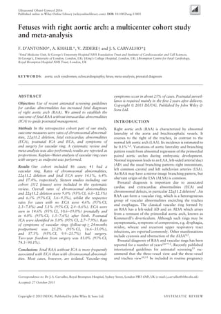
Figure 1 B-mode (a,b) and color Doppler (c,d) ultrasound images of upper mediastinum in Case 22 at 28 weeks of gestation. Note the
right-sided aortic arch, left-sided arterial duct and aberrant origin of left subclavian artery (ALSA) coursing behind the trachea (T). Ao arch,
aortic arch; Inom vein, innominate vein; SVC, superior vena cava.
were considered suitable for inclusion. Pediatrics series
were excluded on the basis that mainly symptomatic
patients are included, thus potentially overestimating
the rate of some of the outcomes explored in this
review. Cohort and case series were included. Editorials,
conference abstracts, case reports and cases series of fewer
than three patients were excluded (Appendix S2). The
outcomes analyzed were chromosomal abnormalities,
22q11.2 deletion, associated ECA detected prenatally,
pressure symptoms and surgery for vascular ring,
additional ICA and ECA diagnosed postnatally only.
For chromosomal abnormalities and 22q11.2 deletion,
analysis was restricted to cases for which karyotype or
phenotype was known, either pre- or postnatally. Anal-
ysis of pressure symptoms and surgery was restricted to
cases with a vascular ring (RAA, left AD and ALSA) and
that, if asymptomatic, had a minimum follow-up time of
24 months.
Two authors (F.D., A.K.) reviewed all abstracts
independently. Agreement about potential relevance was
reached by consensus, and full-text copies of those
papers were obtained. The two reviewers independently
extracted relevant data regarding study characteristics and
pregnancy outcome. Inconsistencies were discussed by the
reviewers and consensus reached.
Risk of bias, summary measures and synthesis of results
Quality assessment of the included studies was performed
using the Newcastle–Ottawa Scale (NOS) for cohort
studies18
.
Statistical analysis
We used meta-analyses of proportions to combine
data19,20
. Funnel plots displaying the outcome rate
from individual studies vs their precision (1/standard
error) were constructed with an exploratory aim.
Tests for funnel-plot asymmetry were not used when
the total number of publications included for each
outcome was less than 10. In this case, the power of
the tests was too low to distinguish chance from real
asymmetry21
. Between-study heterogeneity was explored
using the I2
statistic, which represents the percentage
of between-study variation that is due to heterogeneity
rather than chance. A value of I2 of 0% indicates no
Copyright © 2015 ISUOG. Published by John Wiley & Sons Ltd. Ultrasound Obstet Gynecol 2016.
- 4. 4 D’Antonio et al.
Potentially relevant citations
identified by searching
MEDLINE, EMBASE, CINAHL and
The Cochrane Library
(n = 2167)
Additional records identified
through other sources
(n = 3)
Total citations screened
(n = 2170)
IdentificationScreeningEligibilityIncluded
Citations retrieved for detailed evaluation of full manuscript
(n = 66)
Citations excluded (n = 2104)
• Not relevant
• No data on isolated cases or prenatal diagnosis
• Conference abstracts
• Duplicates
• Reports of fewer than three cases
• Not in English
Full text articles excluded with reasons detailed in
Appendix S2 (n = 51)
Studies included in systematic review
(n = 15)
Studies included in the quantitative analysis (meta-analysis)
(n = 16; including current study)
Figure 2 Flowchart of studies included in systematic review of fetuses with right aortic arch.
observed heterogeneity, whereas values of ≥ 50% indicate
a substantial level of heterogeneity. A fixed-effects model
was used if substantial statistical heterogeneity was not
present; if there was evidence of significant heterogeneity
between included studies, a random-effects model
was used.
A time-to-event (Kaplan–Meier) analysis was carried
out in order to evaluate the time of occurrence of
symptoms requiring surgery related to the presence of
vascular rings. For this analysis, only cases forming a
vascular ring and with known individual follow-up times
were included. Studies reporting only median or mean
follow-up time were not included.
Statistical analysis was performed using Stats Direct
version 2.7.8, (Stats Direct Ltd, Altrincham, UK) and
GraphPad Prism 6 (GraphPad Software, San Diego, CA,
USA) statistical software.
RESULTS
Cohort study
There were 86 cases in the cohort study. Maternal and
fetal characteristics are reported in Table 1. Individual pre-
and postnatal data for all cases are shown in Table S1.
The overall rate of chromosomal abnormalities
was 14.1% (11/78 cases with known karyotype or
phenotype). Of these 11 cases, the nuchal translucency
thickness was measured in 10 and found to be > 2.5 mm
in three cases. The rate of 22q11.2 deletion was 6.4%
(5/78). Associated ECA were identified at the time of
prenatal diagnosis in 15/86 (17.4%) cases. Six of the
11 fetuses with chromosomal abnormalities had normal
anatomical surveys. There were three intrauterine deaths,
two due to complications of twin pregnancies. Eight
pregnancies were terminated (seven with chromosomal
abnormalities, one with spina bifida). Six cases were lost
to follow-up. Of the 69 livebirths, there was one case
each of anal atresia, anorectal malformation and hyper-
trophic pyloric stenosis diagnosed postnatally. Three
children had small ventricular septal defects on postnatal
assessment and another was found to have a double
aortic arch.
Forty-one fetuses had an RAA, a left AD and an
ALSA (Figure 1). Respiratory symptoms occurred in seven
(21.2%) of the 33 known live births. Five had surgery
or had a planned operation to divide the vascular
ring. Another two had mild symptoms, which improved.
Computed tomographic angiography and barium swallow
at 2 and 5 years of age showed no significant obstruction
Copyright © 2015 ISUOG. Published by John Wiley & Sons Ltd. Ultrasound Obstet Gynecol 2016.
- 5. Fetal right aortic arch 5
Table 2 General characteristics of 16 studies included in systematic review of fetuses with right aortic arch
Study Country
Cases
(n)
ALSA
(n) Outcome(s) observed
Follow-up
(months)
Current study
(2016)
UK 86 42 Chromosomal abnormalities, 22q11.2 deletion, associated ECA detected
prenatally, symptoms and surgery related to vascular ring, associated
ICA and ECA detected postnatally
0–165
Razon24
(2014)
Israel 50 23 Chromosomal abnormalities, 22q11.2 deletion, symptoms and surgery
related to vascular ring, associated ICA detected postnatally
NS
Miranda28
(2014)
UK 27 12 Chromosomal abnormalities, 22q11.2 deletion, associated ECA detected
prenatally, pressure symptoms and surgery related to vascular ring,
associated ICA and ECA detected postnatally
NS
Gul29
(2012)
Turkey 7 NS Chromosomal abnormalities, 22q11.2 deletion, ECA detected prenatally,
associated ICA and ECA detected postnatally
NS
Bronshtein30
(2011)
Israel 3 0 Chromosomal abnormalities, 22q11.2 deletion, associated ECA detected
prenatally, associated ICA and ECA detected postnatally
NS
Li31
(2011)
China/USA 35 29 Chromosomal abnormalities, associated ECA detected prenatally,
pressure symptoms and surgery related to vascular ring, associated ICA
and ECA detected postnatally
1–42
Hsu32
(2011)
Taiwan 3 3 Chromosomal abnormalities, 22q11.2 deletion, associated ECA detected
prenatally, pressure symptoms and surgery related to vascular ring,
associated ICA and ECA detected postnatally
9–42
Galindo11
(2009)
Spain 15 14 Chromosomal abnormalities, 22q11.2 deletion, associated ECA detected
prenatally, pressure symptoms and surgery related to vascular ring,
associated ICA and ECA detected postnatally
12–55
Tuo9
(2009)
Italy 6 6 Chromosomal abnormalities, 22q11.2 deletion, associated ECA detected
prenatally, pressure symptoms and surgery related to vascular ring,
associated ICA and ECA detected postnatally
14–33
Turan12
(2009)
USA 3 3 Chromosomal abnormalities, 22q11.2 deletion, associated ECA detected
prenatally, pressure symptoms and surgery related to vascular ring,
associated ICA and ECA detected postnatally
NS
Zidere7
(2006)
UK 25 NS Chromosomal abnormalities, 22q11.2 deletion, ECA detected prenatally,
associated ICA and ECA detected postnatally
NS
Berg8
(2006)
Germany 23 20 Chromosomal abnormalities, 22q11.2 deletion, associated ECA detected
prenatally, associated ICA detected postnatally
NS
Patel10
(2006)
USA 3 2 Chromosomal abnormalities, 22q11.2 deletion, associated ECA detected
prenatally, pressure symptoms and surgery related to vascular ring,
associated ICA and ECA detected postnatally
24–72
Achiron2
(2002)
Israel 18 1 Chromosomal abnormalities, 22q11.2 deletion, associated ECA detected
prenatally, associated ICA detected postnatally
12–80
Chaoui25
(2002)
Germany 3 NS 22q11.2 deletion NS
Bronshtein6
(1998)
Israel 5 5 Associated ECA detected prenatally, pressure symptoms and surgery
related to vascular ring, associated ICA and ECA detected postnatally
NS
Only the first author of each study is given. All studies were retrospective in design. ALSA, aberrant origin of left subclavian artery; ECA,
extracardiac abnormality; ICA, intracardiac abnormality; NS, not stated.
Table 3 Pooled proportions for outcomes observed in this systematic review of fetuses with right aortic arch (RAA)
Outcome
Studies
(n)
Fetuses
(n/N)
I2
(%)
Raw proportion
(% (95% CI))
Pooled proportion
(% (95% CI))
RAA with normal intracardiac anatomy
Chromosomal abnormalities 15 24/284 0 8.5 (5.5–12.3) 9.0 (6.0–12.5)
22q11.2 deletion 14 13/257 0 5.1 (2.7–8.5) 6.1 (3.6–9.3)
Associated ECA diagnosed prenatally 14 37/259 12.3 14.2 (10.3–19.1) 14.6 (10.6–19.0)
Symptoms of vascular rings 11 18/74 20.1 24.3 (15.1–35.7) 25.2 (16.6–35.0)
Surgery for vascular ring 11 12/74 28.3 16.2 (8.7–26.6) 17.1 (9.9–25.7)
RAA with normal intra- and extracardiac anatomy
Chromosomal abnormalities 14 8/204 0 3.9 (1.7–7.6) 4.6 (2.3–7.8)
22q11.2 deletion 13 7/178 0 3.9 (1.6–7.9) 5.1 (2.4–8.6)
Additional ICA diagnosed postnatally 15 12/257 0 4.7 (2.4–8.0) 5.0 (2.7–7.9)
Additional ECA diagnosed postnatally 11 4/148 0 2.7 (0.7–6.8) 4.0 (1.5–7.6)
ECA, extracardiac abnormalities; ICA, intracardiac abnormalities.
Copyright © 2015 ISUOG. Published by John Wiley & Sons Ltd. Ultrasound Obstet Gynecol 2016.
- 6. 6 D’Antonio et al.
0.0
Combined 0.0903 (0.0605–0.1253)
0.0000 (0.0000–0.7076)
0.0000 (0.0000–0.1853)
0.0000 (0.0000–0.7076)
0.0870 (0.0107–0.2804)
0.1200 (0.0255–0.3122)
0.0000 (0.0000–0.4593)
0.0667 (0.0017–0.3195)
0.0000 (0.0000–0.7076)
0.0000 (0.0000–0.7076)
0.0000 (0.0000–0.1482)
0.0000 (0.0000–0.7076)
0.1429 (0.0036–0.5787)
0.0370 (0.0009–0.1897)
0.1064 (0.0355–0.2310)
0.1410 (0.0726–0.2383)
Chaoui (2002)25
Achiron (2002)2
Patel (2006)10
Berg (2006)8
Zidere (2006)7
Tuo (2009)9
Galindo (2009)11
Turan (2009)12
Hsu (2011)32
Li (2011)31
Bronshtein (2011)30
Gul (2012)29
Miranda (2014)28
Razon (2014)24
Chaoui (2002)25
Achiron (2002)2
Patel (2006)10
Berg (2006)8
Zidere (2006)7
Tuo (2009)9
Galindo (2009)11
Turan (2009)12
Hsu (2011)32
Bronshtein (2011)30
Gul (2012)29
Miranda (2014)28
Razon (2014)24
Current study
(a)
(b)
0.2 0.4
Proportion (95% CI)
0.6 0.8
0.0
Combined 0.061 (0.036–0.093)
0.000 (0.000–0.708)
0.056 (0.001–0.273)
0.000 (0.000–0.708)
0.087 (0.011–0.280)
0.080 (0.010–0.260)
0.000 (0.000–0.459)
0.000 (0.000–0.708)
0.000 (0.000–0.218)
0.000 (0.000–0.708)
0.000 (0.000–0.708)
0.000 (0.000–0.708)
0.000 (0.000–0.128)
0.064 (0.013–0.175)
0.064 (0.021–0.143)Current study
0.2 0.4
Proportion (95% CI)
0.6 0.8
(c)
(d)
0.0
Combined 0.146 (0.106–0.190)
0.000 (0.000–0.522)
0.000 (0.000–0.185)
0.333 (0.008–0.906)
0.087 (0.011–0.280)
0.160 (0.045–0.361)
0.000 (0.000–0.459)
0.000 (0.000–0.708)
0.333 (0.118–0.616)
0.000 (0.000–0.708)
0.000 (0.000–0.708)
0.171 (0.066–0.336)
0.143 (0.004–0.579)
0.111 (0.024–0.292)
0.174 (0.101–0.271)Current study
0.2 0.4
Proportion (95% CI)
0.6 0.8 1.0
0.0
Combined 0.046 (0.023–0.078)
0.000 (0.000–0.708)
0.000 (0.000–0.185)
0.000 (0.000–0.842)
0.000 (0.000–0.161)
0.095 (0.012–0.304)
0.000 (0.000–0.708)
0.000 (0.000–0.336)
0.000 (0.000–0.459)
0.000 (0.000–0.148)
0.000 (0.000–0.708)
0.000 (0.000–0.708)
0.167 (0.004–0.641)
0.000 (0.000–0.142)
0.081 (0.027–0.178)Current study
0.3 0.6
Proportion (95% CI)
0.9
Achiron (2002)2
Patel (2006)10
Berg (2006)8
Zidere (2006)7
Tuo (2009)9
Galindo (2009)11
Turan (2009)12
Hsu (2011)32
Li (2011)31
Bronshtein (2011)30
Bronshtein (1998)6
Gul (2012)29
Miranda (2014)28
Achiron (2002)2
Patel (2006)10
Berg (2006)8
Zidere (2006)7
Tuo (2009)9
Galindo (2009)11
Turan (2009)12
Hsu (2011)32
Li (2011)31
Bronshtein (2011)30
Chaoui (2002)25
Gul (2012)29
Miranda (2014)28
(e)
0.0
Combined 0.051 (0.024–0.086)
0.000 (0.000–0.708)
0.056 (0.001–0.273)
0.000 (0.000–0.842)
0.000 (0.000–0.161)
0.095 (0.012–0.304)
0.000 (0.000–0.708)
0.000 (0.000–0.336)
0.000 (0.000–0.459)
0.000 (0.000–0.708)
0.000 (0.000–0.708)
0.333 (0.008–0.906)
0.000 (0.000–0.142)
0.048 (0.010–0.135)
Chaoui (2002)25
Achiron (2002)2
Patel (2006)10
Berg (2006)8
Zidere (2006)7
Tuo (2009)9
Galindo (2009)11
Turan (2009)12
Hsu (2011)32
Bronshtein (2011)30
Gul (2012)29
Miranda (2014)28
Current study
0.2 0.4
Proportion (95% CI)
0.6 0.8 1.0
Figure 3 Pooled proportions (forest plot) of prevalence of chromosomal abnormalities (a), 22q11.2 deletion (b) and associated extracardiac
anomalies detected prenatally (c), in fetuses with right aortic arch (RAA) without intracardiac anomalies, and pooled proportions of the
prevalence of chromosomal abnormalities (d) and 22q11.2 deletion (e), in fetuses with isolated RAA. Only the first author of each study is
given.
to airway or esophagus. At the time of data collection
they remained well, with no surgery. The child with a
postnatal diagnosis of double aortic arch (Case 19) was
thought prenatally to have normal origin of the LSA.
Surgery was undertaken at 6 months of age.
Systematic review and meta-analysis of published
studies
Study selection and characteristics
A total of 2170 articles were identified, of which 66
were assessed with respect to their eligibility for inclusion
(Figure 2). Sixteen studies (15 from previously published
literature plus the current cohort study) were included
in the systematic review. Table 2 shows the general
characteristics of the included studies. Appendix S2
shows the studies excluded from the analysis and reasons
for exclusion.
The quality assessment performed using NOS is shown
in Table S2. Almost all studies showed an overall good
rate with regard to the selection and comparability of the
study groups and for the ascertainment of the outcome of
interest. The main weaknesses of these studies were their
small sample size, being series from high-risk populations,
Copyright © 2015 ISUOG. Published by John Wiley & Sons Ltd. Ultrasound Obstet Gynecol 2016.
- 7. Fetal right aortic arch 7
(a)
0.0
Combined 0.050 (0.027–0.079)
0.000 (0.000–0.602)
0.056 (0.001–0.273)
0.000 (0.000–0.708)
0.000 (0.000–0.168)
0.000 (0.000–0.161)
0.000 (0.000–0.232)
0.000 (0.000–0.708)
0.000 (0.000–0.459)
0.000 (0.000–0.154)
0.000 (0.000–0.708)
0.000 (0.000–0.708)
0.000 (0.000–0.132)
0.000 (0.000–0.522)
0.156 (0.065–0.295)
0.063 (0.017–0.152)
Bronshtein (1998)6
Achiron (2002)2
Patel (2006)10
Berg (2006)8
Zidere (2006)7
Tuo (2009)9
Galindo (2009)11
Turan (2009)12
Hsu (2011)32
Li (2011)31
Bronshtein (2011)30
Gul (2012)29
Miranda (2014)28
Razon (2014)24
Current study
0.2 0.4
Proportion (95% CI)
0.6 0.8
(b)
0.0
Combined 0.04 (0.02–0.08)
0.00 (0.00–0.60)
0.00 (0.00–0.71)
0.00 (0.00–0.16)
0.00 (0.00–0.34)
0.00 (0.00–0.71)
0.00 (0.00–0.46)
0.00 (0.00–0.71)
0.00 (0.00–0.52)
0.00 (0.00–0.71)
0.00 (0.00–0.14)
0.06 (0.02–0.15)
Bronshtein (1998)6
Patel (2006)10
Zidere (2006)7
Tuo (2009)9
Galindo (2009)11
Turan (2009)12
Hsu (2011)32
Bronshtein (2011)30
Gul (2012)29
Miranda (2014)28
Current study
0.2 0.4
Proportion (95% CI)
0.6 0.8
Figure 4 Pooled proportions (forest plot) of prevalence of
intracardiac anomalies (a) and extracardiac anomalies (b) detected
only postnatally in fetuses with right aortic arch. Only the first
author of each study is given.
(a)
0.0
Combined 0.252 (0.166–0.350)
0.000 (0.000–0.522)
0.667 (0.094–0.992)
0.500 (0.013–0.987)
0.000 (0.000–0.842)
0.333 (0.008–0.906)
0.333 (0.008–0.906)
0.111 (0.003–0.482)
0.083 (0.002–0.385)
0.667 (0.094–0.992)
0.154 (0.019–0.454)
0.368 (0.163–0.616)
Bronshtein (1998)6
Patel (2006)10
Zidere (2006)7
Tuo (2009)9
Galindo (2009)11
Turan (2009)12
Hsu (2011)32
Li (2011)31
Miranda (2014)28
Razon (2014)24
Current study
0.2 0.60.4
Proportion (95% CI)
0.8 1.0
(b)
0.0
Combined 0.171 (0.099–0257)
0.000 (0.000–0.522)
0.667 (0.094–0.992)
0.500 (0.013–0.987)
0.000 (0.000–0.842)
0.333 (0.008–0.906)
0.000 (0.000–0.708)
0.000 (0.000–0.336)
0.083 (0.002–0.385)
0.333 (0.008–0.906)
0.077 (0.002–0.360)
0.263 (0.091–0.512)
Bronshtein (1998)6
Patel (2006)10
Zidere (2006)7
Tuo (2009)9
Galindo (2009)11
Turan (2009)12
Hsu (2011)32
Li (2011)31
Miranda (2014)28
Razon (2014)24
Current study
0.2 0.60.4
Proportion (95% CI)
0.8 1.0
Figure 5 Pooled proportions (forest plot) of incidence of pressure
symptoms (a) and surgery for vascular rings (b) in infants with
right aortic arch and normal intracardiac anatomy. Only the first
author of each study is given.
0
0
20
40
60
Freedomfromsurgery(%)
80
100
12 24 36
Time of follow-up (months)
No. at risk 86 66 43 25 14 7 4 2
48 60 72 84
Figure 6 Kaplan–Meier analysis of postnatal pressure symptoms
requiring surgery in cases with right-sided aortic arch forming a
vascular ring. Survival curve ( ) with 95% CI ( ) is
shown.
and lack of ascertainment of all individual outcomes.
Furthermore, most studies had a relatively short period of
follow-up after birth.
Synthesis of results
There were 312 fetuses included in the 16 studies, with a
sample size ranging between 3 and 86. The overall rates
of chromosomal abnormalities, 22q11.2 deletion and
associated ECA detected prenatally in fetuses with RAA
without ICA were 9.0% (95% CI, 6.0–12.5%), 6.1%
(95% CI, 3.6–9.3%) and 14.6% (95% CI, 10.6–19.0%),
respectively (Table 3 and Figure 3a–c). The rates of chro-
mosomal abnormalities and 22q11.2 deletion in fetuses
with isolated RAA were 4.6% (95% CI, 2.3–7.8%) and
5.1% (95% CI, 2.4–8.6%) (Table 3 and Figure 3d and
e). These rates were lower in fetuses with normal first-
and second-trimester ultrasound examinations (pooled
proportions, 2.8% (95% CI, 0.9–5.8%) and 2.9%
(95% CI, 0.8–6.2%), respectively). Associated ICA and
ECA detected only postnatally were present in 5.0%
(95% CI, 2.7–7.9%) and 4.0% (95% CI, 1.5–7.6%),
respectively (Table 3 and Figure 4a and b). The incidence
of symptoms related to vascular rings occurring within 24
months after delivery was 25.2% (95% CI, 16.6–35.0%),
while the corresponding value for surgery for vascular
ring was 17.1% (95% CI, 9.9–25.7%) (Table 3 and
Figure 5a and b).
Data from 87 newborns with RAA forming a vas-
cular ring were included in the time-to-event analysis.
Figure 6 shows the Kaplan–Meier curve illustrating the
freedom from symptoms requiring surgery over time.
In most cases, symptoms occurred within 24 months.
The 2-year freedom from surgery was 83.0% (95% CI,
74.3–90.1%).
Copyright © 2015 ISUOG. Published by John Wiley & Sons Ltd. Ultrasound Obstet Gynecol 2016.
- 8. 8 D’Antonio et al.
Prenatal diagnosis of right-sided aortic arch
Fetal echocardiography and
assessment of extracardiac structures
Normal intracardiac anatomy
Offer karyotyping – lower risk
(review first-trimester screening)
Follow-up scans, monitor for polyhydramnios
Birth
RAA, ALSARAA, normal LSA
Echo: confirm origin of LSA, exclude double aortic arch
Discharge Follow-up at 6–12-month intervals (at least for 2 years)
Make family aware of type of symptoms that may develop
If symptomatic: further investigations/surgery
Neonatal assessment
• Exclude ECA (e.g. gastrointestinal obstruction)
• If aberrant or unknown origin of LSA:
Check for signs of airway compression (stridor, unlikely in neonate)
Check for signs of left arm ischemia, brachial pulses (low risk)
• Elective referral to pediatric cardiologist
Associated ECA
Cardiac abnormality
Offer karyotyping – higher risk
(review first-trimester screening)
Follow-up scans and
management according to the
specific cardiac diagnosis
No ECA = isolated RAA
Figure 7 Proposed algorithm for management of right aortic arch (RAA) diagnosed prenatally. ALSA, aberrant origin of left subclavian
artery; ECA, extracardiac anomaly; LSA, left subclavian artery.
DISCUSSION
Main findings
Data from this study and meta-analysis suggest that the
majority of fetuses with RAA and normal intracardiac
anatomy do not have associated chromosomal abnor-
malities, the risk being approximately 10%, and 5% in
the absence of ECA. Similarly, most children with RAA
and ALSA are asymptomatic. Pressure symptoms occur
in approximately 25% of cases with the majority being
free from surgery at the age of 2 years. The association of
RAA with ECA was slightly higher. This was documented
prenatally in about 15% of cases and, additionally, in
about 5% after birth. In our series, two fetuses had
unilateral renal agenesis and three neonates presented
with malformations of the gastrointestinal system.
Strengths and weaknesses
This is the first systematic review and meta-analysis
exploring the significance of fetal RAA with normal
intracardiac anatomy. We have reported rates of different
fetal outcomes.
The relatively small number of patients, different
periods of follow-up, differences in prenatal and postnatal
imaging protocols and reporting of symptoms related to
vascular ring represent the main weaknesses of this review.
Furthermore, the scarce number of studies did not permit
meaningful stratified meta-analyses to explore the test
performance in subgroups of patients that may be less or
more susceptible to bias. The assessment of the potential
publication bias was also problematic, both because of
the outcome nature (rates with the left side limited to the
value zero), which limits the reliability of funnel plots,
and because of the scarce number of individual studies,
which strongly limits the reliability of formal tests. Funnel
plots displaying the outcome rate from individual studies
versus their precision (1/standard error) were constructed
with an exploratory aim and did not show substantial
heterogeneity for the large majority of the outcomes
observed in this review (Appendix S3). Most of the studies
included were small series reporting only a few cases of
Copyright © 2015 ISUOG. Published by John Wiley & Sons Ltd. Ultrasound Obstet Gynecol 2016.
- 9. Fetal right aortic arch 9
RAA; smaller series tend to report greater intervention
effects than larger studies22. In the present meta-analysis,
the degree of heterogeneity of the smaller series was lower
than that of the large studies and this was mainly due to the
fact these studies were not adequately powered to detect
any size effect, thus apparently lowering the degree of
heterogeneity23
.
In view of these limitations, large prospective studies
are still needed in order to further narrow the
confidence intervals reported here, especially regarding
symptoms related to airway and esophageal compression
(95% CI, 16.6–35.0%), and to confirm the relatively
low incidence of associated chromosomal abnormalities
including 22q11.2 deletion (95% CI, 6.0–12.5%). This
study also highlights the association between RAA and
ECA, some of which can only be diagnosed with
certainty postnatally, such as anal atresia and pyloric
stenosis.
Implications for clinical practice and future research
Based on data from our cohort and previous studies,
we propose an algorithm for management of fetuses and
infants with prenatal diagnosis of RAA (Figure 7). Upon
prenatal identification of RAA, a detailed fetal cardiac
assessment is indicated. The position of the AD and the
course of the LSA should be noted to determine whether a
vascular ring is present. Attempts should be made to rule
out the possibility of a double aortic arch. Razon et al.24
highlighted recently the fact that a double arch may be
overlooked on prenatal scans due to the presence of a
small, or even atretic, left arch. The remainder of the fetal
anatomy should be assessed thoroughly by a fetal medicine
specialist. Current status of prenatal ultrasound allows
investigation of extracardiac defects, which may increase
the suspicion of 22q11.2 deletion syndrome, such as thy-
mus agenesis25,26
and isolated defects in the palate. Fur-
ther studies are needed to evaluate if assessment of chro-
mosomal abnormalities could be improved by looking at
these specific markers, thus reducing the number of inva-
sive tests. First-trimester combined risk of chromosomal
abnormalities should be reviewed to evaluate pre-existing
individual risks. We observed three cases of trisomy 21 in
our series of which maternal age was 36, 38 and 44 years.
One fetus had an isolated RAA, the other two had abnor-
mal first- and/or second-trimester scans. Nevertheless,
in the presence of an isolated RAA with normal
first-trimester scan, the risk of associated chromosomal
abnormalities is low (< 5%). This information may help
parents make an informed decision regarding the option of
an invasive procedure to assess fetal karyotype. However,
there still remains a relatively small risk (∼ 5%) of an ECA
being diagnosed postnatally. Abnormalities of the gas-
trointestinal tract, such as esophageal atresia, have been
reported in neonates with RAA27. However, due to the
small number of papers considering this outcome, it was
not included in the meta-analysis. We did not observe any
case of esophageal atresia in our cohort series, but three
neonates had gastrointestinal malformations diagnosed
postnatally. It is unlikely that these conditions will be diag-
nosed at the time of the routine mid-trimester pregnancy
scan, which highlights the importance of a follow-up fetal
medicine assessment to assess direct or indirect signs of
gastrointestinal obstruction later in pregnancy. Similarly,
abnormalities of arterial supply to the left arm have also
been documented in neonates with RAA and an ALSA5
.
Thus, the newborn with known diagnosis of RAA
should be assessed carefully for possible additional
abnormalities. We recommend that esophageal atresia
be ruled out prior to establishing oral feeds as this can
be performed easily by insertion of a nasogastric tube.
Additionally, whilst compromise of vascular supply to
the left arm may be uncommon if there is an ALSA,
we recommend that normality of brachial as well as
femoral pulses be checked prior to neonatal discharge
from hospital. This provides family reassurance until the
neonate has an elective cardiac assessment. Symptoms
related to airway or esophageal compression are unlikely
to occur in the neonatal period. Later manifestation and
severity of such symptoms are linked to the tightness of
the vascular ring itself and cannot be determined prena-
tally. Initial postnatal cardiac assessment should consist
of transthoracic echocardiography. In the presence of
subclinical or clinical symptoms, other imaging modalities
such as barium swallow, cardiac magnetic resonance
imaging, computed tomography and bronchoscopy
should be considered to rule out airway compression and
reduce potential morbidity. Our Kaplan–Meier analysis
shows that if symptoms requiring surgery develop, they
are more likely to occur within the first 24 months after
delivery. Thus, parents should be aware of potential
symptoms and be able to contact the cardiology team if
symptoms such as feeding difficulty/dysphagia, stridor,
wheeze and recurrent upper respiratory tract infections
occur.
Conclusions
The data from this study and review of the literature
show that the risk of aneuploidy in prenatally diagnosed
cases of isolated RAA is low, but significant enough
for families to consider the option of invasive prenatal
testing. There remains a small risk of postnatal diagnosis
of associated malformations, some of which can only
be diagnosed with certainty after birth. Serial follow-up,
both before and after birth, is required in order to
look for associated abnormalities and for signs of
tracheoesophageal compression that may require surgical
intervention.
ACKNOWLEDGMENTS
We thank Prof C. Berg, Prof Z. Blumenfeld, Prof
M. Bronshtein, Dr Y. Razon, Dr G. Sharland, Dr S.
Turan and Prof S. Yagel for their contribution to this
systematic review in terms of additional data supplied and
support.
Copyright © 2015 ISUOG. Published by John Wiley & Sons Ltd. Ultrasound Obstet Gynecol 2016.
- 10. 10 D’Antonio et al.
REFERENCES
1. Hastreiter AR, D’Cruz IA, Cantez T, Namin EP, Licata R. Right-sided aorta. I.
Occurrence of right aortic arch in various types of congenital heart disease. II. Right
aortic arch, right descending aorta, and associated anomalies. Br Heart J 1966; 28:
722–739.
2. Achiron R, Rotstein Z, Heggesh J, Bronshtein M, Zimand S, Lipitz S, Yagel S.
Anomalies of the fetal aortic arch: a novel sonographic approach to in-utero diagnosis.
Ultrasound Obstet Gynecol 2002; 20: 553–557.
3. McElhinney DB, Clark BJ 3rd, Weinberg PM, Kenton ML, McDonald-McGinn D,
Driscoll DA, Zackai EH, Goldmuntz E. Association of chromosome 22q11 deletion
with isolated anomalies of aortic arch laterality and branching. J Am Coll Cardiol
2001; 37: 2114–2119.
4. Bonnard A, Auber F, Fourcade L, Marchac V, Emond S, Revillon Y. Vascular
ring abnormalities: a retrospective study of 62 cases. J Pediatr Surg 2003; 38:
539–543.
5. Tschirch E, Chaoui R, Wauer RR, Schneider M, Rudiger M. Perinatal management
of right aortic arch with aberrant left subclavian artery associated with critical
stenosis of the subclavian artery in a newborn. Ultrasound Obstet Gynecol 2005;
25: 296–298.
6. Bronshtein M, Lorber A, Berant M, Auslander R, Zimmer EZ. Sonographic diagnosis
of fetal vascular rings in early pregnancy. Am J Cardiol 1998; 81: 101–103.
7. Zidere V, Tsapakis EG, Huggon IC, Allan LD. Right aortic arch in the fetus.
Ultrasound Obstet Gynecol 2006; 28: 876–881.
8. Berg C, Bender F, Soukup M, Geipel A, Axt-Fliedner R, Breuer J, Herberg U,
Gembruch U. Right aortic arch detected in fetal life. Ultrasound Obstet Gynecol
2006; 28: 882–889.
9. Tuo G, Volpe P, Bava GL, Bondanza S, De Robertis V, Pongiglione G, Marasini M.
Prenatal diagnosis and outcome of isolated vascular rings. Am J Cardiol 2009; 103:
416–419.
10. Patel CR, Lane JR, Spector ML, Smith PC. Fetal echocardiographic diagnosis of
vascular rings. J Ultrasound Med 2006; 25: 251–257.
11. Galindo A, Nieto O, Nieto MT, Rodriguez-Martin MO, Herraiz I, Escribano D,
Granados MA. Prenatal diagnosis of right aortic arch: associated findings, pregnancy
outcome, and clinical significance of vascular rings. Prenat Diagn 2009; 29: 975–981.
12. Turan S, Turan OM, Maisel P, Gaskin P, Harman CR, Baschat AA.
Three-dimensional sonography in the prenatal diagnosis of aortic arch abnormalities.
J Clin Ultrasound 2009; 37: 253–257.
13. Carvalho JS, Allan LD, Chaoui R, Copel JA, DeVore GR, Hecher K, Lee W,
Munoz H, Paladini D, Tutschek B, Yagel S. ISUOG Practice Guidelines (updated):
sonographic screening examination of the fetal heart. Ultrasound Obstet Gynecol
2013; 41: 348–359.
14. Yoo SJ, Lee YH, Kim ES, Ryu HM, Kim MY, Choi HK, Cho KS, Kim A. Three-vessel
view of the fetal upper mediastinum: an easy means of detecting abnormalities of the
ventricular outflow tracts and great arteries during obstetric screening. Ultrasound
Obstet Gynecol 1997; 9: 173–182.
15. Yagel S, Arbel R, Anteby EY, Raveh D, Achiron R. The three vessels and trachea view
(3VT) in fetal cardiac scanning. Ultrasound Obstet Gynecol 2002; 20: 340–345.
16. Chaoui R, Heling KS, Sarioglu N, Schwabe M, Dankof A, Bollmann R. Aberrant
right subclavian artery as a new cardiac sign in second- and third-trimester fetuses
with Down syndrome. Am J Obstet Gynecol 2005; 192: 257–263.
17. Stroup DF, Berlin JA, Morton SC, Olkin I, Williamson GD, Rennie D, Moher
D, Becker BJ, Sipe TA, Thacker SB. Meta-analysis of observational studies in
epidemiology: a proposal for reporting. Meta-analysis Of Observational Studies in
Epidemiology (MOOSE) group. JAMA 2000; 283: 2008–2012.
18. Newcastle-Ottawa Scale for assessing the quality of nonrandomised studies
in meta-analyses. http://www.ohri.ca/programs/clinical_epidemiology/oxford.asp.
[Accessed 1 March 2015]
19. Manzoli L, De Vito C, Salanti G, D’Addario M, Villari P, Ioannidis JP. Meta-analysis
of the immunogenicity and tolerability of pandemic influenza A 2009 (H1N1)
vaccines. PloS One 2011; 6: e24384.
20. Hunter JP, Saratzis A, Sutton AJ, Boucher RH, Sayers RD, Bown MJ. In meta-analyses
of proportion studies, funnel plots were found to be an inaccurate method of assessing
publication bias. J Clin Epidemiol 2014; 67: 897–903.
21. Higgins JP, Thompson SG, Deeks JJ, Altman DG. Measuring inconsistency in
meta-analyses. BMJ 2003; 327: 557–560.
22. Sterne JA, Gavaghan D, Egger M. Publication and related bias in meta-analysis:
power of statistical tests and prevalence in the literature. J Clin Epidemiol 2000; 53:
1119–1129.
23. Turner RM, Bird SM, Higgins JP. The impact of study size on meta-analyses:
examination of underpowered studies in Cochrane reviews. PloS One 2013; 8:
e59202.
24. Razon Y, Berant M, Fogelman R, Amir G, Birk E. Prenatal diagnosis and outcome
of right aortic arch without significant intracardiac anomaly. J Am Soc Echocardiogr
2014; 27: 1352–1358.
25. Chaoui R, Kalache KD, Heling KS, Tennstedt C, Bommer C, Korner H. Absent or
hypoplastic thymus on ultrasound: a marker for deletion 22q11.2 in fetal cardiac
defects. Ultrasound Obstet Gynecol 2002; 20: 546–552.
26. Paladini D. How to identify the thymus in the fetus: the thy-box. Ultrasound Obstet
Gynecol 2011; 37: 488–492.
27. Wood JA, Carachi R. The right-sided aortic arch in children with oesophageal atresia
and tracheo-oesophageal fistula. Eur J Pediatr Surgery 2012; 22: 3–7.
28. Miranda JO, Callaghan N, Miller O, Simpson J, Sharland G. Right aortic arch
diagnosed antenatally: associations and outcome in 98 fetuses. Heart 2014; 100:
54–59.
29. Gul A, Gungorduk K, Yildirim G. Perinatal outcomes and anomalies associated with
fetal right aortic arch. J Turk Ger Gynecol Assoc 2012; 13: 184–186.
30. Bronshtein M, Zimmer EZ, Blazer S, Blumenfeld Z. Right ductus arteriosus: facts
and theory. Eur J Obstet Gynecol Reprod Biol 2011; 159: 282–288.
31. Li S, Luo G, Norwitz ER, Wang C, Ouyang S, Yao Y, Chen C, Wen H, Chen
X, Bi J. Prenatal diagnosis of congenital vascular rings and slings: sonographic
features and perinatal outcome in 81 consecutive cases. Prenat Diagn 2011; 31:
334–346.
32. Hsu KC, Tsung-Che Hsieh C, Chen M, Tsai HD. Right aortic arch with aberrant left
subclavian artery–prenatal diagnosis and evaluation of postnatal outcomes: report
of three cases. Taiwan J Obstet Gynecol 2011; 50: 353–358.
SUPPORTING INFORMATION ON THE INTERNET
The following supporting information may be found in the online version of this article:
Appendix S1 Search strategy
Appendix S2 Excluded studies
Appendix S3 Funnel plots with the assessment of publication bias for the outcomes ascertained in the
systematic review
Table S1 Individual pre- and postnatal data for all cases of the cohort study
Table S2 Quality assessment of the studies included in the systematic review
Copyright © 2015 ISUOG. Published by John Wiley & Sons Ltd. Ultrasound Obstet Gynecol 2016.