SlideShare a Scribd company logo
1 of 7
Diabetic Ketoacidosis
ICD-10 CODE E10.1
DEFINITION
Diabetic ketoacidosis (DKA) is a life-threatening complication of diabetes mellitus resulting from
severe insulin deficiency and manifested by severe dehydration, hyperglycemia, ketonemia,
ketonuria, and metabolic acidosis.
PHYSICAL FINDINGS & CLINICAL PRESENTATION
• Evidence of dehydration (tachycardia, hypotension, dry mucous membranes, sunken eyeballs,
poor skin turgor)
• Clouding of mental status
• Tachypnea with air hunger (Kussmaul’s respiration)
• Fruity breath odour (caused by acetone)
• Lipemia retinalis in some patients
• Possible evidence of precipitating factors (infected wound, pneumonia)
• Abdominal tenderness in some patients.
ETIOLOGY
• Metabolic decompensation in diabetics usually precipitated by an infectious process (up to 40%
of cases).
• Poor compliance with insulin therapy and severe medical illness (e.g., CVA, MI) are other
common causes.
• Cocaine abuse has been reported as a risk factor for DKA in adult and teenage patients,
particularly in patients with multiple admissions.
DIFFERENTIAL DIAGNOSIS
• Hyperosmolar Nonketotic state
• Alcoholic ketoacidosis
• Uremic acidosis
• Metabolic acidosis caused by methyl alcohol or ethylene glycol
• Salicylate poisoning
WORKUP
• Laboratory evaluation to confirm diagnosis and evaluate precipitating factors
• ECG to evaluate electrolyte abnormalities and rule out myocardial ischemia or infarction as a
contributing factor
LABORATORY TESTS
• Glucose level reveals severe hyperglycemia (serum glucose generally >14 mmol/l); urine/serum
ketones positive.
• Arterial blood gases reveal acidosis: arterial pH usually <7.30 with PCO2 <40 mm Hg.
• Serum electrolytes:
1. Serum bicarbonate is usually <18 mmol/l.
2. Serum potassium may be low, normal, or high. There is always significant total body
potassium depletion regardless of the initial potassium level.
3. Serum sodium is usually decreased as a result of hyperglycemia, dehydration, and
lipemia.
• Complete blood count with differential, urinalysis, and urine and blood cultures to rule out
infectious precipitating factor.
• Serum calcium, magnesium, and phosphorus; the plasma phosphate and magnesium levels may
be significantly depressed and should be rechecked within 24 hr because they may decrease
further with correction of DKA.
• Blood urea nitrogen and creatinine generally reveal significant dehydration.
• Amylase and liver enzymes should be checked in patients with abdominal pain.
IMAGING STUDIES
Chest radiographs are helpful to rule out infectious process.
NONPHARMACOLOGIC THERAPY
• Monitor mental status, vital signs, and urine output hourly until improved, then monitor q2-4h.
• Monitor electrolytes, ph, renal function, and ketonuria and glucose level.
ACUTE GENERAL Rx
Fluid replacement (usual deficit is 6 to 8 L):
1. Do not delay fluid replacement until laboratory results have been received. Fluid deficits are
typically 100 ml/kg of body weight.
2. The initial fluid replacement should be with 0.9% NS until blood pressure and organ perfusion
are restored (usually ≥1 L). In patients with severe hypernatremia (serum sodium >160 mEq/L),
0.45% saline infusion can be used. Careful monitoring for fluid overload is necessary in elderly
patients and those with a history of congestive heart failure.
3. The rate of fluid replacement varies with the age of the patient and the presence of significant
cardiac or renal disease.
• The usual rate of infusion is 1 L over the first hour and 300-500 ml/hr for the next 12 hr.
• Continue the infusion using NS until the serum glucose level is <14mmol/l, then change
the hydrating solution to D5W to prevent hypoglycemia, replenish free water, and
introduce additional glucose substrate (necessary to suppress lipolysis and ketogenesis).
Insulin administration:
1. Insulin replacement should not be started until serum potassium is >3.3 mEq/L to
prevent life-threatening hypokalemia. (Check ABG Potassium level)
The patient should be given an initial loading IV bolus of 0.15 to 0.2 U/kg of regular insulin
followed by a constant infusion at a rate of 0.1 U/kg/hr (e.g., 50 U of regular insulin in 200 ml of
0.9% saline solution at 20 ml/hr equals 5 U/hr for a 50-kg patient).
2. Monitor serum glucose hourly for the first 2 hr, and then monitor q2-4h.
3. The goal is to decrease serum glucose level by 5 mmol/l/hr (after an initial drop because of
rehydration); if the serum glucose level is not decreasing at the expected rate, increase infusion
rate by 1 units/hr (4ml/hr).
4. When the serum glucose level approaches 14mmol/l, decrease the rate of insulin infusion by
halve and continue this rate until the patient has received adequate fluid replacement, ph and
HCO-
3 are normal, and ketones have cleared.
5. Approximately 120 min before stopping the IV insulin infusion, administer a SC dose of regular
insulin.
Electrolyte replacement:
• Potassium replacement:
1. As a rule of thumb, potassium replacement may be started when there is no ECG
evidence of hyperkalemia.
2. In patients with normal renal function, potassium replacement can be started by adding
20 mmol KCl/L of IV hydrating solution if serum potassium is 3, 5 to 5, 5 mEq/L. In
patients with severe hypokalemia (potassium <3,5mmol/L), give 40 mEq of potassium/hr
until potassium is >3,5mmol/L.
3. Monitor serum potassium level hourly for the first 2 hr, and then monitor q2-4h.
• Phosphate replacement is indicated only in the presence of significant hypophosphatemia.
• Magnesium replacement is indicated only in the presence of significant hypomagnesemia or
refractory hypokalemia.
Bicarbonate therapy:
• Give 50 mmol/l NaHCO3 in 200 ml 0.9% saline over 1 h
Until pH increases to >7.
Patients with DKA should be admitted to a high care unit. Intensive care admition must be
considered in patients with altered level of consciousness, severe metabolic acidosis not
responding well to treatment and in those requiring support of vital organs.
COMMENTS
• Although DKA occurs more commonly in type 1 diabetes mellitus, a significant proportion
(>20%) occurs in patients with type 2 diabetes.
• 20% of DKA admissions involve newly diagnosed diabetics.
• Potential complications of DKA therapy include hypoglycemia, cerebral edema, and cardiac
arrhythmias.
Recommended fluids
0.9% Sodium chloride 1L Over 1 hour. Next
0.9% Sodium chloride 1L Over 2 hours. Next
0.9% Sodium chloride 1L Over 3 hour. Next
0.9% Sodium chloride 1L Over 4 hours. Next
0.9% Sodium chloride 1L Every 6
hours
Bicarbonate therapy
FLUIDS AND ELECTROLYTES
Hypotensive patient (<90mmHg)
- Severely depleted – give 500ml of
0.9% sodium chloride IV over 15 to 20
minutes
- Repeat until SBP >90mmHg (Maximum
of 3 doses)
- Do not give potassium chloride in first
litre or if serum potassium (K+) > 5.5
mmol/L
- All subsequent fluid for the next 24
hours should contain KCl unless urine
output is <30ml/hr or serum potassium
remains in excess of 5.5mmol/l
Not Hypotensive (= >90mmHg)
Recommended
potassium replacement
Potassium Level Replacement Litre Fluid
> 5,5 mmol/l nil
3,5 – 5,5 mmol/l 20 mmol/l
< 3,5 mmol /l 40 mmol/l
When BG less than 14mmol/L
Start: 5% de Glucose 1l over 6 hours.
Start: 10% glucose 1 l over 6 hours.
-Insulin replacement should not be-Insulin replacement should not be
started until serum potassium is >3.3
mmol/L to prevent life-threateningmmol/L to prevent life-threateningmmol/L to prevent life-threatening
hypokalemia. (Check ABG Potassium
- Initial IV bolus of 0.2 U/kg of regular
50
units Actrapid or Humulin Rid or Humulin R,, made uup
to 200 ml with 0.9% Sodium chloride.e.
kg/h.
@20mls /hr =5u/hr for a 50kgs patient
- sulin
- ol/h.
its/h
-When BG level approaches 14mmol/l,
decrease the rate of insulin infusion bydecrease the rate of insulin infusion by
halve
When Stable (pH>7.3)
- Patient is eating and drinking,
Change to S/C insulin regimen.
- Stop IV insulin pump after 2 hours
after the first sc insulin dose
Monitoring
Level of consciousness, Pulse, BP, O2
sats, urine output and capillary
glucose should be monitored and
recorded in the notes
- Measure glucose hourly
- Blood gases (0, 2, 4, 8 and 12
hrs.)
And before stopping fixed rate
insulin regime.
- Monitor hourly urine output
- Check magnesium level
Remember
CXR and ECG
- Look for infections.
- Treat precipitant factors.
- Use of prophylactic anticoagulation in
comatose, obese and hyperosmolar
patients
- Please use the patient data
flowsheet
MANAGEMENT OF DIABETIC KETOACIDOSIS.DEPARTMENT
OF INTERNAL MEDICINE.KIMBERLEY HOSPITAL
COMPLEX.2011
DKA Patient data flow sheet
Patient name: _________________________ Folder No: _______________
Date: __________
Hour
Vital signs
Chemistries
Blood Gases
Insulin (units in past hour)
Fluid/Metabolites (Past hours)
Output
Other
Level of consciousness
Temperature
Pulse
Respiratory rate
Blood pressure
Capillary Glucose
Urine Ketones
Serum Na+
Serum K +
Serum Cl -
Serum HCO3
Urea
pH Specify venous (V) or Arterial (A)
pO2
O2 sat
pCO2
Route
IV
SC
IM
0.45 percent NaCl (ml)
0.9 percent NaCl (ml)
5 percent dextrose (ml)
Others
Urine (ml)

More Related Content

What's hot (20)

JOURNAL diabetic ketoacidosis
JOURNAL  diabetic ketoacidosisJOURNAL  diabetic ketoacidosis
JOURNAL diabetic ketoacidosis
 
Aspiration Pneumonia
Aspiration PneumoniaAspiration Pneumonia
Aspiration Pneumonia
 
Hyperglycemic hyperosmolar
Hyperglycemic hyperosmolarHyperglycemic hyperosmolar
Hyperglycemic hyperosmolar
 
Toxidromes.pptx
Toxidromes.pptxToxidromes.pptx
Toxidromes.pptx
 
Celphos poisoning
Celphos poisoningCelphos poisoning
Celphos poisoning
 
Dka
DkaDka
Dka
 
Sepsis, SIRS & Septic Shock
Sepsis, SIRS & Septic ShockSepsis, SIRS & Septic Shock
Sepsis, SIRS & Septic Shock
 
TURP SYNDROME.pptx
TURP SYNDROME.pptxTURP SYNDROME.pptx
TURP SYNDROME.pptx
 
Propofol infusion syndrome.
Propofol infusion syndrome.Propofol infusion syndrome.
Propofol infusion syndrome.
 
Sodium correction
Sodium correctionSodium correction
Sodium correction
 
Diabetic ketoacidosis/DKA
Diabetic ketoacidosis/DKADiabetic ketoacidosis/DKA
Diabetic ketoacidosis/DKA
 
Perioperative Diabetes mellitus management
Perioperative Diabetes mellitus managementPerioperative Diabetes mellitus management
Perioperative Diabetes mellitus management
 
Deep venous thrombosis dvt
Deep venous thrombosis dvtDeep venous thrombosis dvt
Deep venous thrombosis dvt
 
Hyperosmolar hyperglycemic state
Hyperosmolar hyperglycemic stateHyperosmolar hyperglycemic state
Hyperosmolar hyperglycemic state
 
Dka presentation
Dka presentationDka presentation
Dka presentation
 
ANAESTHESIA FOR PATIENT WITH DIABETES MELLITUS
ANAESTHESIA FOR PATIENT WITH DIABETES MELLITUSANAESTHESIA FOR PATIENT WITH DIABETES MELLITUS
ANAESTHESIA FOR PATIENT WITH DIABETES MELLITUS
 
DVT( Deep Vein Thrombosis)
DVT( Deep Vein Thrombosis)DVT( Deep Vein Thrombosis)
DVT( Deep Vein Thrombosis)
 
Antibiotics in icu
Antibiotics in icuAntibiotics in icu
Antibiotics in icu
 
Breathing circuits
Breathing circuitsBreathing circuits
Breathing circuits
 
Thiopentone and propofol
Thiopentone and propofolThiopentone and propofol
Thiopentone and propofol
 

Similar to Diabetic Ketoacidosis Management Protocol _Internal Medicine KHC

Diabetic keto acidosis
Diabetic keto acidosisDiabetic keto acidosis
Diabetic keto acidosisAliya Emil
 
DIABETIC KETO ACIDOSIS
DIABETIC KETO ACIDOSISDIABETIC KETO ACIDOSIS
DIABETIC KETO ACIDOSISAliya Emil
 
Diabetic Ketoacidosis
Diabetic KetoacidosisDiabetic Ketoacidosis
Diabetic KetoacidosisSof2050
 
Diabetic Ketoacidosis Management Guideline.pptx
Diabetic Ketoacidosis Management Guideline.pptxDiabetic Ketoacidosis Management Guideline.pptx
Diabetic Ketoacidosis Management Guideline.pptxRana Shankor Roy
 
DKA diabetes ketoacidosis in children.ppt
DKA diabetes ketoacidosis in children.pptDKA diabetes ketoacidosis in children.ppt
DKA diabetes ketoacidosis in children.pptssuser69abc5
 
MANAGEMENT OF dka.pptx
MANAGEMENT OF dka.pptxMANAGEMENT OF dka.pptx
MANAGEMENT OF dka.pptxAnkit Kumar
 
DIABETIC MELLITUS -MANAGEMENT OF DIABETIC KETOACIDOSIS 'PHARM'.pptx
DIABETIC MELLITUS -MANAGEMENT OF DIABETIC KETOACIDOSIS 'PHARM'.pptxDIABETIC MELLITUS -MANAGEMENT OF DIABETIC KETOACIDOSIS 'PHARM'.pptx
DIABETIC MELLITUS -MANAGEMENT OF DIABETIC KETOACIDOSIS 'PHARM'.pptxKeerthikavaradhan2
 
Endocrine Emergencies.pptx
Endocrine Emergencies.pptxEndocrine Emergencies.pptx
Endocrine Emergencies.pptxmunriz
 
diabetic ketoacidosis
diabetic ketoacidosisdiabetic ketoacidosis
diabetic ketoacidosisTarek Sallam
 
Acute complications of Diabetes Mellitus
Acute complications of Diabetes MellitusAcute complications of Diabetes Mellitus
Acute complications of Diabetes MellitusVishnu Achievers
 
DIABETIC KETOACIDOSIS (DKA)
DIABETIC KETOACIDOSIS (DKA)DIABETIC KETOACIDOSIS (DKA)
DIABETIC KETOACIDOSIS (DKA)pankaj rana
 
Management of diabetic ketoacidosis
Management of diabetic ketoacidosisManagement of diabetic ketoacidosis
Management of diabetic ketoacidosisNgọc Anh Lương
 

Similar to Diabetic Ketoacidosis Management Protocol _Internal Medicine KHC (20)

DKA.pptx
DKA.pptxDKA.pptx
DKA.pptx
 
Diabetic keto acidosis
Diabetic keto acidosisDiabetic keto acidosis
Diabetic keto acidosis
 
DIABETIC KETO ACIDOSIS
DIABETIC KETO ACIDOSISDIABETIC KETO ACIDOSIS
DIABETIC KETO ACIDOSIS
 
Diabetic Ketoacidosis
Diabetic KetoacidosisDiabetic Ketoacidosis
Diabetic Ketoacidosis
 
Diabetic emergencies.pdf
Diabetic emergencies.pdfDiabetic emergencies.pdf
Diabetic emergencies.pdf
 
Diabetic Emergencies
Diabetic EmergenciesDiabetic Emergencies
Diabetic Emergencies
 
Diabetic Ketoacidosis Management Guideline.pptx
Diabetic Ketoacidosis Management Guideline.pptxDiabetic Ketoacidosis Management Guideline.pptx
Diabetic Ketoacidosis Management Guideline.pptx
 
DKA
DKADKA
DKA
 
DKA diabetes ketoacidosis in children.ppt
DKA diabetes ketoacidosis in children.pptDKA diabetes ketoacidosis in children.ppt
DKA diabetes ketoacidosis in children.ppt
 
MANAGEMENT OF dka.pptx
MANAGEMENT OF dka.pptxMANAGEMENT OF dka.pptx
MANAGEMENT OF dka.pptx
 
DIABETIC MELLITUS -MANAGEMENT OF DIABETIC KETOACIDOSIS 'PHARM'.pptx
DIABETIC MELLITUS -MANAGEMENT OF DIABETIC KETOACIDOSIS 'PHARM'.pptxDIABETIC MELLITUS -MANAGEMENT OF DIABETIC KETOACIDOSIS 'PHARM'.pptx
DIABETIC MELLITUS -MANAGEMENT OF DIABETIC KETOACIDOSIS 'PHARM'.pptx
 
DKA.pptx
DKA.pptxDKA.pptx
DKA.pptx
 
Endocrine Emergencies.pptx
Endocrine Emergencies.pptxEndocrine Emergencies.pptx
Endocrine Emergencies.pptx
 
Dka mgt
Dka mgtDka mgt
Dka mgt
 
Dka+hhs
Dka+hhsDka+hhs
Dka+hhs
 
diabetic ketoacidosis
diabetic ketoacidosisdiabetic ketoacidosis
diabetic ketoacidosis
 
Acute complications of Diabetes Mellitus
Acute complications of Diabetes MellitusAcute complications of Diabetes Mellitus
Acute complications of Diabetes Mellitus
 
Diabetes type 1
Diabetes type 1Diabetes type 1
Diabetes type 1
 
DIABETIC KETOACIDOSIS (DKA)
DIABETIC KETOACIDOSIS (DKA)DIABETIC KETOACIDOSIS (DKA)
DIABETIC KETOACIDOSIS (DKA)
 
Management of diabetic ketoacidosis
Management of diabetic ketoacidosisManagement of diabetic ketoacidosis
Management of diabetic ketoacidosis
 

More from Dr. Lazaro Nicanor Rodriguez Gonzalez

Pulmonary Embolism diagnostic approach.Deparrtment of Internal Medicine
Pulmonary Embolism diagnostic approach.Deparrtment of Internal MedicinePulmonary Embolism diagnostic approach.Deparrtment of Internal Medicine
Pulmonary Embolism diagnostic approach.Deparrtment of Internal MedicineDr. Lazaro Nicanor Rodriguez Gonzalez
 

More from Dr. Lazaro Nicanor Rodriguez Gonzalez (15)

Interpretation of common ecg abnormalities
Interpretation of common ecg  abnormalitiesInterpretation of common ecg  abnormalities
Interpretation of common ecg abnormalities
 
Heart failure august 2019 .....to be reviewed.
Heart failure august 2019 .....to be reviewed.Heart failure august 2019 .....to be reviewed.
Heart failure august 2019 .....to be reviewed.
 
Acute stroke 2019
Acute stroke 2019Acute stroke 2019
Acute stroke 2019
 
Acute pancreatitis
Acute pancreatitisAcute pancreatitis
Acute pancreatitis
 
Too fast or too slow.A bit about life threatening arrhythmias
Too fast or too slow.A bit about life threatening arrhythmiasToo fast or too slow.A bit about life threatening arrhythmias
Too fast or too slow.A bit about life threatening arrhythmias
 
Crimean congo hemorrhagic fever(CCHF)
Crimean congo hemorrhagic fever(CCHF)Crimean congo hemorrhagic fever(CCHF)
Crimean congo hemorrhagic fever(CCHF)
 
Transient Ischemic Attack.An Update.2020
Transient Ischemic Attack.An Update.2020Transient Ischemic Attack.An Update.2020
Transient Ischemic Attack.An Update.2020
 
Solitary pulmonary nodule
Solitary pulmonary noduleSolitary pulmonary nodule
Solitary pulmonary nodule
 
Meningitis 2010
Meningitis 2010 Meningitis 2010
Meningitis 2010
 
Status epilepticus 2017
Status epilepticus 2017Status epilepticus 2017
Status epilepticus 2017
 
Atrial Fibrillation 2016
Atrial Fibrillation 2016Atrial Fibrillation 2016
Atrial Fibrillation 2016
 
Pulmonary Embolism diagnostic approach.Deparrtment of Internal Medicine
Pulmonary Embolism diagnostic approach.Deparrtment of Internal MedicinePulmonary Embolism diagnostic approach.Deparrtment of Internal Medicine
Pulmonary Embolism diagnostic approach.Deparrtment of Internal Medicine
 
CNS Infections.Protocol Internal Medicine KHC
CNS Infections.Protocol Internal Medicine KHCCNS Infections.Protocol Internal Medicine KHC
CNS Infections.Protocol Internal Medicine KHC
 
Status Epilepticus _Management_Department of Internal Medicine
Status Epilepticus _Management_Department of Internal MedicineStatus Epilepticus _Management_Department of Internal Medicine
Status Epilepticus _Management_Department of Internal Medicine
 
Fist Seizure.Department of Internal Medicine
Fist Seizure.Department of Internal MedicineFist Seizure.Department of Internal Medicine
Fist Seizure.Department of Internal Medicine
 

Diabetic Ketoacidosis Management Protocol _Internal Medicine KHC

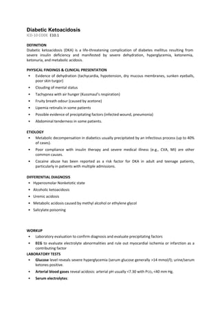
  • 1. Diabetic Ketoacidosis ICD-10 CODE E10.1 DEFINITION Diabetic ketoacidosis (DKA) is a life-threatening complication of diabetes mellitus resulting from severe insulin deficiency and manifested by severe dehydration, hyperglycemia, ketonemia, ketonuria, and metabolic acidosis. PHYSICAL FINDINGS & CLINICAL PRESENTATION • Evidence of dehydration (tachycardia, hypotension, dry mucous membranes, sunken eyeballs, poor skin turgor) • Clouding of mental status • Tachypnea with air hunger (Kussmaul’s respiration) • Fruity breath odour (caused by acetone) • Lipemia retinalis in some patients • Possible evidence of precipitating factors (infected wound, pneumonia) • Abdominal tenderness in some patients. ETIOLOGY • Metabolic decompensation in diabetics usually precipitated by an infectious process (up to 40% of cases). • Poor compliance with insulin therapy and severe medical illness (e.g., CVA, MI) are other common causes. • Cocaine abuse has been reported as a risk factor for DKA in adult and teenage patients, particularly in patients with multiple admissions. DIFFERENTIAL DIAGNOSIS • Hyperosmolar Nonketotic state • Alcoholic ketoacidosis • Uremic acidosis • Metabolic acidosis caused by methyl alcohol or ethylene glycol • Salicylate poisoning WORKUP • Laboratory evaluation to confirm diagnosis and evaluate precipitating factors • ECG to evaluate electrolyte abnormalities and rule out myocardial ischemia or infarction as a contributing factor LABORATORY TESTS • Glucose level reveals severe hyperglycemia (serum glucose generally >14 mmol/l); urine/serum ketones positive. • Arterial blood gases reveal acidosis: arterial pH usually <7.30 with PCO2 <40 mm Hg. • Serum electrolytes:
  • 2. 1. Serum bicarbonate is usually <18 mmol/l. 2. Serum potassium may be low, normal, or high. There is always significant total body potassium depletion regardless of the initial potassium level. 3. Serum sodium is usually decreased as a result of hyperglycemia, dehydration, and lipemia. • Complete blood count with differential, urinalysis, and urine and blood cultures to rule out infectious precipitating factor. • Serum calcium, magnesium, and phosphorus; the plasma phosphate and magnesium levels may be significantly depressed and should be rechecked within 24 hr because they may decrease further with correction of DKA. • Blood urea nitrogen and creatinine generally reveal significant dehydration. • Amylase and liver enzymes should be checked in patients with abdominal pain. IMAGING STUDIES Chest radiographs are helpful to rule out infectious process. NONPHARMACOLOGIC THERAPY • Monitor mental status, vital signs, and urine output hourly until improved, then monitor q2-4h. • Monitor electrolytes, ph, renal function, and ketonuria and glucose level. ACUTE GENERAL Rx Fluid replacement (usual deficit is 6 to 8 L): 1. Do not delay fluid replacement until laboratory results have been received. Fluid deficits are typically 100 ml/kg of body weight. 2. The initial fluid replacement should be with 0.9% NS until blood pressure and organ perfusion are restored (usually ≥1 L). In patients with severe hypernatremia (serum sodium >160 mEq/L), 0.45% saline infusion can be used. Careful monitoring for fluid overload is necessary in elderly patients and those with a history of congestive heart failure. 3. The rate of fluid replacement varies with the age of the patient and the presence of significant cardiac or renal disease. • The usual rate of infusion is 1 L over the first hour and 300-500 ml/hr for the next 12 hr. • Continue the infusion using NS until the serum glucose level is <14mmol/l, then change the hydrating solution to D5W to prevent hypoglycemia, replenish free water, and introduce additional glucose substrate (necessary to suppress lipolysis and ketogenesis). Insulin administration: 1. Insulin replacement should not be started until serum potassium is >3.3 mEq/L to prevent life-threatening hypokalemia. (Check ABG Potassium level)
  • 3. The patient should be given an initial loading IV bolus of 0.15 to 0.2 U/kg of regular insulin followed by a constant infusion at a rate of 0.1 U/kg/hr (e.g., 50 U of regular insulin in 200 ml of 0.9% saline solution at 20 ml/hr equals 5 U/hr for a 50-kg patient). 2. Monitor serum glucose hourly for the first 2 hr, and then monitor q2-4h. 3. The goal is to decrease serum glucose level by 5 mmol/l/hr (after an initial drop because of rehydration); if the serum glucose level is not decreasing at the expected rate, increase infusion rate by 1 units/hr (4ml/hr). 4. When the serum glucose level approaches 14mmol/l, decrease the rate of insulin infusion by halve and continue this rate until the patient has received adequate fluid replacement, ph and HCO- 3 are normal, and ketones have cleared. 5. Approximately 120 min before stopping the IV insulin infusion, administer a SC dose of regular insulin. Electrolyte replacement: • Potassium replacement: 1. As a rule of thumb, potassium replacement may be started when there is no ECG evidence of hyperkalemia. 2. In patients with normal renal function, potassium replacement can be started by adding 20 mmol KCl/L of IV hydrating solution if serum potassium is 3, 5 to 5, 5 mEq/L. In patients with severe hypokalemia (potassium <3,5mmol/L), give 40 mEq of potassium/hr until potassium is >3,5mmol/L. 3. Monitor serum potassium level hourly for the first 2 hr, and then monitor q2-4h. • Phosphate replacement is indicated only in the presence of significant hypophosphatemia. • Magnesium replacement is indicated only in the presence of significant hypomagnesemia or refractory hypokalemia. Bicarbonate therapy: • Give 50 mmol/l NaHCO3 in 200 ml 0.9% saline over 1 h Until pH increases to >7. Patients with DKA should be admitted to a high care unit. Intensive care admition must be considered in patients with altered level of consciousness, severe metabolic acidosis not responding well to treatment and in those requiring support of vital organs. COMMENTS • Although DKA occurs more commonly in type 1 diabetes mellitus, a significant proportion (>20%) occurs in patients with type 2 diabetes. • 20% of DKA admissions involve newly diagnosed diabetics.
  • 4. • Potential complications of DKA therapy include hypoglycemia, cerebral edema, and cardiac arrhythmias.
  • 5. Recommended fluids 0.9% Sodium chloride 1L Over 1 hour. Next 0.9% Sodium chloride 1L Over 2 hours. Next 0.9% Sodium chloride 1L Over 3 hour. Next 0.9% Sodium chloride 1L Over 4 hours. Next 0.9% Sodium chloride 1L Every 6 hours Bicarbonate therapy FLUIDS AND ELECTROLYTES Hypotensive patient (<90mmHg) - Severely depleted – give 500ml of 0.9% sodium chloride IV over 15 to 20 minutes - Repeat until SBP >90mmHg (Maximum of 3 doses) - Do not give potassium chloride in first litre or if serum potassium (K+) > 5.5 mmol/L - All subsequent fluid for the next 24 hours should contain KCl unless urine output is <30ml/hr or serum potassium remains in excess of 5.5mmol/l Not Hypotensive (= >90mmHg) Recommended potassium replacement Potassium Level Replacement Litre Fluid > 5,5 mmol/l nil 3,5 – 5,5 mmol/l 20 mmol/l < 3,5 mmol /l 40 mmol/l When BG less than 14mmol/L Start: 5% de Glucose 1l over 6 hours. Start: 10% glucose 1 l over 6 hours. -Insulin replacement should not be-Insulin replacement should not be started until serum potassium is >3.3 mmol/L to prevent life-threateningmmol/L to prevent life-threateningmmol/L to prevent life-threatening hypokalemia. (Check ABG Potassium - Initial IV bolus of 0.2 U/kg of regular 50 units Actrapid or Humulin Rid or Humulin R,, made uup to 200 ml with 0.9% Sodium chloride.e. kg/h. @20mls /hr =5u/hr for a 50kgs patient - sulin - ol/h. its/h -When BG level approaches 14mmol/l, decrease the rate of insulin infusion bydecrease the rate of insulin infusion by halve When Stable (pH>7.3) - Patient is eating and drinking, Change to S/C insulin regimen. - Stop IV insulin pump after 2 hours after the first sc insulin dose Monitoring Level of consciousness, Pulse, BP, O2 sats, urine output and capillary glucose should be monitored and recorded in the notes - Measure glucose hourly - Blood gases (0, 2, 4, 8 and 12 hrs.) And before stopping fixed rate insulin regime. - Monitor hourly urine output - Check magnesium level Remember CXR and ECG - Look for infections. - Treat precipitant factors. - Use of prophylactic anticoagulation in comatose, obese and hyperosmolar patients - Please use the patient data flowsheet MANAGEMENT OF DIABETIC KETOACIDOSIS.DEPARTMENT OF INTERNAL MEDICINE.KIMBERLEY HOSPITAL COMPLEX.2011
  • 6. DKA Patient data flow sheet Patient name: _________________________ Folder No: _______________ Date: __________ Hour Vital signs Chemistries Blood Gases Insulin (units in past hour) Fluid/Metabolites (Past hours) Output Other Level of consciousness Temperature Pulse Respiratory rate Blood pressure Capillary Glucose Urine Ketones Serum Na+ Serum K + Serum Cl - Serum HCO3 Urea pH Specify venous (V) or Arterial (A) pO2 O2 sat pCO2 Route IV SC IM 0.45 percent NaCl (ml) 0.9 percent NaCl (ml) 5 percent dextrose (ml) Others Urine (ml)
  • 7. DKA Patient data flow sheet Patient name: _________________________ Folder No: _______________ Date: __________ Hour Vital signs Chemistries Blood Gases Insulin (units in past hour) Fluid/Metabolites (Past hours) Output Other Level of consciousness Temperature Pulse Respiratory rate Blood pressure Capillary Glucose Urine Ketones Serum Na+ Serum K + Serum Cl - Serum HCO3 Urea pH Specify venous (V) or Arterial (A) pO2 O2 sat pCO2 Route IV SC IM 0.45 percent NaCl (ml) 0.9 percent NaCl (ml) 5 percent dextrose (ml) Others Urine (ml)