
Recommended
More Related Content
What's hot
What's hot (20)
Similar to Histopathology of granulomatous liver disease
Similar to Histopathology of granulomatous liver disease (20)
Recently uploaded
Recently uploaded (20)
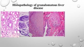
Histopathology of granulomatous liver disease
- 1. Histopathology of granulomatous liver disease
- 2. Presentation outlines Introduction Differential diagnosis Primary biliary cirrhosis Sarcoidosis Infection Drug-induced liver injury Lipogranuloma
- 3. Granuloma Defined as A histologic pattern of tissue reaction which appears following cell injury. Granulomatous inflammation is caused by a variety of conditions including infection, autoimmune, toxic, allergic, drug, and neoplastic conditions. The tissue reaction pattern narrows the pathologic and clinical differential diagnosis and subsequent clinical management From a morphological standpoint, granulomas may be either loose or well-formed (epithelioid) , caseating or noncaseating. Sometimes the histiocytic will fuse together forming multinucleated giant cells. Granulomas are relatively common in liver samples, identified in 2% to 10% of cases. Clinically, they may be restricted to the liver or reflect hepatic involvement by a systemic process. Histologically, they may be seen in the portal tracts, hepatic lobules, or both.
- 4. Differential diagnosis of liver granulomatous disease
- 8. Primary biliary cholangitis Primary biliary cholangitis (PBC), previously known as primary biliary cirrhosis Definition Is an autoimmune disease of the liver. It results from a slow, progressive destruction of the small bile ducts of the liver, causing bile and other toxins to build up in the liver, a condition called cholestasis. Further slow damage to the liver tissue can lead to scarring, fibrosis, and eventually cirrhosis. Causes: most people >90 % have anti-mitochondrial antibodies (AMAS) against pyruvate dehydrogenase complex (pdc-e2), an enzyme complex that is found in the mitochondria
- 9. Primary biliary cholangitis Signs and symptoms:- fatigue dry skin and eyes Itching (pruritus) jaundice in severe PBC. Impairs bone density with increased risk of fracture. Xanthelasma may be present as a result of increased cholesterol levels. Complications :- (progress to cirrhosis) ascites, enlarged spleen, oesophageal varices and hepatic encephalopathy, including coma in extreme cases in more advanced disease.
- 10. Primary biliary cholangitis Pathogenesis Evidence for autoimmune etiology • Aberrant expression MHC class II molecules on bile duct epithelial cells • Autoreactive T cells around bile ducts • Hypergammaglobulinemia with complement activation and circulating immune complexes Associated extrahepatic autoimmune diseases • Sjogren syndrome, scleroderma, thyroiditis, rheumatoid arthritis, membranous glomerulonephritis
- 11. Primary biliary cholangitis Diagnose • Elevated gamma-glutamyl transferase and alkaline phosphatase (alp) are found in early disease. Elevations in bilirubin occur in advanced disease. • Antimitochondrial antibodies are the characteristic serological marker for pbc, being found In 90-95 percent of patients. • Abdominal ultrasound, MRI scanning (mrcp) or a CT scan is usually performed to rule out blockage to the bile ducts. An endoscopic investigation of the bile duct, may be performed.
- 12. Primary biliary cholangitis Morphology • Most patients can be diagnosed without invasive investigation however, a liver biopsy is needed to determine the stage of disease. The florid duct lesion, chronic nonsuppurative destructive cholangitis, is the pathological hallmark. • Granulomas are essential component of the florid duct lesion. Although they may rarely be epithelioid, a more typical appearance is that of a loose collection of histiocytes surrounding an injured bile duct. • There is disruption of the bile duct basement membrane and infiltration by lymphocytes. • Presence of florid duct lesions varies with the stage of the disease. They are more common early in the course and diminish in frequency as PBC progresses and burns out.
- 13. Histopathology stages of PBC:- Stage 1 – portal stage: • normal sized triads; portal inflammation, subtle bile duct damage. Granulomas are often detected in this stage. Stage 2 – periportal stage: • enlarged triads; periportal fibrosis and/or inflammation. Typically characterized by the finding of a proliferation of small bile ducts. Stage 3 – septal stage • active and/or passive fibrous septa. Stage 4 – biliary cirrhosis: • nodules present; garland or jigsaw puzzle pattern.
- 14. Heavily inflamed portal tract containing a typical florid duct lesion. The bile duct (white arrow) is encircled by a loose aggregate of histiocytes (black arrows). Dense lymphoplasmacytic inflammation is also present in the portal tract histiocytes and lymphocytes of the florid duct lesion are directly injuring the bile duct, with disruption of the basement membrane and nuclear irregularity.
- 15. PBC: EARLY HISTOPATHOLOGY Early stage PBC (florid duct lesion): portal mononuclear inflammation destroying bile duct with granulomatous response (arrowheads) Fig. 18-35, PBFD 9th ed, 2015 Residual bile duct epithelium
- 16. Granulomatous cholangitis in primary biliary cirrhosis (PBC). Detail from a portal tract with dense lymphoplasmacytic infiltrate and lymphoid aggregate (left). The interlobular bile duct (center) shows a focal rupture of its cholangiocytic lining, at the site of an adjacent epithelioid granuloma. (H&E)
- 17. Granulation tissue with a poorly formed granuloma to the left of centre. Within this area there is a multinucleate giant cell of the Langhans type
- 18. (A) Primary biliary cirrhosis. Note the significant inflammatory response and expansion of portal areas in a granulomatous reaction. (B) note the inflammatory cells spilling over into the adjacent liver lobule resulting in an interface hepatitis (arrow).
- 19. Sarcoidosis is a multisystem condition of unknown etiology thought to represent a reaction to one or more antigens. Worldwide prevalence is 2 to 60/100,000 people. It is defined by the presence of noncaseating epithelioid granulomas in affected organs or sites. Most frequent sites of involvement are the lung (>90%), lymph nodes, and liver. Although liver involvement is common (50%-70% of sarcoid livers histologically contain granulomas), only 20% to 40% of patients will have elevated lfts. Less than 20% of patients will have clinically significant liver disease.
- 20. Sarcoidosis Noncaseating epithelioid granulomas are present in both the portal tracts and hepatic lobules. Roughly one-third of liver biopsies of hepatic sarcoidosis may have changes similar to those of pbc (19%) or primary sclerosing cholangitis (13%).However, in the majority of cases, the granulomatous infiltrates of PBC and sarcoidosis are sufficiently different to allow one to distinguish them. Sarcoid granulomas are typically larger, more numerous, better defined, not directed at the bile duct, and often associated with multinucleated giant cells.
- 21. Large, noncaseating granuloma from patient with sarcoid.
- 22. Tight, well-formed, noncaseating epithelioid granuloma of sarcoidosis. There is a cuff of lymphocytes (black arrows) and Langhans giant cell (white arrow). Note how the nuclei of the giant cell are arranged in a ring along the periphery, in contrast with foreign body type giant cells. Although not specific, Langhans type giant cells are characteristic of sarcoidosis
- 23. In contrast with PBC, the epithelioid granuloma of sarcoidosis (open arrow) is well formed and usually not directed at the bile duct (closed arrow).
- 24. In some cases, the epithelioid granulomas (white arrows) fuse to become tumefactive nodules. Note the numerous giant cells that are present (black arrows)
- 25. (A) Sarcoidosis. Note the portal distribution and excessive fibrosis around granuloma (arrow; trichrome stain) (B) sarcoidosis, extensive fibrosis (arrow; trichrome stain
- 26. Histological and clinical features of PBC and sarcoidosis Diagnosis Granuloma Location Bile Duct Destruction Granuloma Type Confluent Granulomas Giant Cells AMA Systemic Disease PBC Portal based Present Poorly formed Absent Absent to rare Positive Absent Sarcoidosis Portal and lobular Absent to rare Well formed Occasional Common Negative Present
- 27. Infection A variety of bacterial, viral, fungal, and parasitic organisms may induce a granulomatous reaction in the liver. From a practical, clinical perspective, special stains for acid-fast bacilli and fungal organisms (GMS or periodic acid–schiff) are performed anytime a newly diagnosed granulomatous process is encountered in the liver. Granulomas with central, caseating necrosis are considered infectious until proven otherwise. Conditions such as PBC and sarcoidosis are not associated with caseating necrosis.
- 28. Caseating epithelioid granuloma: typically associated with infections, most notably tuberculosis. Suppurative granuloma: (granulomatous inflammation with stellate abscess formation or suppurative inflammation): bartonellosis, yersinosis, listerosis, actinomycosis and fungal infection. Fibrin ring granuloma: (also known as doughnut granuloma; central fat globule surrounded by a circumferential rim of fibrin and histiocytes)could be associated with mycobacterial infection, staphylococcal bacteremia, leishmaniasis, toxoplasmosis, cytomegalovirus, epstein-barr virus, acute hepatitis a, systemic lupus erythematosus, allopurinol toxicity and lymphoma.
- 29. The characteristic histologic lesion of hepatic cat scratch disease features irregular, stellate microabscesses surrounded by an inner layer of palisading histiocytes, a surrounding rim of lymphocytes, and an outermost thick layer of fibrous tissue
- 30. (A) Granulomatous hepatitis associated with AIDS. Note the ill-defined, nature of granulomas (arrows) (B) example of a better-defined granuloma in granulomatous hepatitis of AIDS. (C) large numbers of Mycobacterium avium intracellulare within and around loosely arranged aggregates of epithelioid histiocytes (arrow). (D) clusters of acid fast bacilli (arrow; Fite acid fast stain)
- 31. Q fever is characterized by hepatitis with granulomas showing a central clearing surrounded by a fine hyaline ring. However, not all granulomas in Q fever show this feature. Conversely, other processes, such as allopurinol toxicity and cytomegalovirus mononucleosis, may also have such “donut-hole” granulomas
- 32. Fibrin ring granuloma central fat globule surrounded by a circumferential rim of fibrin and histiocytes
- 33. Liver biopsy showing a granulomatous reaction against the ovum. Schistosomiasis of liver. Note Schistosoma hematobium egg with terminal spine in the center
- 34. Drug-induced liver injury Drug-induced liver injury (DILI) most commonly manifests histologically as cholestasis. Granulomas may be identified in a minority of cases. One study demonstrated granulomatous hepatitis in 3.6% of patients with documented DILI. Granulomas are not tumefactive (as seen in sarcoidosis) or typically directed at the bile ducts (as seen in PBC). Instead, they are usually small and present within the hepatic lobule, accompanied by a portal and lobular inflammatory infiltrate (i.E. Granulomatous hepatitis). Eosinophils are typically present. Numerous drugs and medications have been implicated in granulomatous dili, with the most common being antibiotics, nonsteroidal anti-inflammatory and antirheumatic agents, antituberculous medications, and antiepileptics.
- 35. small, lobular microgranuloma. Microgranulomas such as this are typically less than seven cells in diameter. Note its presence next to a central vein in zone 3 of the hepatic lobule.
- 36. (A) Granulomatous hepatitis, hypersensitivity to medication. Notice the lobular distribution of granuloma (arrow). (B) extensive eosinophilic reaction in the inflammatory cuff of the granuloma (arrow).
- 37. Lipogranuloma The lipogranuloma is a special type of granuloma composed of lipid-containing macrophages with associated lymphocytes. Initial studies suggested that these formed as a reaction to mineral oil in some foods and medications. However, it has subsequently been demonstrated that they may also form when histiocytes ingest fat droplets released by ruptured hepatocytes in the setting of fatty liver disease.
- 38. central fat globule surrounded by histiocytes
- 39. (A) Granulomatous response in liver (black arrow) associated with a metastatic malignant neoplasm, an islet cell carcinoma, a neuroendocrine neoplasm (red arrow) (B) higher power view of the epithelioid granuloma (arrow)
- 40. SUMMARY
- 41. References • STERNBERG’S SURGICAL PATHOLOGY • ROSAI AND ACKERMAN‘ S SURGICAL PATHOLOGY • LEWIS, J., 2018. HISTOPATHOLOGY OF GRANULOMATOUS LIVER DISEASE. CLINICAL LIVER DISEASE, 11(3), PP.77–80. AVAILABLE AT: HTTP://DX.DOI.ORG/10.1002/CLD.695.