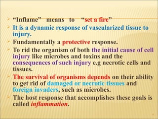
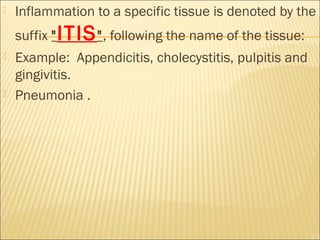
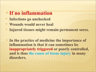
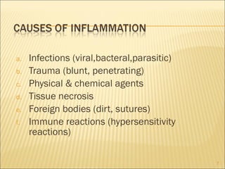
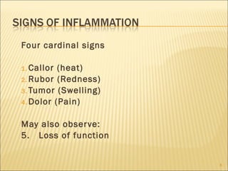
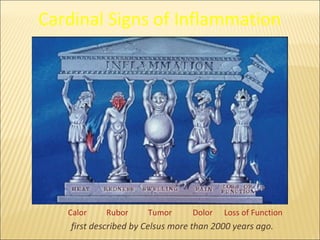
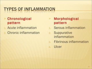
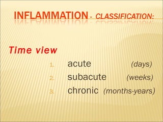
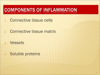
By

Dr Mohammad Manzoor
Mashwani
   What is inflammation?
   Causes
   Signs
   Types
   Components
   Functions of inflammation
   Overview of inflammation
   Specific host defences
   Summary


                                2
3
   “Inflame” means to “set a fire”
   It is a dynamic response of vascularized tissue to
    injury.
   Fundamentally a protective response.
   To rid the organism of both the initial cause of cell
    injury like microbes and toxins and the
    consequences of such injury e.g necrotic cells and
    tissues.
   The survival of organisms depends on their ability
    to get rid of damaged or necrotic tissues and
    foreign invaders, such as microbes.
   The host response that accomplishes these goals is
    called inflammation.
                                                        4
   Inflammation to a specific tissue is denoted by the
    suffix " ITIS ", following the name of the tissue:
   Example: Appendicitis, cholecystitis, pulpitis and
    gingivitis.
   Pneumonia .
 If   no inflammation
   Infections go unchecked
   Wounds would never heal
   Injured tissues might remain permanent sores.

   In the practice of medicine the importance of
    inflammation is that it can sometimes be
    inappropriately triggered or poorly controlled,
    and is thus the cause of tissue injury in many
    disorders.

                                                      6
a.   Infections (viral,bacteral,parasitic)
b.   Trauma (blunt, penetrating)
c.   Physical & chemical agents
d.   Tissue necrosis
e.   Foreign bodies (dirt, sutures)
f.   Immune reactions (hypersensitivity
     reactions)

                                             7
Four cardinal signs

1. Callor (heat)
2. Rubor (Redness)
3. Tumor (Swelling)
4. Dolor (Pain)


May also obser ve:
5. Loss of function


                      8
Cardinal Signs of Inflammation


   Calor    Rubor      Tumor      Dolor   Loss of Function
    first described by Celsus more than 2000 years ago.
    Chronological              Morphological
     pattern                     pattern
1.   Acute inflammation     1.   Serous inflammation
2.   Chronic inflammation   2.   Suppurative
                                 inflammation
                            3.   Fibrinous inflammation
                            4.   Ulcer


                                                      10
Time view
       1.   acute          (days)
       2.   subacute      (weeks)
       3.   chronic (months-years)
1.   Connective tissue cells

2.   Connective tissue matrix

3.   Vessels

4.   Soluble proteins


                                12
1. Destroy and remove pathogens.
2. If destruction is not possible, to limit effects by
  confining the pathogen and its products.
3. Repair and replace tissue damaged by
  pathogen and its products.


                                                         13
    Inflammation is a complex reaction of responses of blood
     vessels and leukocytes.
    Principal defenders are:
1.   Plasma proteins
2.   Leukocytes &
3.   Tissue phagocytes derived from circulation.
     Being in blood they can home any site where they may be
      needed.
    As invaders such as microbes and necrotic cells are
     typically present in tissues, outside the circulation, it
     follows that the circulating cells and proteins have to be
     rapidly recruited to these extravascular sites.
    The inflammatory response coordinates to achieve this
     goal.
                                                                  14
   Inflammator y response
    repair
   It destroys, dilutes, and walls of f the
    injurious agent, series of events that tr y
    to heal the damaged tissue.
   Repair begins during inflammation but
    reaches completion usually af ter the
    injurious influence has been
    neutralized.
   In the process of repair the injured
    tissue is replaced through regeneration
    of native parenchymal cells, by filling of
    the defect with fibrous tissue (scarring)
    or, most commonly, by a combination of        15
   Inflammation may be harmful in some
    situations.
   When inappropriately directed against self tissues or is
    not adequately controlled, it becomes the cause of injury
    and disease.
   Inflammatory reactions underlie common chronic
    diseases, such as rheumatoid arthritis, atherosclerosis,
    and lung fibrosis, as well as life-threatening
    hypersensitivity reactions to insect bites, drugs, and
    toxins.
   Anti-inflammatory drugs, control the harmful sequelae
    of inflammation yet not interfere with its beneficial
    effects.
   Inflammation may contribute to a variety of diseases that
    are not thought to be primarily due to abnormal host
    responses.
                                                                16
Host
Defenses
           17
Introduction
Resistance: Ability to ward off (prevent)
  disease.
  1.   Nonspecific Resistance: Defenses that protect
       against all pathogens.
  2.   Specific Resistance: Protection against
       specific pathogens.


Susceptibility: Vulnerability or lack of
  resistance.
                                                       18
1.   Mechanical Defenses
2.   Chemical Defenses
3.   Cellular elements of blood


                                  19
20
I.Mechanical Defenses
Skin and Mucous Membranes


A. Skin Layers
Epidermis: Thin outer epithelial layer.
 Contains Langerhans cells, dead cells, and keratin.
Dermis: Thick inner layer of connective tissue.

Infections are rare in intact skin. Exceptions…….


                                                       21
   B. Mucous Membranes:
Lining of gastrointestinal, genitourinary, and
respiratory tracts.

   Epithelial layer secretes mucus which maintains moist surfaces.
   Although they inhibit microbial entry, they offer less protection
    than skin.
   Several microorganisms are capable of penetrating mucous
    membranes:
        Papilloma-virus
        Treponema pallidum
        Enteroinvasive E. coli
        Entamoeba histolytica

                                                                    22
   Lacrimal apparatus: Continual washing and
    blinking prevents microbes from settling on the
    eye surface.
    Saliva: Washes microbes from teeth and
    mouth mucous membranes.
    Mucus: Thick secretion that traps many
    microbes.
   Nose Hair: Coated with mucus, filter dust,
    pollen, and microbes.
   Ciliar y Escalator: Cilia on mucous
    membranes of lower respiratory tract move
    upwards towards throat at 1-3 cm/hour.
   Coughing and sneezing: Expel foreign
    objects.
                                                      23
II.Chemical Defenses:
    Sebum: Oily substance produced by sebaceous
     glands that forms a protective layer over skin.
     Contains unsaturated fatty acids which inhibit
     growth of certain pathogenic bacteria and fungi.
    pH: Low, skin pH usually between 3 and 5.
     Caused by lactic acid and fatty acids.
    Perspiration: Produced by sweat glands.
     Contains lysozyme and acids.
    Lysozyme: Enzyme that breaks down gram-
     positive cell walls. Found in nasal secretions,
     saliva, and tears.
                                                        24
   Gastric Juice: Mixture of hydrochloric acid,
    enzymes, and mucus. pH between 1.2 to 3 kills
    many microbes and destroys most toxins. Many
    enteric bacteria are protected by food particles.
        Helicobacter pylori neutralizes stomach acid and can
         grow in the stomach, causing gastritis and ulcers.


   Transferrins: Iron-binding proteins in blood
    which inhibit bacterial growth by reducing
    available iron.


                                                                25
Cell Type                           # Cells/mm 3                    Function

Erythrocytes (RBC)       4.8-5.4 million     Transport O2 and CO2

Leukocytes (WBC          5000-11000                Various
   A. Granulocytes:
   1. Neutrophils                                     Phagocytosis
   2. Basophils                                       Produce histamine
   3. Eosinophils                                     Toxins against parasites
                                                                          some
   phagocytosis
   B. Monocytes/Macrophages                          Phagocytosis
   C. Lymphocytes                                     Antibody production (B cells)
                                                     Cell mediated immunity (T
   cells)
Platelets             300,000               Blood clotting


                                                                                      26
II. Second Line of Defense
1. Phagocytosis:
 Derived from the Greek words “Eat - cell”.

 Phagocytosis is carried out by white blood cells:
  macrophages, neutrophils, and occasionally
  eosinophils.
 Neutrophils predominate early in infection.

 Wandering macrophages: Originate from monocytes
  that leave blood and enter infected tissue, and develop
  into phagocytic cells.
 Fixed Macrophages (Histiocytes): Located in liver,
  nervous system, lungs, lymph nodes, bone marrow,
  and several other tissues.
                                                       27
Stages of Phagocytosis

1. Chemotaxis: Phagocytes are chemically attracted
  to site of infection.
2. Adherence: Phagocyte plasma membrane attaches
  to surface of pathogen or foreign material.
     Adherence can be inhibited by capsules (S. pneumoniae) or
      M protein (S. pyogenes).
      Opsonization: Coating process with opsonins that
      facilitates attachment.
          Opsonins include antibodies and complement proteins.


                                                                  28
29
Stages of
Phagocytosis
3. Ingestion: Plasma membrane of phagocytes
  extends projections (pseudopods) which engulf the
  microbe. Microbe is enclosed in a sac called
  phagosome.
4. Digestion: Inside the cell, phagosome fuses with
  lysosome to form a phagolysosome.
  Lysosomal enzymes kill most bacteria within minutes
  and include:
     Lysozyme: Destroys cell wall peptidoglycan
     Lipases and Proteases
     RNAses and DNAses
 After digestion, residual body with undigestable
                                                        30
31
Inflammation overview

Inflammation overview

  • 1.
  • 2.
    What is inflammation?  Causes  Signs  Types  Components  Functions of inflammation  Overview of inflammation  Specific host defences  Summary 2
  • 3.
  • 4.
    “Inflame” means to “set a fire”  It is a dynamic response of vascularized tissue to injury.  Fundamentally a protective response.  To rid the organism of both the initial cause of cell injury like microbes and toxins and the consequences of such injury e.g necrotic cells and tissues.  The survival of organisms depends on their ability to get rid of damaged or necrotic tissues and foreign invaders, such as microbes.  The host response that accomplishes these goals is called inflammation. 4
  • 5.
    Inflammation to a specific tissue is denoted by the suffix " ITIS ", following the name of the tissue:  Example: Appendicitis, cholecystitis, pulpitis and gingivitis.  Pneumonia .
  • 6.
     If no inflammation  Infections go unchecked  Wounds would never heal  Injured tissues might remain permanent sores.  In the practice of medicine the importance of inflammation is that it can sometimes be inappropriately triggered or poorly controlled, and is thus the cause of tissue injury in many disorders. 6
  • 7.
    a. Infections (viral,bacteral,parasitic) b. Trauma (blunt, penetrating) c. Physical & chemical agents d. Tissue necrosis e. Foreign bodies (dirt, sutures) f. Immune reactions (hypersensitivity reactions) 7
  • 8.
    Four cardinal signs 1.Callor (heat) 2. Rubor (Redness) 3. Tumor (Swelling) 4. Dolor (Pain) May also obser ve: 5. Loss of function 8
  • 9.
    Cardinal Signs ofInflammation Calor Rubor Tumor Dolor Loss of Function first described by Celsus more than 2000 years ago.
  • 10.
    Chronological  Morphological pattern pattern 1. Acute inflammation 1. Serous inflammation 2. Chronic inflammation 2. Suppurative inflammation 3. Fibrinous inflammation 4. Ulcer 10
  • 11.
    Time view 1. acute (days) 2. subacute (weeks) 3. chronic (months-years)
  • 12.
    1. Connective tissue cells 2. Connective tissue matrix 3. Vessels 4. Soluble proteins 12
  • 13.
    1. Destroy andremove pathogens. 2. If destruction is not possible, to limit effects by confining the pathogen and its products. 3. Repair and replace tissue damaged by pathogen and its products. 13
  • 14.
    Inflammation is a complex reaction of responses of blood vessels and leukocytes.  Principal defenders are: 1. Plasma proteins 2. Leukocytes & 3. Tissue phagocytes derived from circulation.  Being in blood they can home any site where they may be needed.  As invaders such as microbes and necrotic cells are typically present in tissues, outside the circulation, it follows that the circulating cells and proteins have to be rapidly recruited to these extravascular sites.  The inflammatory response coordinates to achieve this goal. 14
  • 15.
    Inflammator y response repair  It destroys, dilutes, and walls of f the injurious agent, series of events that tr y to heal the damaged tissue.  Repair begins during inflammation but reaches completion usually af ter the injurious influence has been neutralized.  In the process of repair the injured tissue is replaced through regeneration of native parenchymal cells, by filling of the defect with fibrous tissue (scarring) or, most commonly, by a combination of 15
  • 16.
    Inflammation may be harmful in some situations.  When inappropriately directed against self tissues or is not adequately controlled, it becomes the cause of injury and disease.  Inflammatory reactions underlie common chronic diseases, such as rheumatoid arthritis, atherosclerosis, and lung fibrosis, as well as life-threatening hypersensitivity reactions to insect bites, drugs, and toxins.  Anti-inflammatory drugs, control the harmful sequelae of inflammation yet not interfere with its beneficial effects.  Inflammation may contribute to a variety of diseases that are not thought to be primarily due to abnormal host responses. 16
  • 17.
  • 18.
    Introduction Resistance: Ability toward off (prevent) disease. 1. Nonspecific Resistance: Defenses that protect against all pathogens. 2. Specific Resistance: Protection against specific pathogens. Susceptibility: Vulnerability or lack of resistance. 18
  • 19.
    1. Mechanical Defenses 2. Chemical Defenses 3. Cellular elements of blood 19
  • 20.
  • 21.
    I.Mechanical Defenses Skin andMucous Membranes A. Skin Layers Epidermis: Thin outer epithelial layer. Contains Langerhans cells, dead cells, and keratin. Dermis: Thick inner layer of connective tissue. Infections are rare in intact skin. Exceptions……. 21
  • 22.
    B. Mucous Membranes: Lining of gastrointestinal, genitourinary, and respiratory tracts.  Epithelial layer secretes mucus which maintains moist surfaces.  Although they inhibit microbial entry, they offer less protection than skin.  Several microorganisms are capable of penetrating mucous membranes:  Papilloma-virus  Treponema pallidum  Enteroinvasive E. coli  Entamoeba histolytica 22
  • 23.
    Lacrimal apparatus: Continual washing and blinking prevents microbes from settling on the eye surface.  Saliva: Washes microbes from teeth and mouth mucous membranes.  Mucus: Thick secretion that traps many microbes.  Nose Hair: Coated with mucus, filter dust, pollen, and microbes.  Ciliar y Escalator: Cilia on mucous membranes of lower respiratory tract move upwards towards throat at 1-3 cm/hour.  Coughing and sneezing: Expel foreign objects. 23
  • 24.
    II.Chemical Defenses:  Sebum: Oily substance produced by sebaceous glands that forms a protective layer over skin. Contains unsaturated fatty acids which inhibit growth of certain pathogenic bacteria and fungi.  pH: Low, skin pH usually between 3 and 5. Caused by lactic acid and fatty acids.  Perspiration: Produced by sweat glands. Contains lysozyme and acids.  Lysozyme: Enzyme that breaks down gram- positive cell walls. Found in nasal secretions, saliva, and tears. 24
  • 25.
    Gastric Juice: Mixture of hydrochloric acid, enzymes, and mucus. pH between 1.2 to 3 kills many microbes and destroys most toxins. Many enteric bacteria are protected by food particles.  Helicobacter pylori neutralizes stomach acid and can grow in the stomach, causing gastritis and ulcers.  Transferrins: Iron-binding proteins in blood which inhibit bacterial growth by reducing available iron. 25
  • 26.
    Cell Type # Cells/mm 3 Function Erythrocytes (RBC) 4.8-5.4 million Transport O2 and CO2 Leukocytes (WBC 5000-11000 Various A. Granulocytes: 1. Neutrophils Phagocytosis 2. Basophils Produce histamine 3. Eosinophils Toxins against parasites some phagocytosis B. Monocytes/Macrophages Phagocytosis C. Lymphocytes Antibody production (B cells) Cell mediated immunity (T cells) Platelets 300,000 Blood clotting 26
  • 27.
    II. Second Lineof Defense 1. Phagocytosis:  Derived from the Greek words “Eat - cell”.  Phagocytosis is carried out by white blood cells: macrophages, neutrophils, and occasionally eosinophils.  Neutrophils predominate early in infection.  Wandering macrophages: Originate from monocytes that leave blood and enter infected tissue, and develop into phagocytic cells.  Fixed Macrophages (Histiocytes): Located in liver, nervous system, lungs, lymph nodes, bone marrow, and several other tissues. 27
  • 28.
    Stages of Phagocytosis 1.Chemotaxis: Phagocytes are chemically attracted to site of infection. 2. Adherence: Phagocyte plasma membrane attaches to surface of pathogen or foreign material.  Adherence can be inhibited by capsules (S. pneumoniae) or M protein (S. pyogenes).  Opsonization: Coating process with opsonins that facilitates attachment.  Opsonins include antibodies and complement proteins. 28
  • 29.
  • 30.
    Stages of Phagocytosis 3. Ingestion:Plasma membrane of phagocytes extends projections (pseudopods) which engulf the microbe. Microbe is enclosed in a sac called phagosome. 4. Digestion: Inside the cell, phagosome fuses with lysosome to form a phagolysosome. Lysosomal enzymes kill most bacteria within minutes and include:  Lysozyme: Destroys cell wall peptidoglycan  Lipases and Proteases  RNAses and DNAses After digestion, residual body with undigestable 30
  • 31.