Bacteriology- Select gram ⊕ and gram ⊝ infections
•
11 likes•3,890 views

Bacteriology- Select gram ⊕ and gram ⊝ infections

Recommended
More Related Content
What's hot
What's hot (20)
Similar to Bacteriology- Select gram ⊕ and gram ⊝ infections
Similar to Bacteriology- Select gram ⊕ and gram ⊝ infections (20)
More from Imhotep Virtual Medical School
More from Imhotep Virtual Medical School (20)
Recently uploaded
Recently uploaded (20)
Bacteriology- Select gram ⊕ and gram ⊝ infections
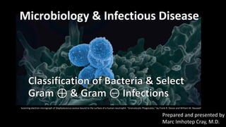
- 1. Microbiology & Infectious Disease Scanning electron micrograph of Staphylococcus aureus bound to the surface of a human neutrophil. “Granulocytic Phagocytes,” by Frank R. DeLeo and William M. Nauseef. Prepared and presented by Marc Imhotep Cray, M.D.
- 2. Marc Imhotep Cray, M.D. host-agent-environment interaction model Fundamental relationships involved in host-agent-environment interaction model In the host, pathogenetic mechanisms extend from level of populations (eg, person-to-person transmission) to level of cellular and molecular processes (eg, genetic susceptibility) Hammer GD and McPhee. Pathophysiology of Disease: An Introduction to Clinical Medicine. McGraw-Hill Education, 2014. The environment includes vectors (insects and other carriers that transmit infectious agents) and zoonotic hosts or reservoirs (animals that harbor infectious agents and often act to amplify the infectious agent)
- 3. Marc Imhotep Cray, M.D. Host Defenses Types= Host defenses include: Nonspecific, antigen-independent mechanisms like anatomic and physiological barriers including skin, lysozyme in tears, stomach acidity, etc. Specific antigen-dependent mechanisms involving humoral and cell-mediated immunity Host defenses can become harmful and contribute to disease process by inducing formation of autoantibodies (e.g. M proteins of S. pyogenes Ag-Ab complexes can damage kidneys and lead to acute poststreptococcal glomerulonephritis)
- 4. Marc Imhotep Cray, M.D. Hierarchy of Microbes: [Smallest Largest] 4 Viruses Bacteria Single-celled eukaryotes (animal-like cells): Parasitic protozoa Fungi/yeast Higher eukaryotes: Parasitic intestinal worms Tissue worms
- 5. Marc Imhotep Cray, M.D. Distribution 5 Microbes are everywhere and in high numbers 1 gram of human feces contains about 1 trillion bacteria
- 6. Marc Imhotep Cray, M.D. The World from the Microbe's Perspective 6 Microbes cannot think they simply exploit the human environment solely for growth and survival MISSION: “SURVIVE AND MULTIPLY TO ENSURE SURVIVAL OF THE SPECIES”
- 7. Marc Imhotep Cray, M.D. Common Clinical Ways of Categorizing Microbes 7 “Avirulent” or “nonpathogenic” - harmless “Virulent” or “pathogenic” - capable of causing disease Normal flora - microbes that colonize the body and usually do not cause disease Opportunistic pathogens - microbes that normally do not cause disease, but may under certain circumstances Frank pathogens - microbes that always cause disease Other - diseases caused mostly by ingestion of preformed toxins or when toxins are produced from bacteria during infection
- 8. Marc Imhotep Cray, M.D. Question What is the term for parasitic relationships between microorganisms and the human body in which the human body is harmed? a. Infectious disease b. Mutual disease c. Communicable disease d. Commensal disease
- 9. Marc Imhotep Cray, M.D. Answer: a RATIONALE: A parasitic relationship is one in which only the infecting organism benefits from the relationship and the host either gains nothing from the relationship or sustains injury from the interaction. If the host sustains injury or pathologic damage in response to a parasitic infection, the process is called an infectious disease. Mutual and commensal relationships do not harm the human body. Communicable diseases can be passed from one human to another; they are not considered parasitic.
- 10. Marc Imhotep Cray, M.D. Question Which of the following sequences accurately describes the stages of an infectious disease? a. Incubation, prodromal, current, recovery, and resolution b. Subacute, prodromal, acute, postacute, and convalescent c. Prodromal, subacute, acute, postdromal, and resolution d. Incubation, prodromal, acute, convalescent, and resolution.
- 11. Marc Imhotep Cray, M.D. Answer: d RATIONALE: The course of any infectious disease can be divided into several distinguishable stages after the point of time in which the potential pathogen enters the host. These stages are: The incubation period The prodromal stage The acute stage The convalescent stage, and The resolution stage There are no postacute, subacute or postdromal stages to an infectious disease.
- 12. Marc Imhotep Cray, M.D. In the figure below, label the areas that represent the course through which an infectious disease progresses: resolution, acute phase, convalescent phase, incubation phase, infection, and prodromal phase. Kipp B. Study Guide for Porth’s Essentials of Pathophysiology, 3rd Ed. Wolters Kluwer Health | Lippincott Williams & Wilkins, 2011.
- 13. Marc Imhotep Cray, M.D. Kipp B. Study Guide for Porth’s Essentials of Pathophysiology, 3rd Ed. Wolters Kluwer Health | Lippincott Williams & Wilkins, 2011.
- 14. Marc Imhotep Cray, M.D. Question There are two criteria that have to be met in order for a diagnosis of an infectious disease to occur. What are these two criteria? a. Recovery of probable pathogen and documentation of signs and symptoms compatible with an infectious process. b. Propagation of a microorganism outside the body and testing to see what destroys it. c. Identification by microscopic appearance and Gram stain reaction d. Serology and an antibody titer specific to the serology
- 15. Marc Imhotep Cray, M.D. Answer: a RATIONALE: The diagnosis of an infectious disease requires two criteria: the recovery of a probable pathogen or evidence of its presence from the infected sites of a diseased host, and accurate documentation of clinical signs and symptoms compatible with an infectious process. Culture and sensitivity (C&S) are the growing of microorganisms outside the body and the testing to see what kills it. Identifying a microorganism by microscopic appearance and Gram stain reaction are not the criteria for diagnosis. Serology, an indirect means of identifying infectious agents by measuring serum antibodies in the diseased host, and the quantification of those antibodies, an antibody titer, are not criteria for diagnosis.
- 16. Marc Imhotep Cray, M.D. Basic Concepts in Bacteriology
- 17. Marc Imhotep Cray, M.D. Bacterial structures Le T and Bhushan V. Microbiology. In: First Aid for the USMLE Step 1 2016. M-H, 2016.
- 18. Marc Imhotep Cray, M.D. Bacterial structures (2) Cell envelope 1. General structure= macromolecular layers that surround bacterium It includes: A cell membrane and a peptidoglycan layer except for mycoplasma An outer membrane layer in Gram-negative bacteria A capsule, a glycocalyx layer, or both (sometimes) Antigens that frequently induce a specific antibody response
- 19. Marc Imhotep Cray, M.D. Bacterial structures (2) Cell envelope 2. Cell wall portion of cell envelope that is external to cytoplasmic membrane and internal to capsule or glycocalyx It confers osmotic protection and Gram-staining characteristics In Gram-positive bacteria it is composed of: o Peptidoglycan o Teichoic acid o Polysaccharides In Gram-negative bacteria, it is composed of: o Peptidoglycan o Lipoprotein o An outer phospholipid membrane that contains lipopolysaccharide
- 20. Marc Imhotep Cray, M.D. Bacterial structures (3) Cell envelope 3. Peptidoglycan (also called mucopeptide or murein) is unique to prokaryotes It is found in all bacterial cell walls except Mycoplasma Structure o This complex polymer consists of a backbone composed of alternating N-acetylglucosamine and N-acetylmuramic acid and a set of identical tetrapeptide side chains Peptidoglycan is site of action of certain antibiotics such as penicillin and cephalosporins In Gram-positive bacteria, it comprises up to 50% of cell wall In Gram-negative bacteria, it comprises only 2% to 10% of cell wall
- 21. Marc Imhotep Cray, M.D. Bacterial structures/ Gram + Cell Envelope Johnson AG, Ziegler RJ & Hawley L. Microbiology and Immunology 5th ed. Baltimore: LLW, 2010.
- 22. Marc Imhotep Cray, M.D. Bacterial structures/Gram - Cell Envelope Johnson AG, Ziegler RJ & Hawley L. Microbiology and Immunology 5th ed. Baltimore: LLW, 2010.
- 23. Marc Imhotep Cray, M.D. Concept of "Pathogenesis" 23 Pathogenesis - the course of the infectious process "Virulence" factors or "pathogenicity" factors Microbes which can cause disease are thought to carry out process by utilizing one or more properties called virulence factors or pathogenicity factors IMPORTANT TO NOTE: The study of microbial virulence factors is a major emphasis In microbiology And infectious disease research. Understanding molecular mechanisms by which pathogens exert their virulence properties leads to new antibiotics and vaccines.
- 24. Marc Imhotep Cray, M.D. Mechanisms of Pathogenesis Bacteria carry a variety of virulence factors that facilitate pathogenesis: These include: factors that help them evade immune system factors that facilitate adhesion factors that facilitate spread through tissues invasion of host cells a variety of different toxins that are toxic to host
- 25. Marc Imhotep Cray, M.D. Virulence factors Features may be genetically encoded on bacterial chromosome or located on plasmids Structural bacterial components virulence factors include: o Antiphagocytic surface proteins and capsules o Adhesins that promote colonization o Endotoxins of Gram-negative bacteria/LPS of outer membrane o Immunoglobulin G (IgG) antibody binding surface proteins o Antigenic switching of surface antigens due to phase variation or antigenic variation processes Extracellular gene products, include: Degradative enzymes like collagenase and hyaluronidase that facilitate tissue invasion IgA antibody-degrading proteases Exotoxins Growth properties=capacity for intracellular growth & ability to form biofilms
- 26. Marc Imhotep Cray, M.D. Toxins Endotoxins consist of lipid-A component of Gram-negative bacteria Endotoxins actions: Induce release of endogenous pyrogens (e.g., interleukin 1 [IL-1], tumor necrosis factor [TNF], prostaglandins, etc.) Increase vascular permeability Initiate complement and blood coagulation cascades Cause fever, hypotension, disseminated intracellular coagulation, and shock Exotoxins are secreted by Gram-positive and Gram-negative bacteria; they may be genetically encoded in bacterial chromosome, a plasmid, or a phage Exotoxins actions: five mechanisms of action: o Alter cellular components o Act as superantigens that cause inappropriate release of cytokines o Inhibit protein synthesis o Increase cAMP o Alter nerve impulse transmission
- 27. Marc Imhotep Cray, M.D. Le T and Bhushan V. Microbiology. In: First Aid for the USMLE Step 1 2016. McGraw-Hill, 2016. Antimicrobial therapy
- 28. Marc Imhotep Cray, M.D. Bacterial taxonomy Le T and Bhushan V. Microbiology. In: First Aid for the USMLE Step 1 2016. McGraw-Hill, 2016.
- 29. Marc Imhotep Cray, M.D. Bacterial taxonomy (2) Le T and Bhushan V. Microbiology. In: First Aid for the USMLE Step 1 2016. McGraw-Hill, 2016.
- 30. Marc Imhotep Cray, M.D. Bacteria Staining Bacteria may be stained by a variety of dyes, including methylene blue, crystal violet, carbol-fuchsin (red), and safranin (red) The two most important methods, the Gram and acid-fast techniques use staining, decolorization, and counterstaining in a manner that helps to classify, as well as, stain organism Gram-positive bacteria stain blue/purple with Gram stain o This is retained when washed with ethanol and acetone Gram-negative bacteria do not retain Gram stain when washed with ethanol and acetone o Counterstain results in pink/red color
- 31. Gram and acid-fast stains Four bacteria and a polymorphonuclear neutrophil are shown at each stage All are initially stained purple by crystal violet and iodine of Gram stain (A1) and red by carbol-fuchsin of acid-fast stain (B1) After decolorization, Gram-positive and acid-fast organisms retain their original stain o Others are unstained (A2, B2) Safranin of Gram counterstain stains o Gram-negative bacteria and makes background red (A3), and methylene blue leaves a blue background for contrasting red acid- fast bacillus (B3) Ryan KJ and Ray CG Eds. Sherris Medical Microbiology, 5th Ed. New York: McGraw-Hill, 2010.
- 32. Marc Imhotep Cray, M.D. Clinical Bacteriology Gram-positive lab algorithm Le T and Bhushan V. Microbiology. In: First Aid for the USMLE Step 1 2016. McGraw-Hill, 2016.
- 33. Marc Imhotep Cray, M.D. Question A 54-year-old man develops a pyogenic infection along the suture line after knee surgery. The laboratory gives a preliminary report of a beta-hemolytic, catalase-positive, coagulase-positive, Gram-positive coccus. The most likely causative agent is (A) Moraxella catarrhalis (B) Staphylococcus aureus (C) Staphylococcus epidermidis (D) Streptococcus agalactiae (E) Streptococcus pyogenes
- 34. Marc Imhotep Cray, M.D. Clinical Bacteriology Gram-negative lab algorithm Le T and Bhushan V. Microbiology. In: First Aid for the USMLE Step 1 2016. McGraw-Hill, 2016.
- 35. Marc Imhotep Cray, M.D. Gram-positive Organisms Staphylococcal infections Staphylococcus aureus causes: Skin infections Osteomyelitis Pneumonia Endocarditis Toxic shock syndrome Virulence factors – remember SET: Surface proteins for adherence Enzymes Toxins Grow in clusters Staphylococcus aureus Sputum sample from pt. with pneumonia shows gram positive cocci in clusters
- 36. Marc Imhotep Cray, M.D. Gram-positive Organisms (2) Streptococcal infections Facultative or obligate anaerobes Streptococcus pneumoniae: acquired pneumonia Enterococci: UTI and endocarditis Virulence factors: Capsules resist phagocytosis M protein inhibits alternative pathway of complement system Pneumolysin destroys membranes of host cells Grow in pairs or chains Streptococcus pneumoniae Sputum sample from a pt. with pneumonia shows gram positive diplococci
- 37. Marc Imhotep Cray, M.D. Gram-positive Organisms (3) Diphtheria Corynebacterium diphtheriae Rod shaped Exotoxin causes damage to heart and nerves Symptoms include pseudomembranous pharyngitis (grayish-white membrane) with lymphadenopathy, myocarditis, and arrhythmias Lab diagnosis based on gram ⊕ rods with metachromatic (blue and red) granules and ⊕ Elek test for toxin Toxoid vaccine prevents diphtheria
- 38. Gram-positive Organisms (4) Anthrax Bacillus anthracis Gram ⊕, spore-forming rod that produces anthrax toxin Only bacterium with a polypeptide capsule (contains d-glutamate) Cutaneous anthrax Painless papule surrounded by vesicles ulcer with black eschar (painless, necrotic) uncommonly progresses to bacteremia and death Pulmonary anthrax Inhalation of spores flu-like symptoms that rapidly progress to fever, pulmonary hemorrhage, mediastinitis, and shock death Also known as woolsorter’s disease Bacillus cereus Gram ⊕ spore-forming rod Causes food poisoning (Reheated rice syndrome) Spores survive cooking rice Keeping rice warm results in germination of spores & enterotoxin formation Emetic type usually seen with rice and pasta N/V within 1–5 hr. Caused by cereulide, a preformed toxin Diarrheal type causes watery, nonbloody diarrhea and GI pain within 8–18 hrs.
- 39. Gram-positive Organisms (5) Nocardia vs Actinomyces: Both are gram ⊕ and form long, branching filaments resembling fungi Nocardia Aerobe Acid fast (weak) Found in soil Causes pulmonary infections in immunocompromised with CNS involvement (can mimic TB but with (- PPD) cutaneous infections after trauma in immunocompetent Treat with sulfonamides (TMP-SMX) Actinomyces Anaerobe Not acid fast Normal oral, reproductive, and Gl flora Causes oral/facial abscesses that drain through sinus tracts, forms yellow "sulfur granules;" can also cause PID with IUDs Treat with penicillin
- 40. Marc Imhotep Cray, M.D. Gram-positive Organisms (6) Listeriosis Listeria monocytogenes Gram ⊕, facultative intracellular rod (bacillus) acquired by ingestion of unpasteurized dairy products & cold deli meats, via transplacental transmission, or by vaginal transmission during birth Forms “rocket tails” (red) via actin polymerization that allow intracellular movement and cell-to- cell spread across cell membranes, thereby avoiding antibody Can cause amnionitis, septicemia, and spontaneous abortion in pregnant women, granulomatosis infantiseptica, neonatal meningitis, meningitis in elderly and immunocompromised patients; mild, self-limited gastroenteritis in healthy individuals Treatment: ampicillin in infants, immunocompromised and elderly as empirical treatment of meningitis
- 41. Gram-negative Organisms Neisserial infections: Gram ⊝ diplococci Neisseria meningitides meningitis and Neisseria gonorrhoeae STI Gonococci No polysaccharide capsule No maltose metabolized No vaccine due to antigenic variation of pilus proteins Sexually or perinatally transmitted Causes gonorrhea, septic arthritis, neonatal conjunctivitis, pelvic inflammatory disease (PID) Condoms decrease sexual transmission, erythromycin eye ointment prevents neonatal blindness Treatment: ceftriaxone + (azithromycin or doxycycline) for possible chlamydial coinfection Meningococci Polysaccharide capsule Maltose fermentation Vaccine (type B vaccine not widely available) Transmitted via respiratory and oral secretions Causes meningococcemia with petechial hemorrhages and gangrene of toes Meningitis Waterhouse-Friderichsen syndrome (adrenal insufficiency, fever, DIC shock) Rifampin, ciprofloxacin, or ceftriaxone prophylaxis in close contacts Treatment: ceftriaxone or penicillin G
- 42. Gram-negative Organisms (2) Pseudomonas infection Pseudomonas aeruginosa is an opportunistic aerobic, motile, gram ⊝ rod (bacillus ) Non-lactose fermenting, oxidase ⊕ Produces pyocyanin (blue-green pigment); has a grape-like odor Produces endotoxin (fever, shock) exotoxin A (inactivates EF-2) phospholipase C (degrades cell membranes) and pyocyanin (generates reactive oxygen species) PSEUDOMONAS is associated with: Pneumonia, pyocyanin Sepsis Ecthyma gangrenosum UTIs Diabetes, drug use Osteomyelitis (e.g., puncture wounds) Mucoid polysaccharide capsule Otitis externa (swimmer’s ear) Nosocomial infections (catheters, equipment) exotoxin A Skin infections (hot tub folliculitis) Ecthyma gangrenosum rapidly progressive, necrotic cutaneous lesion caused by Pseudomonas bacteremia. Immunocompromised pts.
- 43. Marc Imhotep Cray, M.D. Gram-negative Organisms (3) Plague Yersinia pestis = Zoonotic bacteria (Zoonosis: infectious disease transmitted betw. animals and humans) Transmission & Source: Fleas (rats and prairie dogs are reservoirs) Whooping cough Bordetella pertussis Gram ⊝, aerobic coccobacillus Virulence factors include pertussis toxin (disables Gi) and tracheal cytotoxin Causes whooping cough Prevented by Tdap, DTaP vaccines May be mistaken as viral infection due to lymphocytic infiltrate resulting from immune response
- 44. Marc Imhotep Cray, M.D. Gram-negative Organisms (4) Haemophilus influenza Small gram ⊝ (coccobacillary) rod Aerosol transmission Nontypeable (unencapsulated) strains are most common cause of mucosal infections (otitis media, conjunctivitis, bronchitis) as well as invasive infections since vaccine for capsular type b was introduced Produces IgA protease Culture on chocolate agar HaEMOPhilus causes Epiglottitis (endoscopic appearance in (A) , can be “cherry red” in children; “thumbprint sign” on x-ray (B) Meningitis Otitis media, and Pneumonia Treatment: amoxicillin +/− clavulanate for mucosal infections; ceftriaxone for meningitis Rifampin prophylaxis for close contacts (A) (B)
- 45. Marc Imhotep Cray, M.D. Gram-negative Organisms (5) Mycoplasma pneumoniae Pleomorphic (Illust.) Classic cause of atypical “walking” pneumonia (insidious onset, headache, nonproductive cough, patchy or diffuse interstitial infiltrate) X-ray looks worse than patient High titer of cold agglutinins (IgM) Grown on Eaton agar Treatment: macrolides, doxycycline, or fluoroquinolone (N.B. penicillin ineffective since Mycoplasma have no cell wall) o Not seen on Gram stain Bacterial membrane contains sterols for stability Mycoplasmas pneumonia is more common in patients < 30 years old Frequent outbreaks in military recruits and prisons
- 46. Marc Imhotep Cray, M.D. Gram ⊕, spore-forming, obligate anaerobic rods Clostridia (with exotoxins=C tetani, C botulinum, C perfringens & C difficile ) Clostridia tetani Produces tetanospasmin, an exotoxin causing Tetanus Tetanus toxin (and botulinum toxin) are proteases that cleave SNARE proteins for neurotransmitters Blocks release of inhibitory neurotransmitters, GABA and glycine, from Renshaw cells in spinal cord Causes spastic paralysis, trismus (lockjaw), risus sardonicus (raised eyebrows and open grin) Prevent with tetanus vaccine Treat with antitoxin +/− vaccine booster diazepam (for muscle spasms), and wound debridement
- 47. Marc Imhotep Cray, M.D. Gram ⊕, spore-forming, obligate anaerobic rods (2) Clostridia botulinum Produces a heat-labile toxin that inhibits ACh release at neuromuscular junction causing botulism In adults, disease is caused by ingestion of preformed toxin In babies, ingestion of spores (e.g., in honey) leads to disease (floppy baby syndrome) o Botulinum is from bad bottles of food, juice, and honey (causes a descending flaccid paralysis) Treat with antitoxin Note: Local botox injections used to Tx focal dystonia, achalasia, and muscle spasms Also used for cosmetic reduction of facial wrinkles
- 48. Marc Imhotep Cray, M.D. Gram ⊕, spore-forming, obligate anaerobic rods (3) Clostridia perfringens Produces α toxin (lecithinase, a phospholipase) that can cause myonecrosis (gas gangrene) and hemolysis Spores can survive in undercooked food when ingested, bacteria release heat-labile enterotoxin food poisoning Perfringens perforates a gangrenous leg Le T and Bhushan V. Microbiology. In: First Aid for the USMLE Step 1 2016. McGraw-Hill, 2016.
- 49. Gram ⊕, spore-forming, obligate anaerobic rods (4) Clostridia difficile Produces 2 toxins Toxin A, enterotoxin, binds to brush border of gut Toxin B, cytotoxin, causes cytoskeletal disruption via actin depolymerization diarrhea pseudomembranous colitis (Illust.) Difficile causes diarrhea Often 2° to antibiotic use, especially clindamycin or ampicillin and associated with PPI use Diagnosed by detecting one or both toxins in stool by PCR Treatment: metronidazole or oral vancomycin For recurrent cases, consider repeating prior regimen, fidaxomicin, or fecal microbiota transplant Le T and Bhushan V. Microbiology. In: First Aid for the USMLE Step 1 2016. McGraw-Hill, 2016.
- 50. 50 THE END See next slide for further study tools and resources.
- 51. Marc Imhotep Cray, M.D. Further study: 51 eLearning (IVMS Cloud) Infectious Disease Microbial biology & Immune System Textbooks: Ryan KJ and Ray CG Eds. Sherris Medical Microbiology, 5th Ed. New York: McGraw-Hill, 2010. Carroll KC etal. Jawetz, Melnick, & Adelberg’s Medical Microbiology 27th Ed. New York: McGraw-Hill, 2016.