
Recommended
More Related Content
What's hot
What's hot (20)
Similar to Spinal dysraphism
Similar to Spinal dysraphism (20)
Recently uploaded
Recently uploaded (20)
Spinal dysraphism
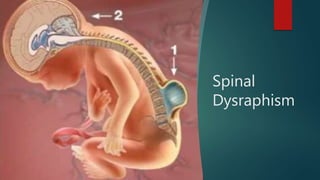
- 2. Spinal Dysraphism – Spina bifida Spectrum of congenital disorders in which there is failure of midline closure of neural, bony, or other mesenchymal tissues.
- 3. Embryology
- 5. Spinal Cord Development Three basic embryologic stages. The first stage is gastrulation 2nd or 3rd week of embryonic development. Gastrulation involves conversion of the embryonic disk from a bilaminar disk to a trilaminar disk composed of ectoderm, mesoderm, and endoderm.
- 6. The second stage - Primary neurulation (weeks 3–4) in which the notochord and overlying ectoderm interact to form the neural plate. The neural plate bends and folds to form the neural tube, which then closes bidirectionally in a zipper like manner
- 7. Neurulation and derangement of neurulation: Four stages of neurulation: 1- Formation of neural tube. 2- Shaping the neural plate. 3- Bending of the neural plate. 4- Fusion. the cranial neuropore closes approximately on day 24 and the caudal neuropore on day 28. Failure of the cranial (superior) and caudal (inferior)
- 8. The final stage - Secondary neurulation (weeks 5–6). During this stage, a secondary neural tube is formed by the caudal cell mass. The secondary neural tube is initially solid and subsequently undergoes cavitation, eventually forming the tip of the conus medullaris and filum terminale by a process called retrogressive differentiation. Abnormalities in any of these steps can lead to spine or spinal cord malformations. The retrogressive differentiation, involves programmed cell death leading to regression of the primitive distal spinal cord to form the fetal conus, filum terminale, and ventriculus terminalis
- 10. Aetiology 0.5-2 per 1,000 pregnancies worldwide. Incidence of spinal dysraphism has significantly decreased over the last few decades, all over the world Familial tendency (2.5% vs. 0.2% risk in general population) Nutritional factors; social class difference Folic acid use preconception and during pregnancy Teratogens e.g., valproate, phenytoin, alcohol etc
- 11. Classification by Tortori- Donati et al in 2000 Spina bifida cystica and aperta - Open Spinal Dysraphism (OSD) Spina bifida occulta - Closed Spinal Dysraphism (CSD) CSD is further subdivided by the presence or absence of a subcutaneous mass
- 12. Open Spinal Dysraphism (95%) (skin not intact) Myelomeningocele - Neural placode protrudes above skin surface Myelocele -Neural placode flush with skin surface Hemimyelomeningocele - Myelocele associated with diastematomyelia Hemimyelocele - Myelomeningocele associated with diastematomyelia
- 13. Closed Spinal Dysraphism (5%) (skin intact) With a subcutaneous mass Lipomyelocele – placode –lipoma interface within spinal cord Lipomyelomeningocele - placode –lipoma interface outside spinal cord Meningocele – herniation of csf filled sac lined by dura Terminal myelocystocele – terminal syrinx herniating into posterior meningocele Myelocystocele – dilated central canal herniating through posterior spina bifida Can be at cervical or lumbosacral region
- 14. Closed Spinal Dysraphism (5%) (skin intact) Without a subcutaneous mass Simple Intradural lipoma – within dural sca Filar lipoma – fibrolipomatous thicking of filum Tight filum terminale – hypertrophy and shortning of filum Persistent terminal ventricle – persistent cavity within conus medullaris Dermal sinus – epithelial lined fistula between neural tissue and skin surface Complex Diastematomyelia / Diplomyelia – separation of cord into two hemi cords Neurenteric cysts – localized form of dorsal enteric fistula Dorsal enteric fistula – connection between bowel and skin surface Caudal regression syndrome – total or partial agenesis of spinal column
- 15. Open Spinal Dysraphisms Myelomeningocele and myelocele Defective closure of the primary neural tube Exposure of the neural placode through a midline skin defect on the back > 98% of open spinal dysraphisms Myeloceles are rare. 99% are myelomeningocele Hemimyelomeningocele and hemimyelocele extremely rare . Occur when a myelomeningocele or myelocele is associated with diastematomyelia (cord splitting) and one hemicord fails to neurulate. Abnormality of gastrulation with superimposed failure of primary neurulation of one hemicord. All patients with OSD have Chiari II
- 17. Meyelomeningocele Incidence - 0.4 per 1000 live births - Racially variable 85% caudal thoraco lumbar spine, 10 % in the thorax and the rest cervical 80-90 % associated with hydrocephalus and Chiari Trisomy 13 and trisomy 18 Associated defects – Brain stem defect includes Medullary kinking, tectal beaking, and intrinsic nuclei abnormalities Supratentorial abnormalities include partial or complete dysgenesis of the corpus callosum, polymicrogyria, gray matter heterotopia. Mesodermal development of the skull small posterior fossa, short clivus, low-lying tentorium and torcular Herophili enlarged foramen magnum.
- 18. Risk factors
- 19. Prenatal diagnosis Maternal serum Alpha feto protein : initial screening test High resolution fetal ultrasonography. Can also demonstrate hydrocephalus and Chiari II abnormality (lemon and banana sign) Amniocentesis : if MSAFP and USG are suggestive Ach esterase levels along with AFP AFP can increase in other developmental anomalies of the gut and kidneys.
- 20. D/D At least 22 other fetal abnormalities besides myelomeningocele increase MSAFP levels. Abdominal abnormalities such as omphalocele, cloacal exstrophy, esophageal atresia, annular pancreas, duodenal atresia, and gastroschisis urologic abnormalities such as congenital nephrosis, polycystic kidneys, urinary tract obstruction, and renal sacrococcygeal cystic teratoma
- 21. Preop evaluation- Enteral feeding avoided to prevent fecal soiling of placode Prone position, saline dressings Neurosurgical – Sensory level determined Motor evaluation – distal most voluntary motion evaluated. Limb abnormalities documented. Anal tone and anal reflex evaluated Ventricular size documented with preop USG and NCCT head. Renal evaluation • 90 % have neurogenic bladder. • All should have preop Renal ultrasound for detecting severe anomalies. CIC if fails to void Cardiac evaluation – associated VSD / ASD
- 22. Investigation Role of MRI: Anatomic characterization. Presurgical evaluation. Identification of cord splitting when present
- 23. Surgical Management To treat or not to treat? Improving the quality of life Effectiveness of early and aggressive intervention Medical ethics and individual rights Education of the parents regarding care of the infant Role of the treating physician – Details of possible outcome The right to health and the right to life is for everyone How ever Withhold extreme measures for those with severe anomalies
- 24. Surgical Repair Timing of repair: performed safely up to 72 hours after birth Delayed repair – Increases chance of ventriculitis by 5 times shunt infection developed in about 75%, and the mortality was 13% In case Delay Cultures from the neural placode – No growth – go ahead n repair If infection +/- external ventricular drainage and appropriate antibiotics until the infection clears – Then repair
- 25. Shunt before repair ? High chance of shunt inf./ Meningitits – IQ impairment (due to inf) PREPARATION Intraoperatively avoid hypothermia, hypovolemia, and hypoglycemia A doughnut-shaped sponge - to protect the myelomeningocele while intubation If severe Hydrocephalus - CSF diversion before closure of the myelomeningocele - to minimize pressure on the myelomeningocele dural closure Entire back and flanks are prepared and draped to facilitate extensive closure if needed. Contact between povidone- iodine solution and the neural placode should be avoided
- 26. AIM : To protect the functional spinal cord tissue, prevent loss of CSF, and minimize the risk for meningitis by reconstructing the neural tube and its coverings. The margin between the arachnoid of the neural placode and the dystrophic epidermis, or the junctional zone, is the site of the initial incision. The goal is to free the neural placode from the surrounding junctional zone circumferentially. Duraplasty with thoracolumbar fascia or another dural substitute is performed when necessary to prevent leakage of CSF.
- 28. Post op care • Post op antibiotics Prevention of fecal contamination of wound Nurse in Trendlenberg’s Observe for Hydrocephalus – shunt if HCP present Complications Superficial wound dehiscence Meningitis Symptomatic chiari Ensure functioning shunt Hindbrain decompression
- 29. Cause of neuro-deterioration symptomatic hydrocephalus, syringomyelia Retethering of cord. Chiari II malformation, Risk factors - mostly due to shunt malfunction resulting in hydrocephalus
- 30. Prognosis 10 to 15% succumb <6yr of age even with Multi specialty aggressive approach >95% Lives >2 years 8 to 17 % will have urinary control rest on Drugs / CIC >87% will have social fecal incontinence L3 function allows one to stand erect, and L4 and L5 function allows ambulation During the first decade, approximately 60% of children with spina bifida are community ambulators, without or with assistive devices (including wheelchairs) – Reduces to 17% in teenagers IQ stays N if no inf. – only <10% economically independent
- 31. Closed Spinal Dysraphism (5%) (skin intact) With a subcutaneous mass Lipomyelocele – placode –lipoma interface within spinal canal Lipomyelomeningocele - placode –lipoma interface outside spinal canal Meningocele – herniation of csf filled sac lined by dura Terminal myelocystocele – terminal syrinx herniating into posterior meningocele Myelocystocele – dilated central canal herniating through posterior spina bifida Can be at cervical or lumbosacral region
- 32. lipomyelocele Lipomas with a dural defect result from a defect in primary neurulation whereby mesenchymal tissue enters the neural tube and forms lipomatous tissue. presence of a subcutaneous fatty mass above the intergluteal crease.
- 34. Chapman’s classification of LMM Type I (dorsal lipoma) Type II (transitional lipoma) Type III ( terminal lipoma)
- 35. Cystocele Myelocystocele Terminal myelocystocele No part of spinal cord enters sac, spinal cord is usually normal but tethering is common
- 36. The mass is clinically evident at birth, the diagnosis is usually made before significant neurological deterioration ensues However, infants not treated before the age of 6 months develop hyposthenia and hypotrophy of the lower limb muscles, gait disturbances, urinary incontinence, and paresthesias. Clinical features progress over time if the child is left untreated.
- 37. Clinical features Subcutaneous masses over the back Stigmata of occult dysraphism Hypertrichosis Hemangioma Hypo/ hyperpigmented patch Dermal pit or sinus Assymmetric gluteal cleft Orthopedic syndrome Limb length discrepancy, high pedal arches, hammer toes, calcaneovarus/ valgus foot deformity. Urologic syndrome Urinary incontinence, post void dribbling, urgency, frequency Intractable pain in the legs, back, pelvis or perineum.
- 38. Indication for surgical repair Asymptomatic infant older than 2 months Presence of orthopedic, pain or urologic syndrome Neurological symptoms
- 39. Principal Goal of surgery Detethering of spinal cord Decompression of the cord by removing as much lipoma as possible Reconstruct the spinal cord and dural sac Preservation of the functional tissue Surgical principles Relationship between the lipoma-cord interface and dorsal roots to be established Conservative excision of the lipoma to avoid injury to the cord/ exiting roots.
- 40. Surgical steps
- 41. Complications of surgical repair Early – CSF leak/ pseudo meningoceles & New Neurological deficit Late – retethering of the cord Mostly presenting between 3-8, 11-22 months after surgery Upto 20% cases may demonstrate retethering Diagnosis primarily clinical. Aseptic meningitis from host-graft inflammation meningitis, Intradural abscess, wound infection, and wound breakdown
- 42. Closed Spinal Dysraphism (5%) (skin intact) Without a subcutaneous mass Simple Intradural lipoma – within dural sca Filar lipoma – fibrolipomatous thicking of filum Tight filum terminale – hypertrophy and shortning of filum Persistent terminal ventricle – persistent cavity within conus medullaris Dermal sinus – epithelial lined fistula between neural tissue and skin surface
- 43. Intra dural lipoma lipoma - contained within the dural sac. No open spinal dysraphism is present. M.C site: lumbosacral - present with tetheredcord syndrome, a clinical syndrome of progressive neurologic abnormalities in the setting of traction on a lowlying conus medullaris.
- 44. Filar lipoma Fibrolipomatous thickening of the filum terminale is referred to as a filar lipoma. On imaging, a filar lipoma Normal variant if there is no clinical evidence of tethered cord syndrome Tight filum terminale is characterized by hypertrophy and shortening of the filum terminale causing tethering of the spinal cord and impaired ascent of the conus medullaris. The conus medullaris is low lying relative to its normal position that is above the L2–L3 disc level.
- 45. persistent terminal ventricle Persistence of a small, ependymal lined cavity within the conus medullaris Location immediately above the filum terminale and lack of contrast enhancement, which differentiate this entity from other cystic lesions of the conus medullaris.
- 46. Closed Spinal Dysraphism (5%) (skin intact) Without a subcutaneous mass Complex Diastematomyelia / Diplomyelia – separation of cord into two hemi cords Neurenteric cysts – localized form of dorsal enteric fistula Dorsal enteric fistula – connection between bowel and skin surface Caudal regression syndrome – total or partial agenesis of spinal column
- 47. Split cord malformations Diastematomyelia—Separation of the spinal cord into two hemicords. The two hemi cords are usually symmetric, although the length of separation is variable. Diastematomyelia can present clinically with scoliosis and Tethering Both types may be present simultaneously at different levels
- 48. Split cord Malformation Exceedingly rare Represent 3.8% to 5% of all congenital spinal anomalies. prevalence of SCM to be 1 in 5000 (0.02%) live births. slight female preponderance, approximately 1.3:1.7 The peak age is 4 to 7 years, second peak between 12 and 16 years - post pubescent growth spurt. Type I SCMs > type II lesions
- 49. Pang’s theory of embryogenesis Formation of abnormal fistula through midline embryonic disc that maintains communication b/w Yolk sac & amniotic cavity contact b/w ecto + Endoderm This fistula causes splitting of Notochord & overlying neural plate
- 50. Associated Anomaly Tethered/low-lying cord (>50%), Kyphoscoliosis (44% to 60%), Syringomyelia (27.5% to 44%), Spina bifida (11% to 26%) “faun’s tail,” consists of a patch of unusually coarse, raised hair - strong association with type I SCM. Capillary hemangioma underlying these hairy patches. 50% of patients have gross (i.e., structural) asymmetry of the lower extremities- - neuro-orthopedic syndrome. characterized by a triad of 1. limb length discrepancy, 2. muscular atrophy (resulting in secondary weakness), and 3. clubfoot deformity (talipes equinovarus). The smaller limb is often ipsilateral to a smaller hemicord.
- 51. management 85% of patients without intervention suffer from a progressive neurological deficit versus only 4.5% after surgical treatment Pang too suported prophylactic surgery in Type 1 malformation.. Whereas in Type 2 both- some – W&W policy Surgery – careful – incision 1 to 2 level more exposure Surgical detethering of cord by excision of the bony spur/ division of the fibrous bands Avoid damage to the hemi cords during excision of the spur. For type I SCM, the initial laminectomies should be limited to adjacent levels while initially avoiding exposure of dura at the level of the midline bony spur. Rongeurs or a high-speed drill (or both) - to perform bilateral paramedian laminectomies ( preserve the midline lamina and spinous process to prevent any torque or lateral force from disrupting the bony spur prematurely.)
- 52. Careful when removing lamina at the level of the SCM in type II malformations because of the frequent presence of transdural adhesions - most commonly attached dorsally but ventrally too. Often, the dura at this level grossly abnormal. All non-neural and non- functional adhesive bands should be transected, beginning dorsally and then gently rolling the hemicords to one side and transecting any ventral attachments Hemicords are typically closely approximated with no clear intervening plane. Resection of the fibrous band within the split spinal cord itself is not indicated. Any associated tethering lesion (sinus tract, fatty filum, or terminal lipoma) should also be addressed
- 53. Neurenteric cysts Persistent neurenteric canal communicating between yolk sac and amniotic cavity Intradural, extramedullary mucosa lined cysts Formed from persistents tracts communicating with respiratory and gut epithelia. These cysts are lined with mucin secreting epithelium similar to the GIT and located in the cervicothoracic spine anterior to the spinal cord Associated with vertebral anomalies MRI- demonstrates non- contrast enhancing intradural extramedullary cyst Presentation usually in late years (50-60 years) • May also present in pediatric age group Complete excision of cyst – long term symptom free survival.
- 54. Dermal sinus tracts Abnormal tracts communicating between the skin and intraspinal compartment. Most common- lumbosacral location May occur anywhere from nasion to coccyx in midline May be accompanied by other cutaneous stigmata. Tract terminates within thecal sac mostly Half may have associated dermoids, epidermoids, teratoma at termination. Potential pathway for spread of infection Repeated episodes of meningitis with atypical organisms Operative repair consists of complete excision of the track under prophylactic antibiotic cover. Gram positive and gram negative coverage
- 55. Dermal sinus
- 56. Recent Advancements • Foetal MMC repair is an advancing development but no definitive data exists The Management of Myelomeningocele Study (MOMS trial)1, a 7-year, multi-center, randomized clinical research trial published in March 2011, was the first to comprehensively describe the outcomes of open fetal surgery for repair of spina bifida, compared to a traditional postnatal repair. Results are favourable in decreasing neurologic deficits and reducing the occurrence of CM II and hydrocephalus, improved motor outcomes at 30 months versus postnatal repair, but was also associated with maternal and fetal risks Done at 23 weeks and 25 weeks 6 days gestation No final consensus or guidelines; still experimental
- 57. Perls Periconceptional folic acid intake results in a 42% relative risk reduction in the incidence of first occurrence of spina bifida Periconceptional folate results in a 72% relative risk reduction in the recurrence of spina bifida in subsequent children In patients with lumbosacral dimples, US exam is more cost effective than MRI in screening for occult spinal dysraphism the anomaly could not be eradicated due to its multifactorial nature
- 58. Approximately 95% of couples that have a fetus affected with ONTD have a negative family history. Whenever the conus lies below the L2-3 interspace in an infant, cord tethering should be considered. Patients with spina bifida occulta may present with scoliosis in later years Spina bifida occulta is characterized by variable absence of several neural arches and various cutaneous abnormalities, such as lipoma, hemangioma, cutis aplasia, dermal sinus, or hairy patch, and it is often associated with a low- lying conus open forms are often associated with hydrocephalus and Arnold-chiari malformation type II – requires surgical intervention after proper counceling
- 59. Neural tube defects (NTDs) exact emotional and economic toll on families and health care systems The tragedy is that NTDs are preventable simply by having women take a folic acid supplement during the 2 months before they become pregnant. 0.4 mg daily before conception and for the first 3 months of pregnancy, reduces the risk of having a baby with spina bifida.