Posttraumatic radio ulnar synostosis
•Download as PPTX, PDF•
0 likes•230 views

Very rare complication for radial head fracture and even other trauma (elbow dislocation, radius/cubitus shaft fracture,…) Lost of range of motion and specifiquely for pronosupine Lot of treatment in litterature as: - preventive irradiation - resection without interposition - resection/interposition - resection wo interposition + irradiation - resection/interposition + irradiation Still no consensus

Recommended
More Related Content
What's hot
What's hot (20)
Similar to Posttraumatic radio ulnar synostosis
Similar to Posttraumatic radio ulnar synostosis (20)
More from ROBERT ELBAUM
More from ROBERT ELBAUM (20)
Recently uploaded
Recently uploaded (20)
Posttraumatic radio ulnar synostosis
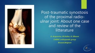
- 1. Post-traumatic synostosis of the proximal radio- ulnar joint: About one case and review of the litterature R. Acquaviva, M.Hallez, R. Elbaum CHIREC orthopaedic group Brussel,Belgium
- 2. The only man who never makes mistakes is the one who does nothing. THEODORE ROOSEVELT (1858 - 1919)
- 3. Introduction Very rare complication for radial head fracture and even other trauma (elbow dislocation, radius/cubitus shaft fracture,…) or after forearm osteotomy Lost of range of motion (Prosupination) Lot of treatment in litterature as: - preventive irradiation - resection without interposition - resection/interposition - resection wo interposition + irradiation - resection/interposition + irradiation Still no consensus
- 4. Case report Zoe 8 years old Radial head fracture at the age of 7 years old (08/2015) Fracture: Salter– Harris 2-3 Conservative treatment started at trauma date
- 5. First visit on 01/09/2016 Pain in the forearm and the elbow Limited ROM Functional disability to draw and write Limitation PS 30/0/30 Extension -30° Elective pain on radial head
- 7. MRI
- 8. Treatment Open surgery: Boyd Approach Resection of synostosis Bank fascia lata interposition AINS post- operative Cast for 3 weeks PT
- 13. Post-operative follow-up 1 w post-op : wound control + BAB 3 w post-op: remove cast + self kine 7 w post-op: • - PS > 60/45 • - Rx showing resection zone • - start kine
- 14. POST OP 12/2016
- 15. CT Scan 27/03/17 Complete resection of the synostosis
- 16. POST OP 04/2017
- 17. Anterior approach (Henry) Abord antérieur du coude selon Henry avec bifurcation dans l’interligne du coude. Mise en évidence du brachial antérieur et du long supinateur. Ligature des différents vaisseaux superficiels. Mise en évidence du nerf radial qui sera laqué. On se dirige progressivement vers l’interligne articulaire radio-ulnaire proximal. Désinsertion du court supinateur et ouverture de la capsule articulaire. Mise en évidence de la tête radiale. On visualise une zone protubérante au niveau de la région sigmoïdienne entre l’ulna et le radius.
- 18. LAST FU 11/2018 No pain Complete RTS (Ballet) No limitation at school PS 45/0/60 Temporary paraparesy in the radial territory (Extensor)
- 19. Post traumatic synostosis of proximal(PPRUS) radio ulnar joint in childhood: How to deal with it?
- 20. PPRUS Classification Vince and Miller classification Vince K. G., Miller J. E. Cross union complicating fracture of the forearm. The Journal of Bone & Joint Surgery—American Volume. 1987;69:640–653.
- 21. PPRUS Etiology Proximal Radial or Ulnar fracture or dislocations After closed or open reduction Periostal interpositions Surgical approach with traumatisation of the tissue Repeated manipulation Excessive callus formation • Isolated fractures of the proximal end of the radius in children epidemiology, treatment and prognosis. Henrikson B Acta Orthop Scand. 1969; 40(2):246-60. • Injuries involving the proximal radial epiphysis. O'Brien PIClin Orthop Relat Res. 1965 Jul-Aug; 41():51-8. • Severe fracture of the neck of the radius in children. Dougall AJ J R Coll Surg Edinb. 1969 Jul; 14(4):220-5. • Radioulnar synostosis following proximal radial fracture in child. Roy DR Orthop Rev. 1986 Feb; 15(2):89-94.
- 22. PPRUS Epidemiology 1969 • Acta Orthop Scand. 1969;40(2):246-60. Isolated fractures of the proximal end of the radius in children epidemiology, treatment and prognosis. Henrikson B. 1982 • Ogden JA. Skeletal injury in the child. Philadelphia: Lea and Febiger 1997 • von Laer L, Pirwitz A, Vocke AK. Posttraumatic problem cases involving the elbow in children. Orthopade 1997;26:1030-6 2012 • Wierer M, Huber-Wagner S, Mutschler W. Post-traumatic proximal radioulnar synostosis. Surgical technique and review of the literature. Unfallchirurgie 2012;115:451-6 1900 • Mouchet A. Les fractures du col du radius. Chirurgie 1900;21:596-622
- 23. Case Rep Orthop. 2018 Feb 19;2018 Posttraumatic Proximal Radioulnar Synostosis after Closed Reduction for a Radial Neck and Olecranon Fracture. Keller PR, Cole HA, Stutz CM, Schoenecker JG Here, we present a pediatric case of PPRUS that developed after a nonoperatively treated minimally displaced radial neck fracture with concomitant olecranon fracture. While more cases are needed to establish the association between this pattern of injury and PPRUS, we recommend that when encountering patients with a minimally displaced radial neck fracture and a concomitant elbow injury, the rare possibility of developing proximal radioulnar synostosis should be considered.
- 24. PPRUS Treatment option Roy DR. Radioulnar synostosis following proximal radial fracture in child. Orthop Rev 1986;15:89-94 Aner A, Singer M, Feldbrin Z, et al. Surgical treatment of posttraumatic radioulnar synostosis in children. J Pediatr Orthop 2002;22:598-600 Kamineni S, Maritz NG, Morrey BF. Proximal radial resection for posttraumatic radioulnar synostosis: a new technique to improve forearm rotation. J Bone Joint Surg Am 2002;84-A:745-51 Proubasta IR, Lluch A. Proximal radioulnar synostosis treated by interpositional silicone arthroplasty. A case report. Int Orthop 1995;19:242-4 Abrams RA, Simmons BP, Brown RA, Botte MJ. Treatment of posttraumatic radioulnar synostosis with excision and low-dose radiation. J Hand Surg Am 1993;18:703-7 Friedrich JB, Hanel DP, Chilcote H, Katolik LI. The use of tensor fascia lata interposition grafts for the treatment of posttraumatic radioulnar synostosis. J Hand Surg Am 2006;31:785-93
- 25. Orthop Res Rev. 2017 Dec Optimal management of post-traumatic radioulnar synostosis. Osterman AL, Arief MS. Post-traumatic radioulnar synostosis is a rare complication after forearm or elbow injury that can result in loss of motion and significant disability. Risk factors include aspects of the initial trauma and of the surgical treatment of that trauma. Surgical intervention for synostosis is the standard of care and is determined based on the location of the bony bridge. Surgical timing is recommended between 6 months and 2 years with recent advocacy for the 6- to 12-month period after radiographs demonstrate bony maturation but early enough to prevent further stiffness and contractures.
- 26. Aktuelle Traumatol. 1986 Feb;16(1):13-6. [Post-traumatic radio-ulnar synostoses in childhood]. Bätz W, Hofmann-v Kap- herr S, Pistor G. This article reports on five children treated surgically during 1973-1975 for posttraumatic radioulnar synostosis. The article presents the technique of surgical treatment of proximal radioulnar synostosis with Lyodura sheathing. Follow-up examination showed good results in two children, a moderate result in one, and poor results of surgery in two cases. Improved results may be expected from further improvement of the surgical method, such as resection of the bicipital tuberosity (tuberositas radii) or from additional partial sheathing of the ulna at the side facing the radius.
- 27. Aust N Z J Surg. 1993 Dec Post-traumatic radio-ulnar synostosis treated by surgical excision and adjunctive radiotherapy. Thurston AJ, Spry NA. The management of three cases of traumatic radio-ulnar synostosis involved surgical excision of the synostotic bone followed by radiotherapy. Irradiation was commenced on the first postoperative day and was continued daily. No acute side effects were observed. All three patients regained a good, functional range of forearm rotation with no evidence of recurrence of the synostosis after 2 years.
- 28. J Bone Joint Surg Am. 2002 May. Proximal radial resection for posttraumatic radioulnar synostosis: a new technique to improve forearm rotation. Kamineni S, Maritz NG, Morrey BF. Resection of a 1-cm-thick section of the proximal part of the radial shaft provides a safe and reliable method of improving forearm rotation in patients with heterotopic ossification of the elbow. A single technical factor that seems to positively influence the result is the application of bone wax at the resection site. This simple procedure is ideally suited for patients who have a proximal radioulnar synostosis that (1) is too extensive to allow a safe and discrete resection, (2) involves the articular surface, and (3) is associated with an anatomical deformity.
- 29. J Pediatr Orthop. 2002 Sep- Oct;22(5):598-600. Surgical treatment of posttraumatic radioulnar synostosis in children. Aner A, Singer M, Feldbrin Z, Rzetelny V, Bar-On E. The authors describe two children who underwent surgical treatment of radioulnar synostosis. One case involved simple excision; the other, excision and interposition of Gore-Tex vascular graft material. In a review of the literature, no other report of the latter type of surgical treatment was found. A discussion of the literature concerning this rare complication in children and the current surgical treatment options are included.
- 30. Conclusion The Posttraumatic Proximal Cross-union of the Forearm in Childhood: What is Recommended? Marcel Dudda, Tobias Fehmer, Thomas A. Schildhauer, and Christiane Kruppa Orthop Rev (Pavia). 2013 Jun. Postoperative cross-unions are rarely seen in the pediatric population and there is no consensus as to treatment. Compared to the adult population, worse results in pediatric diaphyseal (Type 2) cross-unions are reported. We suggest resection within 6-24 months without necessarily an interposition technique. For delayed treatment, resection of the radial head as salvage procedure can be performed. We advocate postoperative oral therapy with NSAID in all patients and irradiation in cases of delayed treatment of cross-unions; All patients should be treated with intensive physiotherapy and continuative postoperative follow up to prevent a loss of range of motion.
- 31. OSCAR WILDE (1854-1900) Experience is the name everyone gives to his mistakes.
Editor's Notes
- Ladies and Gentlemen, Dear Colleague, I would like thanks the organising comitee and especially Dr Zorman and Molenaers for this intersting Congress about Complications in Orthopaedics
- As we know, complications in orthopaedics related to surgery or to the pathology represent a source of anxiety and worry for all the surgons, especially when the lawyers get involved. Especially in Paediatric Orthopaedics where our action have a consequence in the very long term of our patient. But as Theodore Roosvelt have sayed …
- Many complication after elbow fracture are describe but posttraumatic Radio Ulnar synostosis are very rare and often unpredictible. It is caracterised by a loss of motion in the elbow ans specifically by loss pf Prosupination Many treatment are proposed in the littérature but because of this rare condition, there is no consensus
- , Zoe; is a 8 year old girl who present at our clinic 1 year after an radial head fracture on his left elbow.
- She complaint of Pain in the forearm and the elbow and especially when she was writing or drawing She had limitation in Prosupination and in extension The Xray done in Romania showed a proximal coalition of the radioulnar joint Unfortunally I could’nt get this X-Ray
- A Ct Scan with 3D Reconstruction showed this complete coalition between the sigmoidienne fossette of the ulna distally and proximal part of the radius under the epiphysis. There is also a fusion of the epiphyseal nucleus of the radial head, small,
- On the MRI the upper part of the radius appears enlarged and largely fused with the ulna. The fusion between the radius and the ulna is about 12 mm in height. This injury is made proximally to the bicipital tuberosity
- In front of the complain of the patient, the disability and the delay between the first trauma and the coalition ,Surgery was proposed. We perform the resection of the synostosis by a Boyd approach and interposition of Fascia lata. In postop we prescribe NSAI therapy and active mobilisation after cast .
- Here on the Perop view, you can see the radiohumeral joint after detaching the epitrochlean muscle and opening the capsula Boyd approach was choosed to avoid neurovascular damage
- By this approach we could reach almost all the coalition and you can see on the right image the margin of the bony coalition. First the resection was done with muller cissels and then with a little bur
- Here is the bony coalition resected
- On this video you cans see the moblity obtain after resection
- After 3week of immobilisation in a sling ,she start selfkine ans active mobilisation At 7W post op Prosupination was 60/45
- XRAY showed the almost complete resection
- 4mo postop there is a growth disturbance on the radial proximal epiphysis
- Unfortunatly 6 mo lather we assist to partial recurrence of the synostosis especially on tne anterior part . So we decide after discussion a lot with the parent about the neuro vascular risk , to complete the resection by an anterior approach
- On last FU Zoe has improved his mobility. She return to sport. As we could expect ,she present a temporary paraparesy in the radial teritory .
- PPRUS in children is a rare condition and not so easy to manage. Litterature is not so profuse and we have try to do a global review about this complication
- Vince and Miller published in 1987 a classification about posttraumatic coalition of the PRUJ for adult. In our case it was a type 3, the most unfrequent type.
- Concerning etiology ,many condition and agravating factor are proposed. It can occure after Radio Ulnar fracture or dislocation like the Monteggia Type. Closed or openreduction are equally involved but surgical approach with tissue injuries can increase bony formation. If there is a periostal interposition or after repeated manipulation Excessive callus formation that occures frequently in children fracture may also be an etiology
- PPRUS represent 2% of adult population with forearm fracture but there is not a precise incidence in childhood but it is very law The first report of PPRUS in children was performed by Mouchet in 1900 Few case were reported since this in the littérature.
- Recently Keller and coll published one case exactly the same than ours and recommend…
- Concerning management of PPRUS in children , littérature is not so profuse and i will present some article concerning this subject
- In 1986 five case of PPRUS treated with Lyodura interposition. They suggested that resection of bicipital tuberosity or sheating of the ulna may improved the result
- Thurston in 1993 published 3 case of PPRUS treat by adjunctive radiotherapy starting in the first postoperative day. There was no evidence of recurrence after 2 years
- Kamieni and coll describe a technique resecting a 1cm thick section of the radial sahft to improve forearm rotation. According to him this procedure is indicated when the PPRUS is to extensive and involve the articular surface.
- In 2002 Aner and coll describe 2 case using in one , interposition of gore tex between the resected coalition.
- In conclusion