Ultrasound-guided pudendal nerve pulsed radiofrequency in patients with refractory pudendal neuralgia
•
0 likes•16,387 views

Pulsed Radiofrequency Treatment (PRF) of the Pudendal Nerve is discussed.

Recommended
Recommended
More Related Content
What's hot
What's hot (20)
Similar to Ultrasound-guided pudendal nerve pulsed radiofrequency in patients with refractory pudendal neuralgia
Similar to Ultrasound-guided pudendal nerve pulsed radiofrequency in patients with refractory pudendal neuralgia (20)
More from Jason Attaman
More from Jason Attaman (14)
Recently uploaded
Recently uploaded (20)
Ultrasound-guided pudendal nerve pulsed radiofrequency in patients with refractory pudendal neuralgia
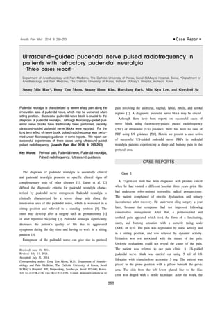
- 1. Ultrasound-guided pudendal nerve pulsed radiofrequency in patients with refractory pudendal neuralgia -Three cases report- Case 1
- 2. Fig. 3. The location of the needle tip checked by fluoroscopy. FH: femoral head, IS: ischial spine. Fig. 2. Doppler image showing the internal pudendal artery at the level of the ischial spine. The open arrow indicates the internal pudendal artery, the closed arrow indicates the pudendal nerve. The arrowheads indicate the needle. SSL: sacrospinous ligament, STL: sacrotuberous ligament, IS: ischial spine, GM: gluteus maximus. Fig. 1. Ultrasound-guided pudendal nerve radiofrequency. The transducer is placed transversely over the ischial spine. The needle electrode is advanced laterally with the in-plane approach. Case 2
- 3. Fig. 4. Ultrasound image of pudendal nerve radiofrequency. The needle is advanced near the pudendal nerve. The open arrow indicates the internal pudendal artery, the closed arrow indicates the pudendal nerve. The arrowheads indicate the needle. SSL: sacrospinous ligament, STL: sacrotuberous ligament, IS: ischial spine, GM: gluteus maximus. Case 3