1. Chronic Lymphocytic Leukemia appearing as skin lesions
Gonçalves Estevens, J.; Rodriguez Vera, J.; Vylchez, J.; Ferreira, Mª L.; Arez, L.
Internal Medicine Service – Sector I
Centro Hospitalar do Barlavento Algarvio - Portugal
We present the case of a man who IDENTIFICATION
consulted to the Emergency Department 75 years old man, caucasian. Agricultural worker, resident in a country area, in Algarve.
for a bleeding lesion with spontaneous
intermittent remission in the frontal
region from 4 months. (fig. 1) HISTORY OF PRESENTING PROBLEM
Bleeding lesion with spontaneous intermittent remission in the frontal region from 4 months.
He also referred having found not enlarged lymph nodes in the axilar and cervical regions from one month..
painful enlarged lymph nodes in the
axilar and cervical regions from one
month..
Blood test taken at admission showed a
leucocytosis of 133.200/ µl with
lymphocytosis; β2-microglobulin and
LDH elevated.
Peripheral blood smear showed
Gumprecht nuclear shadows.
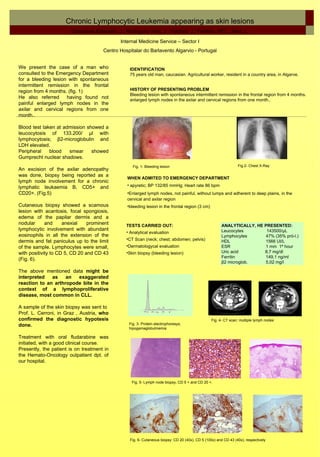
Fig. 1- Bleeding lesion Fig.2- Chest X-Ray
An excision of the axilar adenopathy
was done, biopsy being reported as a
WHEN ADMITED TO EMERGENCY DEPARTMENT
lymph node involvement for a chronic
lymphatic leukaemia B, CD5+ and • apyretic; BP 132/85 mmHg; Heart rate 86 bpm
CD20+. (Fig.5) •Enlarged lymph nodes, not painful, without lumps and adherent to deep plains, in the
cervical and axilar region
Cutaneous biopsy showed a scamous •bleeding lesion in the frontal region (3 cm)
lesion with acantosis, focal spongiosis,
edema of the papilar dermis and a
nodular and anexial prominent TESTS CARRIED OUT: ANALYTICALLY, HE PRESENTED:
lymphocytic involvement with abundant Leucocytes 143500/µL
• Analytical evaluation
eosinophils in all the extension of the Lymphocytes 47% (35% pró-l.)
•CT Scan (neck; chest; abdomen; pelvis)
dermis and fat paniculus up to the limit HDL 1566 UI/L
of the sample. Lymphocytes were small, •Dermatologycal evaluation ESR 1 mm 1ª hour
with positivity to CD 5, CD 20 and CD 43 •Skin biopsy (bleeding lesion) Uric acid 8,7 mg/dl
Ferritin 149,1 ng/ml
(Fig. 6).
β2 microglob. 5,02 mg/l
The above mentioned data might be
interpreted as an exaggerated
reaction to an arthropode bite in the
context of a lymphoproliferative
disease, most common in CLL.
A sample of the skin biopsy was sent to
Prof. L. Cerroni, in Graz , Austria, who
confirmed the diagnostic hypotesis Fig. 4- CT scan: multiple lymph nodes
done. Fig. 3- Protein electrophoresys;
hipogamaglobulinemia
Treatment with oral fludarabine was
initiated, with a good clinical course.
Presently, the patient is on treatment in
the Hemato-Oncology outpatient dpt. of
our hospital.
Fig. 5- Lymph node biopsy, CD 5 + and CD 20 +.
Fig. 6- Cutaneous biopsy: CD 20 (40x), CD 5 (100x) and CD 43 (40x), respectively