Call Girls Horamavu WhatsApp Number 7001035870 Meeting With Bangalore Escorts
Pet mr poster-4-15-2012_v2[1]
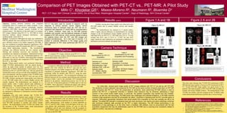
1. Comparison of PET Images Obtained with PET-CT vs.. PET-MR: A Pilot Study
Millo C , Khorjekar GR , Maass-Moreno R , Neumann R , Bluemke D
1 2 3 3 3
PET / CT Dept, NIH Clinical Center1(NIH), Div of Nucl Med, Washington Hospital Center2 , Dept of Radiology, NIH Clinical Center3
Abstract Introduction Results cont’d Figure 1 A and 1B Figure 2 A and 2B
Objective: Hybrid positron emission and computed PET-CT has been used for number of reasons, such as Artifacts causing poor image quality were observed in two
tomography (PET-CT) scanners are widely available. A new diagnosis of various cancers, cancer staging, cancer PET-MR studies due to urinary bladder activity and metallic
hybrid modality, positron emission tomography magnetic recurrence, fever of unknown origin etc. PET can be co- implants.
resonance (PET-MR), became recently available in few registered with diagnostic CT and/or MR for characterization The biodistribution was identical in 11 paired studies,
medical centers. The objective of this pilot study is to compare of a lesion. However, there was no PET-MR scanner while 5 studies showed difference in biodistribution for example
PET images obtained using PET-CT vs.. PET-MR for lesion available until recently, and the National Institutes of Health changes in the GI distribution urinary bladder activity etc. The
detection, artifacts, altered biological distribution, and installed a PET-MR scanner, which recently started enrolling individual SUVs for PET-CT vs.. PET-MR were 23.0 vs.. 21.0 for
standard uptake values (SUV’s). the patients where both the PET and MR images are average max SUV, and 2.7-121.0 vs.. 2.7-90.7 for max SUV
Method: A prospective IRB-approved study was conducted at acquired simultaneously. As a new type of scanner, it is range, tentatively these differences are attributed to differences in
NIH for patients who were referred for PET-CT scan and also prudent to evaluate difference in the image quality and attenuation correction methods and uptake time.
gave informed consent to be further imaged by PET-MR, if lesion detection on PET when performed with PET-CT vs.
this was considered of potential diagnostic significance. PET- PET-MR.
MR was performed within 90 minutes after PET-CT, using the
same initial radioisotope injection. The reconstruction Camera Technique
technique was different between the two scanners (post-
filtering and resolution recovery), but consistent. SUV
Objective
Table I Table II
calculation was identical in all patients. Administered activity To compare PET images obtained using PET-CT vs.. PET-
Specifications for PET-CT Specifications for PET-MR Coronal Coronal
was 9.4 mCi to 16.2 mCi for 18F-fluorodeoxyglucose (18F- MR for lesion detection, artifacts, altered biological distribution,
Acquisition Acquisition and Processing
FDG) and 12.3 mCi for 18F-DOPA. Three physicians evaluated and standard uptake values (SUV’s).
all images with side-by-side comparison of PET images from
PET-CT vs.. PET-MR. Siemens mMR
Results: Fifteen paired studies were performed following a Method Siemens mCT
Acquisition: 3 min/bed Acquisition: 5 min/bed
single (18F-FDG) administration with one patient having a Reconstruction: High Reconstruction: iterative (3/24)
A prospective IRB-approved study was conducted at NIH for
follow up study (Total number of patients were 14). One Definition PSF, 2.65 mm MR: 3T
patients who were referred for PET-CT scan and also gave
patient had 18F-DOPA administration. Total number of lesions isotropic pixels Acquisition: 3D Dixon images,
informed consent to be further imaged by PET-MR, if this was
identified on PET-CT and PET-MR were >80 (one patient had CT: 120keV, 56mAs, axial STIR, SSSFE
considered of potential diagnostic significance. PET-MR was
innumerable lesions, all seen on both of the modalities), and all reconstruction: FBP
performed within 90 minutes after PET-CT, using the same
sites were identical, with the exception of the 18F-DOPA study initial radioisotope injection. The reconstruction technique was
in which lesions seen on the PET-CT were not seen on the different between the two scanners (post-filtering and resolution
PET-MR. Artifacts causing poor image quality were observed
in two PET-MR studies due to urinary bladder activity and
recovery) (Table 1 and 2), but consistent. SUV calculation was Conclusions
metallic implants. The biodistribution was identical in 11
identical in all patients. Administered activity was 9.4 mCi to
16.2 mCi for 18F-fluorodeoxyglucose (18F-FDG) and 12.3 mCi for
Discussion This pilot study indicates that 18F-FDG PET performed on PET-
paired studies, while 5 studies showed some difference in 18
F-DOPA. Three physicians evaluated all images with side-by- MR detected same number of lesions as PET performed on PET-
biodistribution. The individual SUVs for PET-CT vs.. PET- Our initial data showed good image quality of PET images obtained with PET-CT as well as with PET-MR.
side comparison of PET images from PET-CT vs.. PET-MR. CT. Overall PET-MR image quality was good except in few
MR were 23.0 vs.. 21.0 for average max SUV, and 2.7-121.0 The sensitivity of lesion detection was preserved for PET-MR as the same number of lesions were seen on PET-MR
vs.. 2.7-90.7 for max SUV range, tentatively these differences patients with reduced quality of PET from PET-MR secondary to
when compared with PET-CT. Given the preservation of sensitivity, when performing PET/MR without a need to metallic and urinary bladder artifacts. The biodistribution of the
are attributed to differences in attenuation correction methods
and uptake time. Results perform the PET-CT, a significant reduction in radiation dose exposure may be achieved, addressing a major
concern in the current public health arena. In particular, the pediatric population may benefit from this modality.
18
F-FDG was similar, with the exception of the 18F-DOPA study
which demonstrated differences possibly due to lesion biology.
Conclusion: This pilot study indicates that 18F-FDG PET However, as with any new technology, PET/MR poses new challenges and potential limitations. A PET/MR machine
performed on PET-MR detected same number of lesions as Fifteen paired studies were performed following a single involves a high equipment cost that not all facilities can afford. Expertise in operating both the PET and MR gantry is
PET performed on PET-CT. Overall PET-MR image quality (18F-FDG) administration with one patient having a follow up required. Particularly appropriate indications that would benefit from evaluation by this new modality are still to be
was good except in few patients with reduced quality of PET study. The total number of patients was 14. One patient had 18F- defined. The added MR strength in evaluating head and neck structures, liver lesions and pelvic pathology combined References
from PET-MR secondary to metallic and urinary bladder DOPA administration. Total number of lesions identified on with functional metabolic data provided by the PET is highly informative. The MR limited performance in the 1. Eiber M, Martinez-Möller A, Souvatzoglou M, Holzapfel K, Pickhard A, Löffelbein D, Santi I,
artifacts. The biodistribution of the 18F-FDG was similar with PET-CT and PET-MR ( Figure 1A,B and Figure 2A,B) was > 80 assessment of lung pathology is, on the other hand, a limitation. MR artifacts, more so encountered with the fast Rummeny EJ, Ziegler S, Schwaiger M, Nekolla SG, Beer AJ. Value of a Dixon-based MR/PET
the exception of the 18F-DOPA study, which demonstrated (one patient had innumerable lesions, all seen on both the acquisition sequences that may be preferentially used for the PET/MR due to scanning time constraints related to
attenuation correction sequence for the localization and evaluation of PET-positive lesions.
Eur J Nucl Med Mol Imaging 2011;38(9):1691-701.
differences possibly due to lesion biology. modalities), and all sites were identical, with the exception of patient’s comfort, may also be a challenge.
2. N. F. Schwenzer, H. Schmidt and C. D. Claussen. Whole-body MR/PET: applications in
abdominal imaging. Abdominal Imaging 2011; 37 (1): 20-28.
the 18F-DOPA study in which lesions seen on the PET-CT were 3. Delso G, Fürst S, Jakoby B, Ladebeck R, Ganter C, Nekolla SG, Schwaiger M, Ziegler SI.
not seen on the PET-MR. Performance Measurements of the Siemens mMR Integrated Whole-Body PET/MR Scanner. J
Nuc Med 2011; 52:1-9